. 2020 Jan 17;11(7):1800-1807.
doi: 10.7150/jca.39562. eCollection 2020.

Cytokine TNF-α promotes invasion and metastasis of gastric cancer by down-regulating Pentraxin3

Affiliations

Cytokine TNF-α promotes invasion and metastasis of gastric cancer by down-regulating Pentraxin3

Xinye Cui et al. J Cancer. .

Abstract

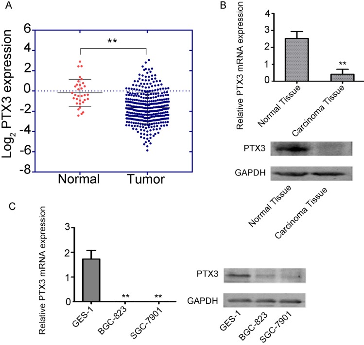
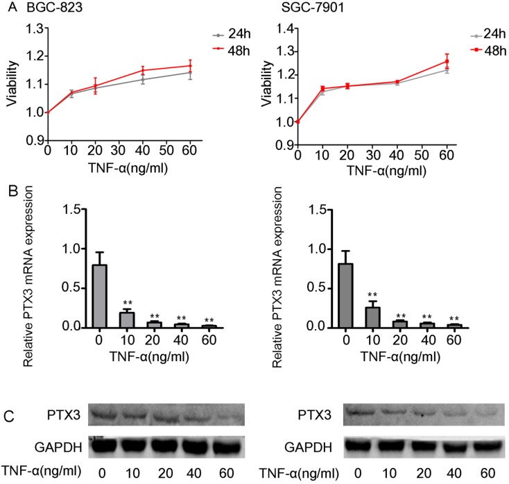
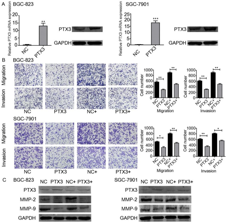
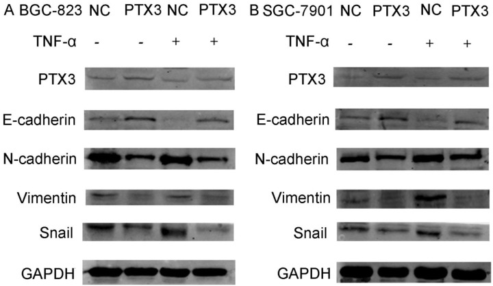
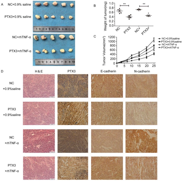
As a novel multifaceted player in cancer, Pentraxin3(PTX3) was recognized to be a possible factor related with tumor development. Recent researches have indicated that PTX3 is involved in immune response, inflammation, as well as cancer, and is greatly controlled by numerous cytokines. Tumor necrosis factor (TNF-α) is an imperative cytokine that demonstrates an extensive array of biological consequences in gastric cancer advancement. Here, we inspected the expression of PTX3 in gastric carcinoma tissues along with gastric cell lines and established that PTX3 was suggestively inferior in gastric cancer tissue and cells. The treatment of the gastric cell lines BGC-823 as well as SGC-7901 with rhTNF-α caused substantial decrease in the expression of PTX3. Furthermore, PTX3 controlled the capability of cell migration, invasion as well as epithelial-mesenchymal transition (EMT) in gastric cancer cell lines mediated by TNF-α. Additionally, PTX3 upregulation inhibited tumorigenicity in vivo and could be reversed by exogenous TNF-α. Conversely, overexpression of PTX3 inhibited progress both in vitro as well as in vivo in gastric cancer mediated by TNF-α. Further studies are necessary to demonstrate the mechanism of interaction between PTX3 and cytokines.

Keywords: EMT; Gastric cancer; Milky spot; PTX3; TNF-α.


Conflict of interest statement

Competing Interests: The authors have declared that no competing interest exists.

Figures

Figure 1
Figure 1
Low expression of PTX3 in human gastric cancer. A. Cancer with PTX3 transcripts evidently had lower manifestation (n = 375) compare to normal gastric tissues (n = 32) from the TCGA database (** P < 0.01). B. PTX3 expression levels were detected in the gastric cancer tissue as well as the adjoining normal tissue samples via qRT-PCR and Western blotting (**P<0.01). C. PTX3 manifestation levels were identified in human normal gastric epithelial cells (GES-1) and human gastric cancer cells (BGC-823 and SGC-7901) via qRT-PCR and western blotting (**P < 0.01).
Figure 2
Figure 2
Effects of exogenous TNF-α on the expression of the PTX3 in gastric cancer cells. A. The viability assay of rhTNF-α on the BGC-823 as well as SGC-7901 cells through the CCK-8 assay.There had no inhibitory and toxic effects on the two cells at 24 hours and 48 hours at different concentrations of rhTNF-α (0,10,20,40,60ng/ml). B.Five different concentrations (0,10,20,40,60ng/ml) of rhTNF-α were added to BGC-823 as well as SGC-7901 cells. Both cells displayed suggestively reduced expression of PTX3 in a concentration-dependent manner (0,10,20,40,60ng/ml) via qRT-PCR and western blotting.
Figure 3
Figure 3
Effect of TNF-α on PTX3 in the migration as well as invasion of gastric cancer cell lines. A. BGC-823 as well as SGC-7901 cells were transfected with PTX3 upregulation or a negative control (NC) plasmid. Western blotting as well as qRT-PCR of PTX3 upregulation efficiency. B. Representative images from cell migration and invasion assay after treated by rhTNF-α(20ng/ml) in PTX3 upregulation cells and NC cells. C. Western blotting investigation of PTX3, MMP-2, MMP-9 in PTX3 upregulation cells and NC cells after treated with rhTNF-α(20ng/ml) for 48h.
Figure 4
Figure 4
Protein levels of PTX3, E-cadherin, N-cadherin, Vimentin and Snail as detected by western blot in gastric cancer cells after the NC group or the PTX3 overexpression group was treated with rhTNF-α(20ng/ml) for 48 h. GAPDH served as a loading control.
Figure 5
Figure 5
TNF-αreversed the inhibitory effect of tumorigenicity mediated by upregulation PTX3 on EMT in vivo. A. Descriptions of tumor xenografts and B tumor weight for tumor xenografts treated with 0.9% saline or rhTNF-α. **P < 0.01. C. Tumor growth curves were sketched up to day 25. D. Immunohistochemical detection of PTX3, E-cadherin, N-cadherin in tumor xenografts.

References

    1. Smith MG. et al. Cellular and molecular aspects of gastric cancer. World J. Gastroenterol. 2006;12:2979–2990. - PMC - PubMed
    1. Liu J, Geng X, Li Y. Milky spots: omental functional units and hotbeds for peritoneal cancer metastasis[J] Tumor Biology. 2016;37(5):5715–5726. - PMC - PubMed
    1. Berberich S, Dahne S, Schippers A, Peters T, Muller W, Kremmer E, Forster R, Pabst O. Differential molecular and anatomical basis for B cell migration into the peritoneal cavity and omental milky spots. J Immunol. 2008;180:2196–2203. - PubMed
    1. Mebius R E. Lymphoid Organs for Peritoneal Cavity Immune Response: Milky Spots[J] Immunity. 2009;30(5):670–672. - PubMed
    1. Rangel-Moreno J, Moyron-Quiroz JE, Carragher DM, Kusser K, Hartson L, Moquin A, Randall TD. Omental milky spots develop in the absence of lymphoid tissue-inducer cells and support B and T cell responses to peritoneal antigens. Immunity. 2009;30:731–743. - PMC - PubMed