. 2020 Feb 15;12(2):592-601.
eCollection 2020.

Fugan Wan alleviates hepatic fibrosis by inhibiting ACE/Ang II/AT-1R signaling pathway and enhancing ACE2/Ang 1-7/Mas signaling pathway in hepatic fibrosis rat models

Affiliations

Fugan Wan alleviates hepatic fibrosis by inhibiting ACE/Ang II/AT-1R signaling pathway and enhancing ACE2/Ang 1-7/Mas signaling pathway in hepatic fibrosis rat models

Shu Li et al. Am J Transl Res. .

Abstract

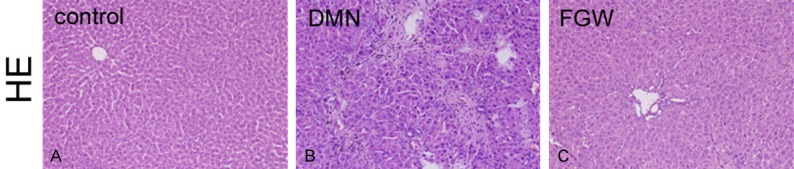
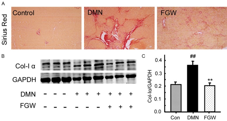
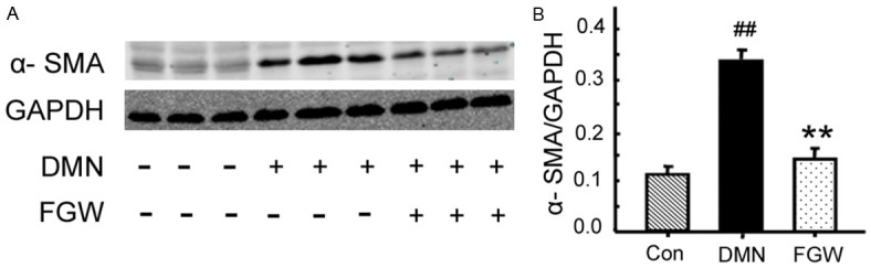
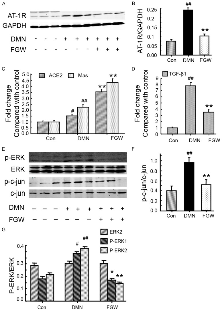
Hepatic fibrosis is a repair and healing reaction for chronic injuries of liver. This study aimed to investigate protective effects of Fugan Wan (FGW) on hepatic fibrosis and clarify associated mechanisms. Hepatic fibrosis model was established by administrating dimethyl nitrosamine (DMN) to rats. Rats were divided into control, DMN and FGW groups. Haematoxylin and eosin (HE) staining was conducted to evaluate inflammatory response in hepatic fibrosis tissues. Sirius red staining was used to assess collagen disposition. Quantitative real-time PCR (qRT-PCR) was employed to detect antiotensin-converting enzyme homologue 2 (ACE2), Mas, transforming growth factor β1 (TGF-β1) mRNA. Western blot was used to examine collagen I, smooth muscle actin α (α-SMA), angiotensin type 1 receptor (AT-1R), extra-cellular regulated protein kinase (ERK), phosphorylated ERK (p-ERK), c-Jun and phosphorylated-c-Jun (p-c-Jun) expression. The results indicated that FGW significantly reduced inflammatory response of hepatic fibrosis tissues. FGW significantly decreased collagen deposition compared to that of DMN group (P < 0.01). FGW significantly down-regulated α-SMA expression compared to that of DMN group (P < 0.01). FGW significantly decreased AT-1R levels compared to that of DMN group (P < 0.01). Comparing with DMN group, ACE2 and Mas mRNA levels were significantly increased in FGW group (P < 0.01). FGW significantly down-regulated p-c-Jun and p-ERK1/2 compared to DMN group (P < 0.01). GFW significantly inhibited compared to DMN group (P < 0.01). In conclusion, FGW alleviated hepatic fibrosis by inhibiting ACE/Ang II/AT-1R signaling and enhancing ACE2/Ang 1-7/Mas signaling pathway in hepatic fibrosis rat models.

Keywords: Fugan Wan; hepatic fibrosis; renin angiotensin system; signaling pathway.


Conflict of interest statement

None.

Figures

Figure 1
Figure 1
FGW alleviated the inflammatory responses in liver tissues of hepatic fibrosis rats. A. HE staining image for inflammatory response in Control group. B. HE staining image for inflammatory response in DMN group. C. HE staining image for inflammatory response in FGW group. Magnification, 100 ×.
Figure 2
Figure 2
Evaluation for the collagen deposition in liver tissues of hepatic fibrosis rat. A. Sirius red staining for the hepatic fibrosis. B. Western blot bands for the collagen I expression. C. Statistical analysis for the collagen I expression. ##P < 0.01 vs. Control group. **P < 0.01 vs. DMN group.
Figure 3
Figure 3
FGW inhibited the α-SMA expression in liver tissues of hepatic fibrosis rat. A. Western blot bands for α-SMA expression. B. Statistical analysis for α-SMA expression. ##P < 0.01 vs. Control group. **P < 0.01 vs. DMN group.
Figure 4
Figure 4
FGW modulated expressions of AT1R, ERK and c-jun in liver tissues of hepatic fibrosis rat. A. Western blot bands for AT-1R expression. B. Statistical analysis for AT-1R expression. C. Statistical analysis for ACE2 and Mas mRNA expression. D. Statistical analysis for TGF-β1 mRNA expression. E. Western blot bands for p-ERK, ERK, p-c-Jun and c-Jun expression. F. Statistical analysis for p-c-Jun expression. G. Statistical analysis for p-ERK expression. ##P < 0.01 vs. Control group. **P < 0.01 vs. DMN group.

Similar articles

Cited by

References

    1. Hu Z, Qin F, Gao S, Zhen Y, Huang D, Dong L. Paeoniflorin exerts protective effect on radiaiton-induced hepatic fibrosis in rats via TGF-beta1/Smads signaling pathway. Am J Transl Res. 2018;10:1012–1021. - PMC - PubMed
    1. Zhu Q, Li N, Li F, Zhou Z, Han Q, Lv Y, Sang J, Liu Z. Therapeutic effect of renin angiotensin system inhibitors on liver fibrosis. J Renin Angiotensin Aldosterone Syst. 2016;17:1470320316628717. - PMC - PubMed
    1. Pereira RM, dos Santos RA, da Costa Dias FL, Teixeira MM, Simões e Silva AC. Renin-angiotensin system in the pathogenesis of liver fibrosis. World J Gastroenterol. 2009;15:2579–2586. - PMC - PubMed
    1. Beljaars L, Meijer DK, Poelstra K. Targeting hepatic stellate cells for cell-specific treatment of liver fibrosis. Front Biosci. 2002;7:e214–e222. - PubMed
    1. Czaja AJ. The prevention and reversal of hepatic fibrosis in autoimmune hepatitis. Aliment Pharmacol Ther. 2014;39:385–406. - PubMed

LinkOut - more resources