. 2020 Apr 6;10(2):20190079.
doi: 10.1098/rsfs.2019.0079. Epub 2020 Feb 14.

Generation of a three-dimensional collagen scaffold-based model of the human endometrium

Affiliations

Generation of a three-dimensional collagen scaffold-based model of the human endometrium

Yassen Abbas et al. Interface Focus. .

Abstract

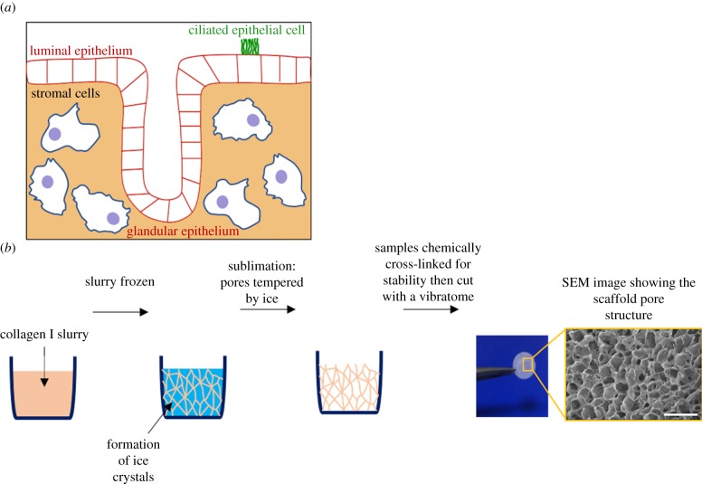
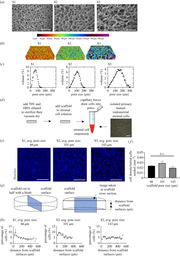
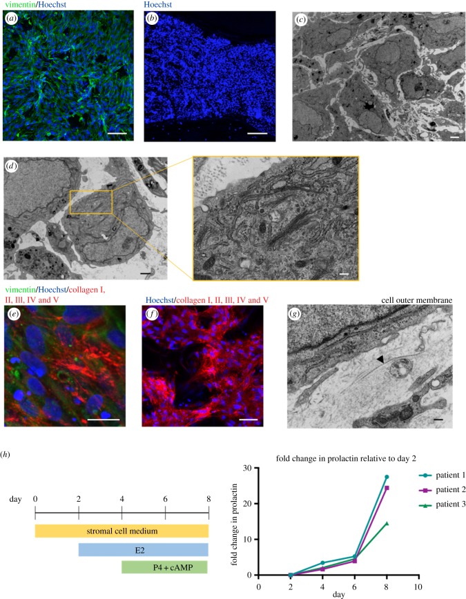
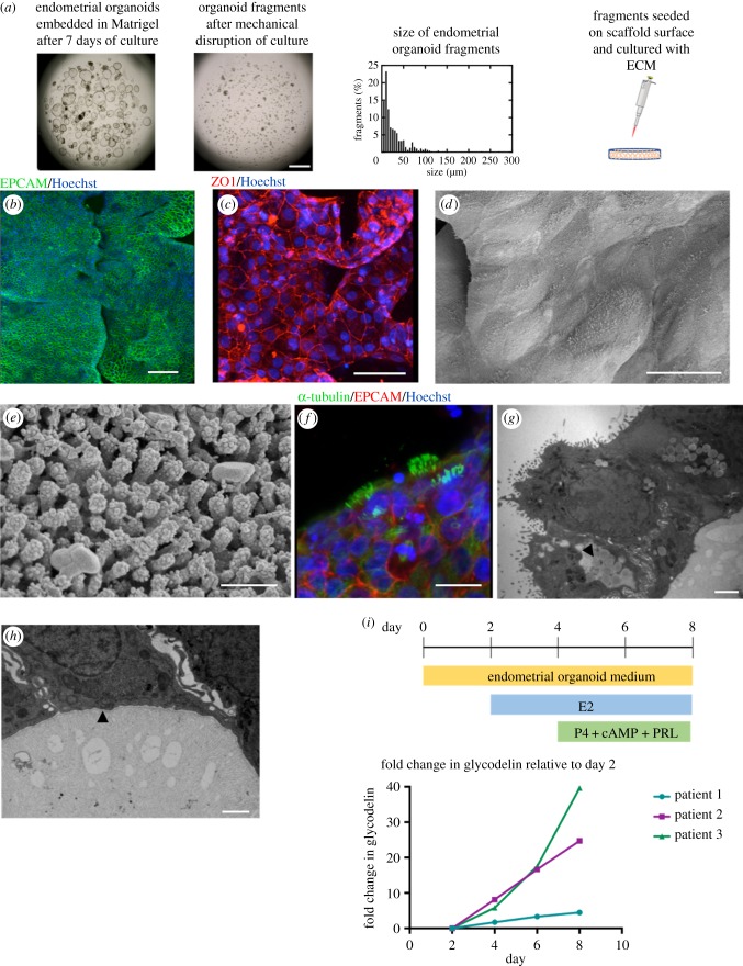
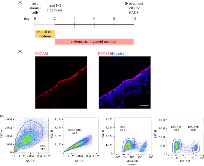
The endometrium is the secretory lining of the uterus that undergoes dynamic changes throughout the menstrual cycle in preparation for implantation and a pregnancy. Recently, endometrial organoids (EO) were established to study the glandular epithelium. We have built upon this advance and developed a multi-cellular model containing both endometrial stromal and epithelial cells. We use porous collagen scaffolds produced with controlled lyophilization to direct cellular organization, integrating organoids with primary isolates of stromal cells. The internal pore structure of the scaffold was optimized for stromal cell culture in a systematic study, finding an optimal average pore size of 101 µm. EO seeded organize to form a luminal-like epithelial layer, on the surface of the scaffold. The cells polarize with their apical surface carrying microvilli and cilia that face the pore cavities and their basal surface attaching to the scaffold with the formation of extracellular matrix proteins. Both cell types are hormone responsive on the scaffold, with hormone stimulation resulting in epithelial differentiation and stromal decidualization.

Keywords: co-culture; collagen scaffolds; endometrium; organoids.


Conflict of interest statement

We declare we have no competing interests.

Figures

Figure 1.
Figure 1.
Porous collagen scaffolds are used to develop a model of the human endometrium. (a) Schematic of the human endometrium comprising epithelial and stromal cells. There are two epithelial cell populations, the luminal that lines the lumen of the uterus and glandular epithelium, which secrete factors required to maintain the conceptus pre- and post-implantation. Epithelial cells are polarized, with the apical surface facing the outside surface and basal inwards. A number of epithelial cells are ciliated. (b) Porous collagen scaffolds used to develop a model of the endometrium are produced by lyophilization. An aqueous slurry of collagen I is frozen. As ice crystals form, the solid material in the slurry is pushed to the boundaries of the ice crystals. The pressure of the chamber is then lowered, and the temperature is raised, inducing sublimation of the ice crystals. The final product is a porous scaffold with solid material lining the spaces previously occupied by the ice crystals. Samples are cross-linked for stability then cut with a vibratome. Scale bar, 400 µm.
Figure 2.
Figure 2.
Optimization of scaffold pore size. (a) SEM images of the surface morphology of the pores within the collagen scaffolds. Three scaffolds of varying pore sizes were made using the following moulds: scaffold 1 (S1), a 316L stainless steel cylindrical well with a diameter of 46.5 mm, scaffold 2 (S2), a polystyrene 48-well cell culture plate, and scaffold 3 (S3), a polystyrene 6-well cell culture plate. Scale bar, 400 µm. (b) Micro-CT visualization of pore structures within scaffolds, with dimensions of 4 mm × 4 mm × 750 µm. (c) Pore size distributions based on micro-CT data. (d) Schematic of the seeding of primary stromal cells onto the scaffolds. Scale bar, 200 µm. (e) Representative images of Hoechst nuclear stain of stromal cells on the top 100 µm of scaffolds with average pore sizes of 66, 101 and 143 µm and percolation diameters of 80, 104 and 136 µm. Images were taken 7 days after seeding 183 k cells per scaffold. Scale bar, 500 µm. (f) Stromal cell density at the top 100 µm of the scaffold surfaces. (g) Schematic showing cross-section taken of scaffold. (h) Stromal cell location throughout the cross-section of each scaffold with average pore sizes of 66, 101 and 143 µm and percolation diameters of 80, 104 and 136 µm. Measurements were taken 7 days after seeding 332 k cells per scaffold.
Figure 3.
Figure 3.
Primary endometrial stromal cells within scaffold. (a) Immunofluorescence of stromal cells stained with vimentin (green) and Hoechst nuclear stain (blue) at scaffold surface. Scale bar, 100 µm. (b) Cross-section of scaffold after 7 days of stromal cell culture, labelled with Hoechst nuclear stain (blue). Scale bar, 200 µm. (c) TEM of stromal cells within scaffold. Scale bar, 2 µm. (d) TEM showing cytoplasm containing plentiful Golgi bodies and secretion of ECM protein. Scale bar, 1 µm with inset: 200 nm. (e) Immunofluorescence of stromal cells showing secretion of human collagen I, II, III, IV and V (red) with vimentin (green) and Hoechst nuclear stain (blue). Scale bar, 20 µm. (f) Low-power immunofluorescence image, showing stromal cells are interconnected on the scaffold surface with secretions of human collagen I, II, III, IV and V (red) with Hoechst nuclear stain (blue). Scale bar, 50 µm. (g) TEM showing the formation of collagen bundles by stroma (see arrowhead). Scale bar, 200 nm. (h) Stromal cells are differentiated over 8 days within the scaffold with the addition of hormones resulting in upregulation of prolactin, measured by ELISA.
Figure 4.
Figure 4.
Endometrial organoid fragments seeded form a luminal-like epithelium on scaffold surface. (a) Endometrial organoids emended in Matrigel® are broken up mechanically to form fragments, the majority of which are less than 50 µm. These fragments are seeded on the surface of the collagen scaffold. Scale bar, 1 mm. (b) Immunofluorescence image of the epithelial marker EPCAM (green) on the scaffold surface with Hoechst nuclear stain (blue). Scale bar, 100 µm. (c) Epithelial cells form tight junctions with positive staining of Zonula occludens-1 (ZO-1, red) with Hoechst nuclear stain (blue). Scale bar, 50 µm. (d) SEM image of epithelial cells on scaffold surface with visible cell boundaries. Scale bar, 20 µm. (e) SEM showing microvilli confirming apical polarization of epithelial cells to the outside surface. Scale bar, 500 nm. (f) Some epithelial cells are ciliated on the scaffold surface, with staining of α-tubulin (green), EPCAM (red) and Hoechst nuclear stain (blue). Scale bar, 15 µm. (g) TEM shows apical polarization to the outside surface and presence of glycocalyx and lipid droplets (see arrowhead). Scale bar, 2 µm. (h) TEM showing a thin and fibrous lining at the basal end of the epithelial cells, indicating the formation of a basement membrane (see arrowhead). Scale bar, 1 µm. (i) Stimulation of epithelial cells with hormones results in increased production of glycodelin, measured by ELISA.
Figure 5.
Figure 5.
Co-culture of stromal and epithelial cells on scaffold. (a) Timeline of co-culture with stromal cells added at day 0, then seeding of organoid fragments at day 2. (b) Immunofluorescence of scaffold cross-section with epithelial cells stained with EPCAM (red) and stromal cells underneath with Hoechst nuclear stain (blue). Scale bar, 200 µm. (c) Cells can be removed from the scaffold for downstream analysis. Here, they were analysed using flow cytometry, showing a population of EPCAM+ (epithelial) and EPCAM– (stromal) cells.

Similar articles

Cited by

References

    1. Burton GJ, Watson AL, Hempstock J, Skepper JN, Jauniaux E. 2002. Uterine glands provide histiotrophic nutrition for the human fetus during the first trimester of pregnancy. J. Clin. Endocrinol. Metab. 87, 2954–2959. (10.1210/jcem.87.6.8563) - DOI - PubMed
    1. Cakmak H, Taylor HS. 2011. Implantation failure: molecular mechanisms and clinical treatment. Hum. Reprod. Update 17, 242–253. (10.1093/humupd/dmq037) - DOI - PMC - PubMed
    1. Ulukus M, Cakmak H, Arici A. 2006. The role of endometrium in endometriosis. J. Soc. Gynecol. Investig. 13, 467–476. (10.1016/j.jsgi.2006.07.005) - DOI - PubMed
    1. Motta PM, Andrews PM. 1976. Scanning electron microscopy of the endometrium during the secretory phase. J. Anat. 122, 315. - PMC - PubMed
    1. Turco MY, et al. 2017. Long-term, hormone-responsive organoid cultures of human endometrium in a chemically defined medium. Nat. Cell Biol. 19, 568–577. (10.1038/ncb3516) - DOI - PMC - PubMed

LinkOut - more resources