. 2020 Mar 17:6:13.
doi: 10.1038/s41421-020-0146-2. eCollection 2020.

A unique DNA-binding mode of African swine fever virus AP endonuclease

Affiliations

A unique DNA-binding mode of African swine fever virus AP endonuclease

Yiqing Chen et al. Cell Discov. .

Abstract

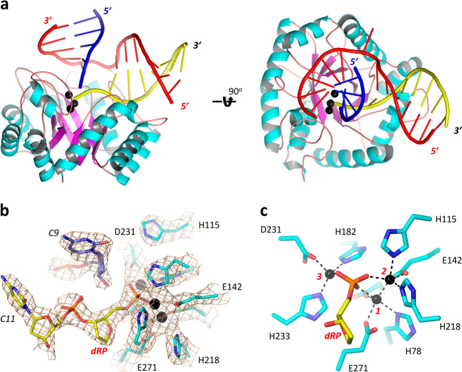
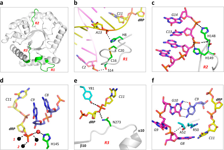
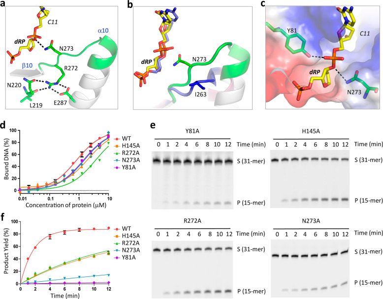
African swine fever virus (ASFV) is highly contagious and can cause lethal disease in pigs. ASFV is primarily replicated in the cytoplasm of pig macrophages, which is oxidative and caused constant damage to ASFV genome. ASFV AP endonuclease (AsfvAP) catalyzes DNA cleavage reaction at the abasic site and is a key enzyme of ASFV base excision repair (BER) system. Although it plays an essential role in ASFV survival in host cells, the basis underlying substrate binding and cleavage by AsfvAP remains unclear. Here, we reported the structural and functional studies of AsfvAP, showing that AsfvAP adopts a novel DNA-binding mode distinct from other APs. AsfvAP possesses many unique structural features, including one narrower nucleotide-binding pocket at the active site, the C16-C20 disulfide bond-containing region, and histidine-rich loop. As indicated by our mutagenesis, in vitro binding and cleavage assays, these features are important for AsfvAP to suit the acidic and oxidative environment. Owing to their functional importance, these unique features could serve as targets for designing small molecule inhibitors that could disrupt the repair process of ASFV genome and help fight against this deadly virus in the future.

Keywords: Base excision repair; X-ray crystallography.


Conflict of interest statement

Conflict of interestThe authors declare that they have no conflict of interest.

Figures

Fig. 1
Fig. 1. Comparison of AP endonuclease activities of AsfvAP and EcNfo.
a Sequence alignment of AsfvAP and homologous proteins. b Impacts of pH on the AP endonuclease activity of AsfvAP. c Comparison of the Kobs values of AsfvAP under different pH. d SDS-PAGE gel analysis showing DNA cleavage by AsfvAP under pH 6.3. e Impacts of pH on the AP endonuclease activity of EcNfo. f Comparison of the Kobs values of EcNfo under different pH. g SDS-PAGE gel analysis showing DNA cleavage by EcNfo under pH 8.0. FAM-labeled DNA-3 (5′-GGTAAGGGCAGCGTCCFCGACGAGGAATGCA-FAM-3′, 3′-CCATTCCCGTCGCAGGGGCTGCTCCTTACGT-5′) was used in all AP endonuclease assays. The substrate and product bands are labeled by S and P, respectively. DNA marker (5′-FCGACGAGGAATGCA-FAM-3′) is labeled as M. In panels b, c and e, f, the data represent the mean of three independent experiments. The standard deviation (±SD) values are indicated by error bars. All SDS-Page gel analysis were repeated for at least three times, the representative gels were shown in panels d, g.
Fig. 2
Fig. 2. Structure of AsfvAP/DNA-1 complex.
a Cartoon view showing the overall fold of AsfvAP/DNA-1 complex. α-helices, β-strands and loops of AsfvAP are colored in cyan, purple, and pink, respectively. The uncleaved strand, the upstream and downstream of the product strand of DNA-1 are colored in red, blue and yellow, respectively. b Conformations of the active site residues and nucleotides. 2Fo–Fc electron density map was contoured at 1.5 sigma level. c Detailed coordination of the Zn2+ ions. The Zn2+ ions are shown as spheres in black in all panels.
Fig. 3
Fig. 3. Protein-DNA interactions observed in the AsfvAP/DNA-1 complex.
a Cartoon view showing the conformations and relative orientations of the R1, R2 and R3 regions, which are colored in green. b Detailed interactions between DNA-1 and the R1 region. DNA-1 and R1 are shown as cartoon. The interacting residues (His8 and Ser14) and nucleotides (A13 and C2) are highlighted by sticks. dRP nucleotide of DNA-1 and Cys16-Cys20 disulfide bond of R1 are also highlighted by sticks. cd Detailed interactions between DNA-1 and the R2 region. Zn2+ ions and water molecule are shown as spheres in black and red, respectively. e Detailed interactions between DNA-1 and the R3 region. Besides Asn273 of the R3 region, the conserved Tyr81 residue is also highlighted by sticks. f Interactions between nick region nucleotides and AsfvAP residues.
Fig. 4
Fig. 4. Structural comparison between AsfvAP and homologous proteins.
a Superposition of AsfvAP, BaNfo, MtEndoIV, and TtNfo structures. b, c Superposition of AsfvAP/DNA-1 and EcNfo/substrate (PDB: 2NQJ) complexes. d Superposition of the DNAs bound in the active sites of AsfvAP/DNA-1, EcNfo/product (PDB: 2NQ9), and EcNfo/substrate complexes. In panels a, b, AsfvAP, BaNfo, MtEndoIV, TtNfo, and EcNfo are colored in white, cyan, yellow, orange, and pink, respectively. The R1–R3 regions are colored in green for AsfvAP, whereas they are colored in blue for all other proteins. In panel c, AsfvAP and EcNfo are colored in white and pink, respectively. For the AsfvAP/DNA-1 complex, the uncleaved strand, upstream and downstream of the cleaved strand are colored in red, blue and yellow, respectively. Zn2+ ions are shown as black spheres. For the EcNfo/substrate complex, DNA strands and Zn2+ ions are all colored in cyan. In panel d, DNA, protein, and Zn2+ ions are colored in atomic colors (C, white; N, blue; O, red; P, orange; Zn2+, black) for the AsfvAP/DNA-1 complex. The EcNfo/product complex is colored in green. For the EcNfo/substrate complex, DNA is colored in cyan, protein and Zn2+ ions are colored in pink.
Fig. 5
Fig. 5. Verification of the functional importance of R1 and R2 regions.
a Comparison of DNA binding by WT and mutated AsfvAP. b, c SDS-PAGE gel analysis and comparison of DNA-3 cleavage by WT and mutated AsfvAP. The substrate and product bands are labeled by S and P, respectively. d Overall fold of the AsfvAP/DNA-2 complex. The two AsfvAP molecules are colored in green and cyan, respectively. The R1 regions are colored in red. dA:dG mispair is highlighted by spheres. e Superposition of the R1 regions observed in the AsfvAP/DNA-1 and AsfvAP/DNA-2 complexes. For the AsfvAP/DNA-1 complex, DNA and the R1 region are colored in orange and yellow, respectively. For the AsfvAP/DNA-2 complex, the R1 regions are colored cyan and green. The Cys16-Cys20 disulfide bonds are highlighted by sticks in both complexes. In panels a and c, the data represent the mean of three independent experiments. The standard deviation (±SD) values are indicated by error bars. All SDS-Page gel analysis were repeated for at least three times, the representative gels were shown in panel b.
Fig. 6
Fig. 6. The unique nucleotide-binding pocket of AsfvAP.
a The H-bond interactions maintained by the R3 region residues in the AsfvAP/DNA-1 complex. DNA and the R3 region are shown as stick and cartoon, respectively. The C atoms of DNA and the R3 region residues are colored in yellow and green, respectively. b Superposition of DNAs and the R3 regions observed in the AsfvAP/DNA-1 and EcNfo/substrate complexes. AsfvAP/DNA-1 complex is colored as in a, the C atoms are colored in blue for both DNA and the R3 region in the EcNfo/substrate complex. c Surface presentation showing the nucleotide-binding pocket of AsfvAP. d Comparison of DNA binding by WT AsfvAP and mutant proteins. e, f SDS-PAGE gel analysis and comparison of DNA-3 cleavage by WT and mutated AsfvAP. The substrate and product bands are labeled by S and P, respectively. In panels d, f, the data represent the mean of three independent experiments. The standard deviation (±SD) values are indicated by error bars. All SDS-Page gel analysis were repeated for at least three times, the representative gels were shown in panels e.

References

    1. Tulman ER, Delhon GA, Ku BK, Rock DL. African swine fever virus. Curr. Top. Microbiol. Immunol. 2009;328:43–87. - PubMed
    1. Vinuela E. African swine fever virus. Curr. Top. Microbiol. Immunol. 1985;116:151–170. - PubMed
    1. Gogin A, Gerasimov V, Malogolovkin A, Kolbasov D. African swine fever in the North Caucasus region and the Russian Federation in years 2007–2012. Virus Res. 2013;173:198–203. doi: 10.1016/j.virusres.2012.12.007. - DOI - PubMed
    1. Costard S, Mur L, Lubroth J, Sanchez-Vizcaino JM, Pfeiffer DU. Epidemiology of African swine fever virus. Virus Res. 2013;173:191–197. doi: 10.1016/j.virusres.2012.10.030. - DOI - PubMed
    1. Zhou X, et al. Emergence of African Swine Fever in China, 2018. Transbound. Emerg. Dis. 2018;65:1482–1484. doi: 10.1111/tbed.12989. - DOI - PubMed