Review
. 2020 Feb 1;10(2):610-629.
eCollection 2020.

The HIPPO pathway in gynecological malignancies

Affiliations
Review

The HIPPO pathway in gynecological malignancies

Dongying Wang et al. Am J Cancer Res. .

Abstract

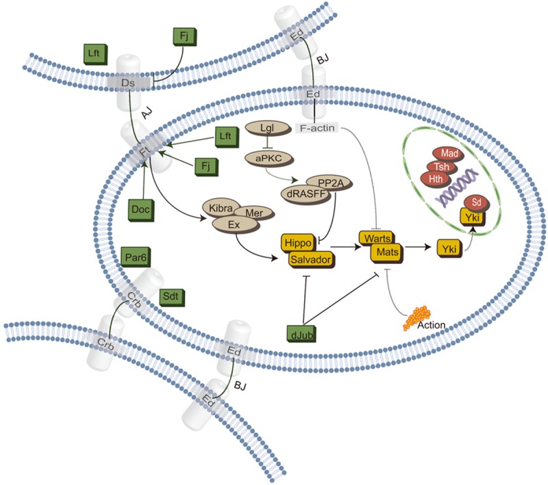
The Hippo pathway has been initially discovered by screening genes that regulate organ size in Drosophila. Recent studies have highlighted the role of the Hippo pathway in controlling organ size, tissue homeostasis and regeneration, and signaling dysregulation, especially the overactivation of the transcriptional coactivator YAP/TAZ, which leads to uncontrolled cell growth and malignant transformation. The core components of the Hippo pathway may initiate tumorigenesis by inducing tumor stem cells and proliferation, ultimately leading to metastasis and drug resistance, which occurs extensively in gynecological malignancies, including cervical cancer, ovarian cancer, and endometrial cancer. In this review, we attempt to systematically summarize recent progress in our understanding of the mechanism of Hippo pathway regulation in tumorigenesis and the mechanisms that underlie alterations during gynecological malignancies, as well as new therapeutic strategies.

Keywords: Hippo pathway; YAP/TAZ; cervical cancer; endometrial cancer; ovarian cancer; therapeutic strategies; tumorigenesis.


Conflict of interest statement

None.

Figures

Figure 1
Figure 1
Schematic diagram of the Hippo pathway in Drosophila. Cells are shown with a blue outline, upstream regulatory proteins are shown in green/gray, intermediate core kinases are shown in yellow and downstream transcriptional proteins are shown in red, with sharp arrows and blunt arrows indicating activation and inhibition interactions, respectively. Continuous lines indicate direct communication, while dashed lines indicate indirect communication. Abbreviations: AJ, adherens junctions; BJ, basolateral junctions; Fj, four-jointed box protein 1; Ds, Dachsous; Ed, echinoid; Ft, FAT; Dco, Discs overgrown; aPKC, atypical protein kinase C; Ljl, Lethal giant larvae; Rassf, Ras-associated factor; PP2A, protein phosphatase 2A; Ex, Expanded; Crb, Crumbs; Mer, Merlin; dJub, Drosophila Ajuba; Sdt, Stardust; Sd, Scalloped; Tsh, Teashirt; Hth, Homothorax.
Figure 2
Figure 2
Schematic diagram of the Hippo pathway in mammals. Cells are shown with a blue outline, upstream regulatory proteins are shown in green/gray, intermediate core kinases are shown in yellow and downstream transcriptional proteins are shown in red, with sharp arrows and blunt arrows indicating activation and inhibition interactions, respectively. Continuous lines indicate direct communication, while dashed lines indicate indirect communication. Abbreviations: AJ, adherens junctions; BJ, basolateral junctions; Ed, echinoid; E-cad, E-cadherin; Ajub, Ajuba LIM Protein; FRMD, FERM Domain Containing; Mer, merlin; Rassf, Ras-associated factor; PP2A, protein phosphatase 2A; Ljl, Lethal giant larvae; Dlg, disks large protein; Scnb, Scribble Planar Cell Polarity Protein; MST1/2, Macrophage Stimulating 1/2; SAV1, Salvador Family WW Domain Containing Protein 1; MOB, MOB kinase activator; LAT1/2, large tumour suppressor 1/2; YAP, Yes-associated protein; TAZ, Tafazzin; RTK, receptor tyrosine kinase; GPCR, G-protein coupled receptor; ECM, extracellular matrix.
Figure 3
Figure 3
Gene-related changes in critical components of the Hippo pathway in patients with gynecological tumors. The genomic profiles examined included the upstream tumor suppressive genes (MST1, LATS1/2, and FAT1/2/3/4, etc.), the downstream tumorigenic effectors (YAP/TAZ & WWTR1), and the mRNA and protein expression of these genes. Genetic alterations of the Hippo pathway occurred in 79% of examined patients with CESE (308 total, A), 38% with UCEC (547 total, B), and 73% with OV (594 total, C). (D) Expression matrix plots of the Hippo pathway in gynecological malignancies and normal tissues deposited in TCGA and GTEx databases. The density of color in each block represents the median expression value of a gene in a specific tissue. (E) The composition of mutations and copy number alterations (CNAs) of gynecologic tumors is presented in the form of stacked histograms. Different colors represent amplification, deletion, mutation, and multiple alterations, among which 3 data sets have the most significant proportion of mutations, and 5 data sets have the most significant portion of amplification. (F) Network view of alterations of HIPPO pathway linker genes in gynecologic malignancies. We analyzed the genomic data of 308 cervical cancer patients, 594 ovarian cancer patients and 547 endometrial cancer patients, which were deposited in TCGA, and the seed genes are indicated by the thick border. (G) Kaplan-Meier curves showed a correlation between overall survival in cervical and ovarian cancer patients and genetic alterations in the Hippo pathway. Patients with TCGA survival information were divided into two groups: patients with high gene expression levels (red line) and patients with low gene expression levels (blue line). Abbreviations: CESC, cervical squamous cell carcinoma and endocervical adenocarcinoma; OV, ovarian serous cystadenocarcinoma; UCEC, uterine corpus endometrial carcinoma.
Figure 4
Figure 4
A schematic diagram shows the proposed mechanism by which the Hippo pathway regulates cervical cancer. In cervical cancer cells, nuclear accumulation of YAP protein stimulates the expression of EGF-like ligands, such as TGF-α and AREG, which in turn activates EGFR and inhibits LATS 1/2 and MOB1 binding and further phosphorylation. Activated YAP activates transcription factors and induces the expression of growth factors, such as TGF-α and AREG, thereby promoting the growth of cervical cancer. HPV E6/E7 oncoproteins maintain YAP protein levels in cervical cancer cells by preventing the degradation of proteasome-dependent YAP. Accordingly, the high expression of YAP further promotes persistent HPV infection by upregulating putative HPV membrane receptor molecules and suppressing innate immunity in host cells.
Figure 5
Figure 5
A schematic diagram showing the proposed mechanism by which the Hippo signaling pathway regulates ovarian cancer. In normal ovarian tissues, deactivated FGF ligands, such as FGFRs, are insufficient to activate YAP, leading to ubiquitin-dependent degradation of YAP proteins. In ovarian cancer tissues, the nuclear accumulation of YAP protein stimulates the expression of FGFs, FGFRs and AREG, thereby activating FGF receptors and EGFR, which in turn interact with downstream signaling pathways, such as the PI3K and MAPK pathways, inhibiting the Hippo pathway and activating the YAP protein. Moreover, the NRSF, UCA1, and LPA-G12/13-RhoA-ROCK-PP1A-YAP signaling pathways can stimulate the expression of YAP, miRNA-129-5p, and miRNA-149-5p and inhibit the expression of YAP, which affects the occurrence and development of ovarian cancer.
Figure 6
Figure 6
A schematic diagram showing the proposed mechanism by which the Hippo signaling pathway regulates endometrial cancer. The Hippo pathway activates the PI3K/AKT pathway via growth factors, including 1-oleoyl-2-hydroxy-sn-glycerol-3-phosphate (LPA), EGF, insulin, and IGF1, resulting in YAP and TAZ dephosphorylation, leading to nuclear translocation and TEAD transcriptional activation. MIR31 significantly inhibits the luciferase activity of mRNA binding to the LATS2 3’-UTR, and the downregulation of LATS2 leads to the dephosphorylation of YAP, promotes the translocation of YAP into the nucleus, and increases the transcription of CCND1. Inactivation of deubiquitinating enzyme USP51 inhibits FAT4, resulting in decreased phosphorylation of LATS1/2 and YAP, while increased YAP nuclear translocation and loss of LSR upregulate TEAD/AREG in EC cells, which promote proliferation and invasion. YAP/TEAD-AP1 cooperation engages SRC1-3 coactivators and drives downstream gene expression to regulate endometrial cancer cell migration and invasion. Verteporfin induces YAP retention in the cytoplasm through increasing levels of 14-3-3 and blocks the transcriptional activation of targets downstream of YAP.
Figure 7
Figure 7
A schematic diagram showing putative targets and small molecules specific to the Hippo pathway. Cells are shown with a blue outline, upstream regulatory proteins are shown in green, and core kinases are shown in yellow, with sharp arrows and blunt arrows indicating activation and inhibition interactions, respectively. Continuous lines indicate direct communication, while dashed lines indicate indirect communication.

References

    1. Tapon N, Harvey KF, Bell DW, Wahrer DC, Schiripo TA, Haber D, Hariharan IK. Salvador promotes both cell cycle exit and apoptosis in drosophila and is mutated in human cancer cell lines. Cell. 2002;110:467–478. - PubMed
    1. Halder G, Johnson RL. Hippo signaling: growth control and beyond. Development. 2011;138:9–22. - PMC - PubMed
    1. Patel SH, Camargo FD, Yimlamai D. Hippo signaling in the liver regulates organ size, cell fate, and carcinogenesis. Gastroenterology. 2017;152:533–545. - PMC - PubMed
    1. Pandurangan AK, Divya T, Kumar K, Dineshbabu V, Velavan B, Sudhandiran G. Colorectal carcinogenesis: insights into the cell death and signal transduction pathways: a review. World J Gastrointest Oncol. 2018;10:244–259. - PMC - PubMed
    1. Maugeri-Sacca M, De Maria R. Hippo pathway and breast cancer stem cells. Crit Rev Oncol Hematol. 2016;99:115–122. - PubMed

LinkOut - more resources