. 2020 Feb 18;2(2):100093.
doi: 10.1016/j.jhepr.2020.100093. eCollection 2020 Apr.

miR-22 inhibition reduces hepatic steatosis via FGF21 and FGFR1 induction

Affiliations

miR-22 inhibition reduces hepatic steatosis via FGF21 and FGFR1 induction

Ying Hu et al. JHEP Rep. .

Abstract

Background & aims: Metabolism supports cell proliferation and growth. Surprisingly, the tumor suppressor miR-22 is induced by metabolic stimulators like bile acids. Thus, this study examines whether miR-22 could be a metabolic silencer.

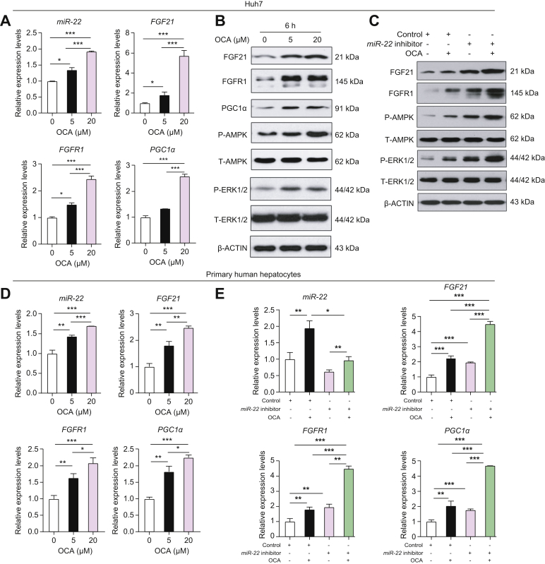
Methods: The relationship between miR-22 and the expression of fibroblast growth factor 21 (FGF21) and its receptor FGFR1 was studied in cells and fatty livers obtained from patients and mouse models. We evaluated the effect of an miR-22 inhibitor alone and in combination with obeticholic acid (OCA) for the treatment of steatosis.

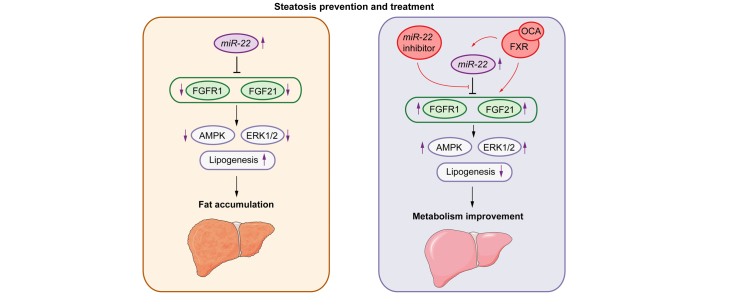
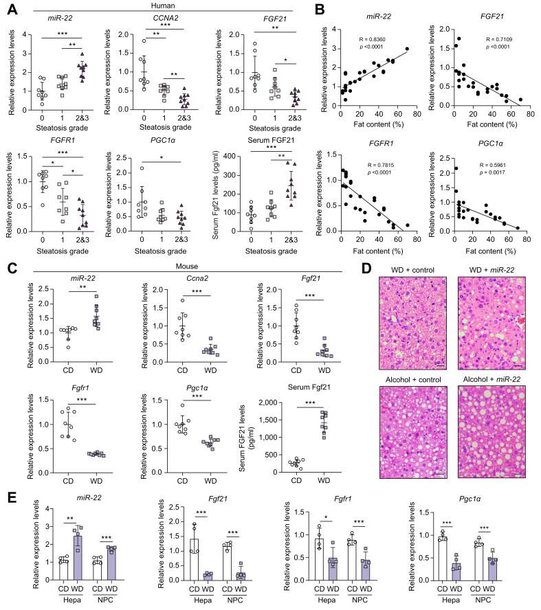
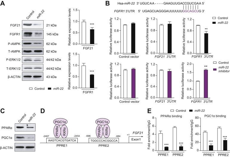
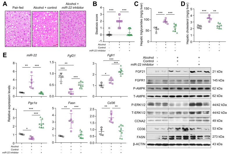
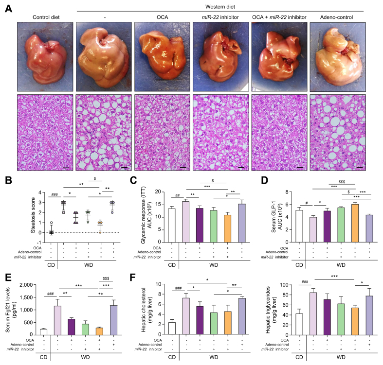
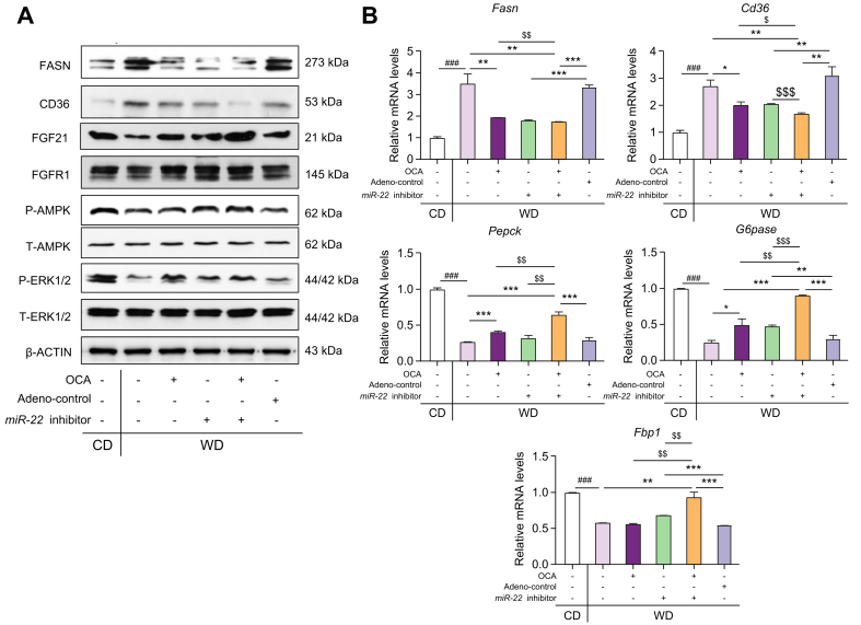
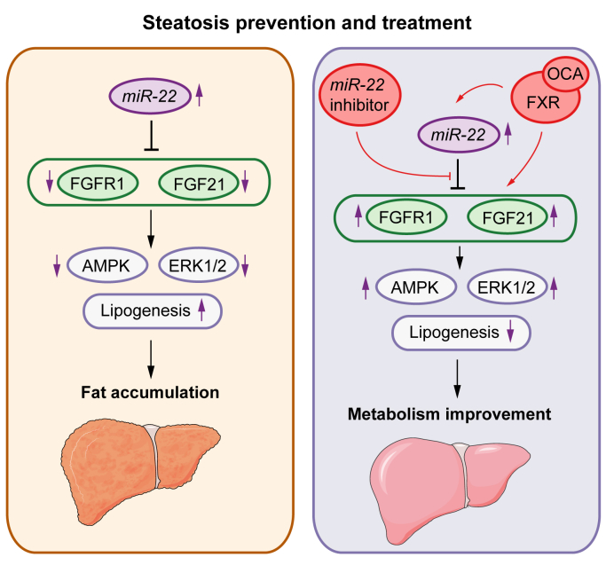
Results: The levels of miR-22 were inversely correlated with those of FGF21, FGFR1, and PGC1α in human and mouse fatty livers, suggesting that hepatic miR-22 acts as a metabolic silencer. Indeed, miR-22 reduced FGFR1 by direct targeting and decreased FGF21 by reducing the recruitment of PPARα and PGC1α to their binding motifs. In contrast, an miR-22 inhibitor increases hepatic FGF21 and FGFR1, leading to AMPK and ERK1/2 activation, which was effective in treating alcoholic steatosis in mouse models. The farnesoid x receptor-agonist OCA induced FGF21 and FGFR1, as well as their inhibitor miR-22. An miR-22 inhibitor and OCA were effective in treating diet-induced steatosis, both alone and in combination. The combined treatment was the most effective at improving insulin sensitivity, releasing glucagon-like peptide 1, and reducing hepatic triglyceride in obese mice.

Conclusion: The simultaneous induction of miR-22, FGF21 and FGFR1 by metabolic stimulators may maintain FGF21 homeostasis and restrict ERK1/2 activation. Reducing miR-22 enhances hepatic FGF21 and activates AMPK, which could be a novel approach to treat steatosis and insulin resistance.

Lay summary: This study examines the metabolic role of a tumor suppressor, miR-22, that can be induced by metabolic stimulators such as bile acids. Our novel data revealed that the metabolic silencing effect of miR-22 occurs as a result of reductions in metabolic stimulators, which likely contribute to the development of fatty liver. Consistent with this finding, an miR-22 inhibitor effectively reversed both alcohol- and diet-induced fatty liver; miR-22 inhibition is a promising therapeutic option which could be used in combination with obeticholic acid.

Keywords: 3'-UTR, 3' untranslated region; ALP, alkaline phosphatase; ALT, alanine aminotransferase; CD, control diet; FGF21, fibroblast growth factor 21; FXR, farnesoid X receptor; GLP-1, glucagon-like peptide; HDAC, histone deacetylase; ITT, insulin tolerance test; LPS, lipopolysaccharide; NPCs, non-parenchymal cells; OCA, obeticholic acid; PFUs, plaque-forming units; PGC1α, PPAR-activated receptor-γ coactivator-1α; PHHs, primary human hepatocytes; PPREs, peroxisome proliferative-response elements; RARβ, retinoic acid receptor β; RT-PCR, reverse transcription PCR; SIRT1, sirtuin 1; Steatosis; WD, Western diet; alcoholic steatosis; insulin sensitivity; metabolic syndrome; non-alcoholic steatohepatitis; obeticholic acid.


Conflict of interest statement

The authors declare no conflicts of interest that pertain to this work. Please refer to the accompanying ICMJE disclosure forms for further details.

Figures

None
Graphical abstract
Fig. 1
Fig. 1
Elevated miR-22 is accompanied by reduced CCNA2, FGF21, FGFR1, and PGC1α in human and mouse steatosis livers. (A) Hepatic miR-22, CCNA2, FGF21, FGFR1, and PGC1α mRNA levels as well as serum FGF21 concentrations in healthy people and patients with fatty liver. Steatosis was graded by a pathologist based on fat content: grade 0 (normal) ≤5%; grade 1 (mild) = 5%∼33%; grade 2 (moderate) = 34%∼66%; grade 3 (severe) ≥67%, n = 8-9 livers per group. Because only 1 patient had a steatosis score >3, the patients with steatosis grade 2 and 3 were grouped together. One-way ANOVA with Tukey’s t test. ∗p <0.05, ∗∗p <0.01, ∗∗∗p <0.001. (B) Relationships between the expression levels of indicated genes and hepatic fat content; (C) Hepatic miR-22, Ccna2, Fgf21, Fgfr1, and Pgc1α mRNA levels as well as serum FGF21 concentrations in CD- or WD-fed male mice after 6 months of WD feeding. n = 8 mice per group. Two-tailed Student’s t test. ∗p <0.05, ∗∗p <0.01, ∗∗∗p <0.001. (D) Liver histology revealed that miR-22 promotes fat deposition in diet- and alcohol-induced fatty liver models. WD-fed mice (3-months old) received adenovirus control, or miR-22 (1×10⁹ PFU, via tail vein, 3 times in 10 days) after 10 weeks of WD feeding. For the alcoholic steatosis model, 3-month-old male mice were fed a Liber DeCarli diet supplemented with and without 5% alcohol for 3 weeks. The mice received adenovirus control or miR-22 (1×10⁹ PFU, via tail vein, 3 times in 10 days). The same diet was given during the interventions. Representative H&E-stained liver sections are presented. Scale bar indicates 100 μm. (E) The expression levels of miR-22, Fgf21, Fgfr1, and Pgc1α mRNA levels in hepatocytes (Hepa) and NPCs of CD- or WD-fed male mice. Data = mean ± SD, n = 4. One-way ANOVA with Tukey’s t test. ∗p <0.05, ∗∗p <0.01, ∗∗∗p <0.001. CD, control diet; NPCs, non-parenchymal cells; PFUs, plaque-forming units; WD, Western diet.
Fig. 2
Fig. 2
The mechanisms by which miR-22 reduces FGFR1 and FGF21. (A) The levels of indicated proteins and mRNAs in Huh7 cells at 48 h post adenoviral-miR-22 (miR-22) or negative adenoviral (control) infection. (B) miR-22, which is conserved between humans and mice, partially pairs with the 3'-UTR of the FGFR1 gene. Adenovirus negative control (control), adenoviral-miR-22 (miR-22), or adenoviral-miR-22 inhibitor (miR-22 inhibitor) were used to infect Huh7 cells for 48 h before transfection with reporter constructs containing the 3'-UTR of the FGF21 or FGFR1 cloned into psiCHECK2. psiCHECK2 without the insert was used as a negative control. Reporter assays revealed that miR-22 reduced the luciferase activity driven by the FGFR1 3'-UTR, but not by the FGF21 3'-UTR. Thus, miR-22 directly silences FGFR1 expression. (C) PPARα and PGC1α protein levels in Huh7 cells at 48 h post-infection of control or miR-22. (D) There are 2 PPREs present in the regulatory region of both the human and mouse FGF21 gene. (E) Huh7 cells were infected with miR-22 or control followed by chromatin isolation. Chromatin immunoprecipitation was performed on cell lysates using the indicated antibodies followed by qPCR using FGF21-specific primers. Binding was expressed relative to the IgG antibody that was used as a negative control. Chromatin immunoprecipitation-qPCR data revealed that miR-22 reduced the recruitment of PPARα and PGC1α to the PPREs. Data are presented as the mean ± SD with ∗∗p <0.01; ∗∗∗p <0.001. Two-tailed Student’s t test. 3'-UTR, 3' untranslated region; PPREs, peroxisome proliferative-response elements.
Fig. 3
Fig. 3
The miR-22 inhibitor treats alcoholic steatosis by inducing FGF21-mediated AMPK activation. Three-month-old C57BL/6 male mice were fed a Liber DeCarli diet supplemented with and without 5% alcohol for 3 weeks. The alcohol-fed mice were treated with miR-22 inhibitor (1×10⁹ PFU, via tail vein, 3 times over 10 days) or adenovirus serving as a negative control. All the mice were euthanized 1 day after the last viral injection. The same diet was given during the interventions. (A) Representative H&E-stained liver sections; (B) steatosis scores; (C) hepatic cholesterol level; (D) hepatic triglyceride level; and (E) hepatic miR-22 as well as the indicated mRNA and protein levels in each group. Hepatic fat content was scored as 0 (<5%), 1 (5–33%), 2 (34–66%), and 3 (>67%). Scale bar in the micrograph of liver section indicates 100 μm. Data are shown as mean ± SD. One-way ANOVA with Tukey’s t est. ∗p <0.05, ∗∗p <0.01, ∗∗∗p <0.001 (n = 4–12 for each group).
Fig. 4
Fig. 4
OCA simultaneously induces miR-22, as well as FGF21, FGFR1, and PGC1α in liver cells. Inhibiting miR-22 enhances OCA-induced FGF21 signaling leading to AMPK and ERK1/2 activation. miR-22, FGF21, FGFR1, and PGC1α mRNA (A) and protein (B) levels in Huh7 cells treated with DMSO or OCA for 6 h. (C) Protein levels in Huh7 cells infected with adenovirus negative control or the miR-22 inhibitor followed by DMSO or OCA (5 μM) treatment for 6 h. (D) miR-22 as well as the indicated mRNA levels in PHHs treated with DMSO or OCA for 6 h. (E) Indicated mRNA levels in PHHs infected with adenovirus negative control or the miR-22 inhibitor followed by DMSO or OCA (5 μM) treatment for 6 h. RT-PCR was performed in triplicate for each sample. PHHs were isolated from 1 donor liver. Data are presented as the mean ± SD (n = 3). One-way ANOVA with Tukey’s t test. ∗p <0.05, ∗∗p <0.01, ∗∗∗p <0.001. OCA, obeticholic acid; PHHs, primary human hepatocytes; RT-PCR, reverse transcription PCR.
Fig. 5
Fig. 5
The miR-22 inhibitor enhances the effect of OCA in reducing steatosis, improving insulin sensitivity, and inducing FGF21 signaling. C57BL/6 male mice were fed a WD after weaning. When those mice were 7 months old, they received OCA (10 μg/g body weight, daily oral gavage), adenovirus negative control, or the miR-22 inhibitor (1×10⁹ PFU, tail vein injection, once a week), or a combination of OCA plus the miR-22 inhibitor for 3 weeks. Age- and sex-matched CD-fed mice without any treatment were used as baseline controls. The same diet was given during the interventions. (A) Representative gross liver morphology and H&E-stained liver sections; (B) steatosis scores; (C) glycemic response measured by ITT; (D) serum GLP-1 secretion; (E) serum FGF21 level; (F) hepatic cholesterol and triglyceride levels; Hepatic fat content was scored as 0 (<5%), 1 (5–33%), 2 (34–66%), and 3 (>67%). Data are shown as mean ± SD (n = 4). One-way ANOVA with Tukey’s t-test. #p <0.05, ##p <0.01, ###p <0.001 between WD and CD; ∗p <0.05, ∗∗p <0.01, ∗∗∗p <0.0001 between treated groups and controls; $p <0.05, $$$p <0.001 between combination and single treatment. CD, control diet; ITT, insulin tolerance test; OCA, obeticholic acid; PFUs, plaque-forming units; WD, Western diet.
Fig. 6
Fig. 6
The miR-22 inhibitor does not have apparent toxicity or a proliferative effect in WD-induced obese mice. C57BL/6 male mice were fed a WD after weaning. When those mice were 7-months old, they received OCA (10 μg/g body weight, daily oral gavage), adenovirus negative control, or the miR-22 inhibitor (1×10⁹ PFU, via tail vein, once a week), or a combination of OCA plus the miR-22 inhibitor for 3 weeks. Age- and sex-matched CD-fed mice without any treatment were used as baseline controls. (A) serum ALT, ALP, and endotoxin (LPS) levels. (B) Representative liver sections of Ki-67 immunohistochemistry staining; Ki-67-positive liver cells were counted in 5 random fields (40X) per liver section. Scale bar indicates 100 μm. Data are shown as mean ± SD (n = 4). One-way ANOVA with Tukey's t-test. ALP, alkaline phosphatase; ALT, alanine aminotransferase; CD, control diet; LPS, lipopolysaccharide; OCA, obeticholic acid; PFUs, plaque-forming units; WD, Western diet.
Fig. 7
Fig. 7
The effects of the miR-22 inhibitor and OCA on regulating the expression of hepatic genes and proteins implicated in metabolism as well as fatty acid synthesis and uptake. C57BL/6 male mice were fed a WD after weaning. When those mice were 7 months old, they received OCA (10 μg/g body weight, daily oral gavage), adenovirus negative control, or the miR-22 inhibitor (1×10⁹ PFU, via tail vein, once a week), or a combination of OCA plus the miR-22 inhibitor for 3 weeks. Age- and sex-matched CD-fed mice without any treatment were used as baseline controls. Hepatic levels of indicated proteins (A) and mRNAs (B) in the CD-fed and WD-fed mice. Data are shown as mean ± SD (n = 4). One-way ANOVA with Tukey's t-test. ###p <0.001 between WD and CD; ∗p <0.05, ∗∗p <0.01, ∗∗∗p <0.001 between treated and their controls; $$$p <0.001 between combination and single treatment. CD, control diet; OCA, obeticholic acid; PFUs, plaque-forming units; WD, Western diet.
Fig. 8
Fig. 8
Metabolic disease development and treatment controlled by miR-22 silenced and FXR activated FGF21 and FGFR1. Left panel: Overexpressed miR-22 likely contributes to the development of steatosis, but it also has a tumor-suppressive effect by limiting ERK1/2 activation. Right panel: Activation of FXR facilitates metabolism and treats steatosis as well as improves inulin sensitivity, but it also induces miR-22, a metabolic inhibitor that silences FGF21 and FGFR1. The induction of miR-22 restricts FGF21-driven growth controlled by ERK1/2 activation. It also maintains FGF21 homeostasis. The miR-22 inhibitor can be used to increase FGF21 and FGFR1 signaling under metabolically compromised conditions.

References

    1. Neely L.A., Patel S., Garver J., Gallo M., Hackett M., McLaughlin S. A single-molecule method for the quantitation of microRNA gene expression. Nat Methods. 2006;3:41–46. - PubMed
    1. Xiong J., Yu D., Wei N., Fu H., Cai T., Huang Y. An estrogen receptor alpha suppressor, microRNA-22, is downregulated in estrogen receptor alpha-positive human breast cancer cell lines and clinical samples. FEBS J. 2010;277:1684–1694. - PubMed
    1. Wang J., Li Y., Ding M., Zhang H., Xu X., Tang J. Molecular mechanisms and clinical applications of miR-22 in regulating malignant progression in human cancer (Review) Int J Oncol. 2017;50:345–355. - PMC - PubMed
    1. Yang F., Hu Y., Liu H.X., Wan Y.J.Y. MiR-22-silenced cyclin A expression in colon and liver cancer cells is regulated by bile acid receptor. J Biol Chem. 2015;290:6507–6515. - PMC - PubMed
    1. Hu Y., French S.W., Chau T., Liu H.X., Sheng L., Wei F. RARbeta acts as both an upstream regulator and downstream effector of miR-22, which epigenetically regulates NUR77 to induce apoptosis of colon cancer cells. FASEB J. 2018 fj201801390R. - PMC - PubMed