. 2020 Mar 1;78(2):ftaa018.
doi: 10.1093/femspd/ftaa018.

The origin of extracellular DNA in bacterial biofilm infections in vivo

Affiliations

The origin of extracellular DNA in bacterial biofilm infections in vivo

Maria Alhede et al. Pathog Dis. .

Abstract

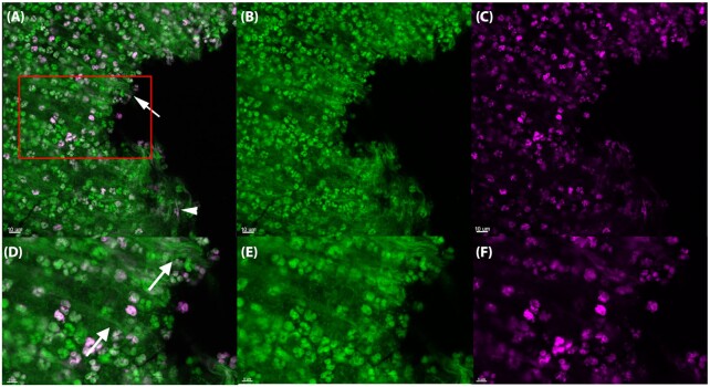
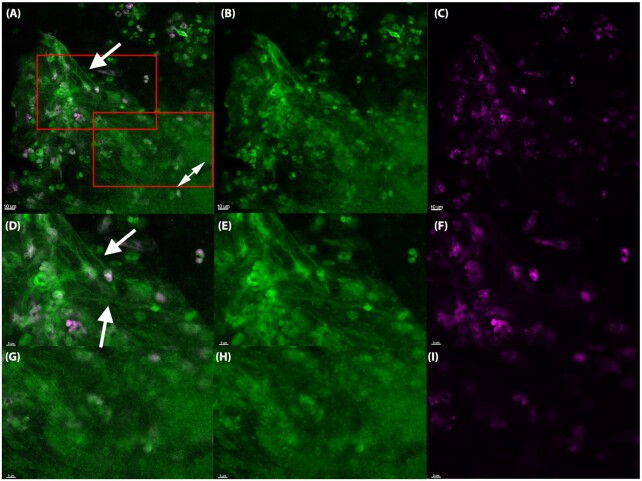
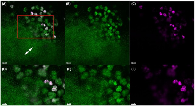
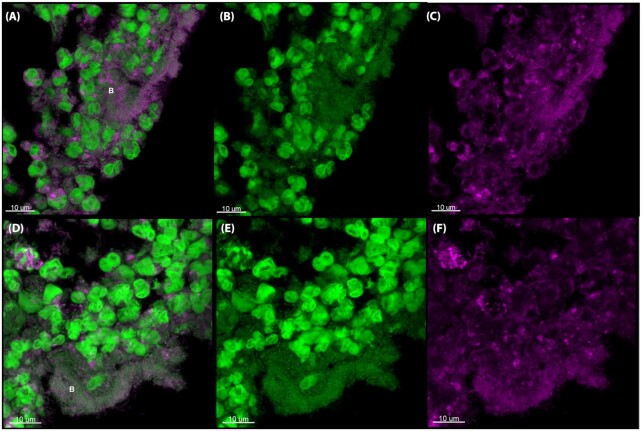
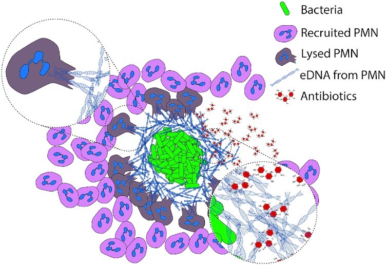
Extracellular DNA (eDNA) plays an important role in both the aggregation of bacteria and in the interaction of the resulting biofilms with polymorphonuclear leukocytes (PMNs) during an inflammatory response. Here, transmission electron and confocal scanning laser microscopy were used to examine the interaction between biofilms of Pseudomonas aeruginosa and PMNs in a murine implant model and in lung tissue from chronically infected cystic fibrosis patients. PNA FISH, DNA staining, labeling of PMN DNA with a thymidine analogue and immunohistochemistry were applied to localize bacteria, eDNA, PMN-derived eDNA, PMN-derived histone H3 (H3), neutrophil elastase (NE) and citrullinated H3 (citH3). Host-derived eDNA was observed surrounding bacterial biofilms but not within the biofilms. H3 localized to the lining of biofilms while NE was found throughout biofilms. CitH3, a marker for neutrophil extracellular traps (NETs) was detected only sporadically indicating that most host-derived eDNA in vivo was not a result of NETosis. Together these observations show that, in these in vivo biofilm infections with P. aeruginosa, the majority of eDNA is found external to the biofilm and derives from the host.

Keywords: NETosis; elastase; histone; neutrophil extracellular traps; polymorphonuclear leukocyte.


Figures

Figure 1.
Figure 1.
(A), Pseudomonas aeruginosa is internalized by PMNs in the murine implant model. Top row: TEM images showing intact and active PMNs containing internalized P. aeruginosa (arrows) 6 h post-insertion of a pre-coated silicone implant (Bar: 2 μm). Bottom row: internalized bacteria are magnified (Bar: 500 nm). Images represents sections obtained from two biological samples (two implants). (B–D), Interaction between PMNs and P. aeruginosa in the murine implant model. Pre-coated silicone implants were inserted into the peritoneal cavity of BALB/c mice. The interaction between PMNs and bacteria was imaged by TEM at 6 h (B), 24 h (C) and 48 h (D) post-insertion. In (B) black arrows points to bacteria and in (C) to a P. aeruginosa biofilm. Arrow heads: PMNs (Bar: 5 μm). Images represents sections obtained from two biological samples (two implants). (E-G), Matrix material surrounding P. aeruginosa on a silicone implant at 24 h post-insertion. TEM images showing matrix material surrounding P. aeruginosa bacteria at different magnifications. Matrix material (black arrows) can be seen between the bacteria. (E) Bar: 2 μm; (F) Bar: 1 μm; and (G) Bar: 200 nm. Images represents sections obtained from two biological samples (two implants).
Figure 2.
Figure 2.
In vivo DNA labeling of PMNs in the murine implant model. PMNs in the murine implant model were labeled with Click-iT®, in which a modified thymidine analogue, EdU (5-ethynyl-2'-deoxyuridine), is incorporated into DNA during active DNA synthesis in murine immune cells in vivo, and hence will label only DNA originating from murine PMNs. DNA is labeled ex vivo with Alexa Fluor® 647 (pink) and counterstained with SYTO9 (green) on an implant 24 h post-insertion in the peritoneal cavity. The number of PMNs stained with SYTO9 were estimated to 370 whereas the EdU labeled (pink) PMNs were estimated to 165. Thereby 45% PMNs in the images were EdU labeled. In the PMN accumulations SYTO9 (green)-stained DNA strings (white arrows) were observed as well as a few pink DNA strings (white arrowheads). (A, D), Merged images showing both EDU (pink) and SYTO9 (green) staining. (B, E), Images showing only the SYTO9 staining. (C, F), images showing only the EDU staining. Red square in A indicates magnified area in D–F. Images represents staining obtained from four biological samples (four implants).
Figure 3.
Figure 3.
The in vivo biofilm lack PMN-derived DNA 24 h post-insertion in the murine implant model. PMNs in the murine implant model were labeled with Click-iT®, in which a modified thymidine analogue, EdU (5-ethynyl-2'-deoxyuridine), is incorporated into DNA during active DNA synthesis in murine immune cells in vivo, and hence will label only DNA originating from murine PMNs. DNA is labeled ex vivo with Alexa Fluor® 647 (pink) and counterstained with SYTO9 (green) on an implant 24 h post-insertion in the peritoneal cavity. The number of PMNs stained with SYTO9 were estimated to 66 whereas the EdU-labeled (pink) PMNs were estimated to 28. Thereby 42% PMNs in the images were EdU labeled. In the PMN accumulations SYTO9 (green)-stained DNA strings (white arrows) were observed. EdU labeling was observed in PMNs (pink), but was absent from biofilms (double headed arrows), suggesting that PMNs are not a source of eDNA in biofilms. (A, D), merged images showing both EDU (pink) and SYTO9 (green) staining. (B, E), Images showing only the SYTO9 staining. (C, F), images showing only the EDU staining. Red squares in A indicates magnified area in D-F and G-I. Images represents staining obtained from four biological samples (four implants).
Figure 4.
Figure 4.
The in vivo biofilm lack PMN-derived DNA 48 h post-insertion in the murine implant model. PMNs in the murine implant model were labeled with Click-iT®, in which a modified thymidine analogue, EdU (5-ethynyl-2'-deoxyuridine), is incorporated into DNA during active DNA synthesis in murine immune cells in vivo, and hence will label only DNA originating from murine PMNs. DNA is labeled ex vivo with Alexa Fluor® 647 (pink) and counterstained with SYTO9 (green) on an implant 48 h post-insertion in the peritoneal cavity. The number of PMNs stained with SYTO9 were estimated to 36 whereas the EdU labeled (pink) PMNs were estimated to 32. Thereby 89% PMNs in the images were EdU labeled. Labeling was observed in PMNs (pink) but was absent from biofilms (double-headed arrows), suggesting that PMNs are not a source of eDNA in biofilms. (A, D), Merged images showing both EDU (pink) and SYTO9 (green) staining. (B, E), Images showing only the SYTO9 staining. (C, F), images showing only the EDU staining. Red square in A indicates magnified area in D-F. Images represents staining obtained from four biological samples (four implants).
Figure 5.
Figure 5.
PNA FISH and DAPI staining of CF lung tissue. Deparaffinated CF lung tissue section stained with (A), PNA FISH to show a P. aeruginosa (red) and (B), DAPI (blue) to show DNA of PMNs and eDNA. (C), Shows an overlay of A and B. (D), A deparaffinated CF lung tissue section stained with the DNA stain propidium iodide (PI) to illustrate both biofilm and eDNA. White arrows point to P. aeruginosa biofilm and black arrows point to eDNA as strings. (Bars: 9 μm).
Figure 6.
Figure 6.
citH3 antibody staining of the murine implant model. CSLM images of deparaffinized sections of silicone implants from the murine implant model 24 h post-insertion. The sections were stained with primary antibodies specific for citrullinated histone H3 (citH3). The secondary antibody was conjugated to Alexa Fluor 647 (pink). SYTO9 (green) was used as a counterstain. SYTO9 stains DNA in bacteria and eukaryotic cells. (A, D), Merged images of both the antibody (pink) and SYTO9 (green) staining. (B, E), Are only the SYTO9 staining. (C, F), are only the antibody staining. The images represent two implants. The letter B indicates biofilm. Images represents staining of 2–4 sections obtained from two biological samples (two implants).
Figure 7.
Figure 7.
H3 antibody staining of the murine implant model. CSLM images of deparaffinized sections of silicone implants from the murine implant model 24 h post-insertion. The sections were stained with primary antibodies specific for histone H3 (H3). The secondary antibody was conjugated to Alexa Fluor 647 (pink). SYTO9 (green) was used as a counterstain. SYTO9 stains DNA in bacteria and eukaryotic cells. (A, D), Merged images of both the antibody (pink) and SYTO9 (green) staining. (B, E), Is only the SYTO9 staining. (C, F), is only the antibody staining. The images represent two implants. The letter B indicates biofilm. Images represents staining of 2–4 sections obtained from two biological samples (two implants).
Figure 8.
Figure 8.
NE antibody staining of the murine implant model. CSLM images of deparaffinized sections of silicone implants from the murine implant model 24 h post-insertion. The sections were stained with primary antibodies specific for NE. The secondary antibody was conjugated to Alexa Fluor 647 (pink). SYTO9 (green) was used as a counterstain. SYTO9 stains DNA in bacteria and eukaryotic cells. (A, D), Merged images of both the antibody (pink) and SYTO9 (green) staining. (B, E), Are only the SYTO9 staining. (C, F), Are only the antibody staining. The images represent two implants. The letter B indicates biofilm. Images represents staining of 2–4 sections obtained from two biological samples (two implants).
Figure 9.
Figure 9.
Immunostaining of CF lung tissue. CSLM images of deparaffinized sections of CF lung tissue. The sections were stained with primary antibodies specific for citrullinated histone H3 (citH3) (A-C). Histone H3 (D-F) or NE (G-I). The secondary antibody was conjugated to Alexa Fluor 647 (pink). SYTO9 (green) was used as a counterstain. SYTO9 stains DNA in bacteria and eukaryotic cells. (A, D, G), Merged images of both the antibody (pink) and SYTO9 (green) staining. (B, E, H), Are only the SYTO9 staining. (C, F, I), Are only the antibody staining. The images represent staining of 2–4 sections from three different CF lungs. B indicates biofilm. J: Co-localization of biofilm with antibodies in CF lungs shown as Manders’ co-localization coefficient. ‘Outside biofilm’ are areas containing PMNs and ‘biofilms’ are ROI defined biofilms. Sample size (n): NE outside biofilms (16), NE biofilms (20), H3 outside biofilms (18), H3 biofilms (17), citH3 outside biofilms (15), citH3 biofilms (18). There was significant difference between H3 outside biofilms and H3 biofilms (P < 0.0001) and citH3 outside biofilms and biofilms (P < 0.0007). No significant difference was found between NE outside biofilms and biofilms (P < 0.25). P < 0.05 was considered significant.
Figure 10.
Figure 10.
Hypothesis for formation of an eDNA shield in chronic bacterial infections in vivo. Early during biofilm formation, PMNs are able to phagocytose and destroy single bacteria or very small particles of bacterial cells.  As the biofilm develops, bacterial aggregates evade phagocytosis and induce a necrotic cell death of PMNs. In chronic bacterial infections PMNs are continuously recruited to the site of biofilms where they release eDNA via necrosis. This released eDNA does not become incorporated into the biofilm itself. The PMN-derived layer of eDNA, which constitutes a secondary matrix, may provide a passive physical shield for the biofilm against cationic antibiotics such as tobramycin and additional phagocytes.

References

    1. Ahmed K, Dai TC, Ichinose Aet al. .. Neutrophil response to Pseudomonas aeruginosa in respiratory infection. Microbiol Immunol. 1993;37:523–9. - PubMed
    1. Alhede M, Bjarnsholt T, Jensen PØet al. .. Pseudomonas aeruginosa recognizes and responds aggressively to the presence of polymorphonuclear leukocytes. Microbiology. 2009;155:3500–8. - PubMed
    1. Alhede M, Kragh KN, Qvortrup Ket al. .. Phenotypes of non-attached Pseudomonas aeruginosa aggregates resemble surface attached biofilm. PLoS One. 2011;6:e27943. - PMC - PubMed
    1. Allesen-Holm M, Barken KB, Yang Let al. .. A characterization of DNA release in Pseudomonas aeruginosa cultures and biofilms. Mol Microbiol. 2006;59:1114–28. - PubMed
    1. Barliya T, Dardik R, Nisgav Yet al. .. Possible involvement of NETosis in inflammatory processes in the eye: evidence from a small cohort of patients. Mol Vis. 2017;23:922–32. - PMC - PubMed

Publication types