. 2020 Jul 21;29(11):1900-1921.
doi: 10.1093/hmg/ddaa050.

Novel truncating mutations in CTNND1 cause a dominant craniofacial and cardiac syndrome

Affiliations

Novel truncating mutations in CTNND1 cause a dominant craniofacial and cardiac syndrome

Reham Alharatani et al. Hum Mol Genet. .

Abstract

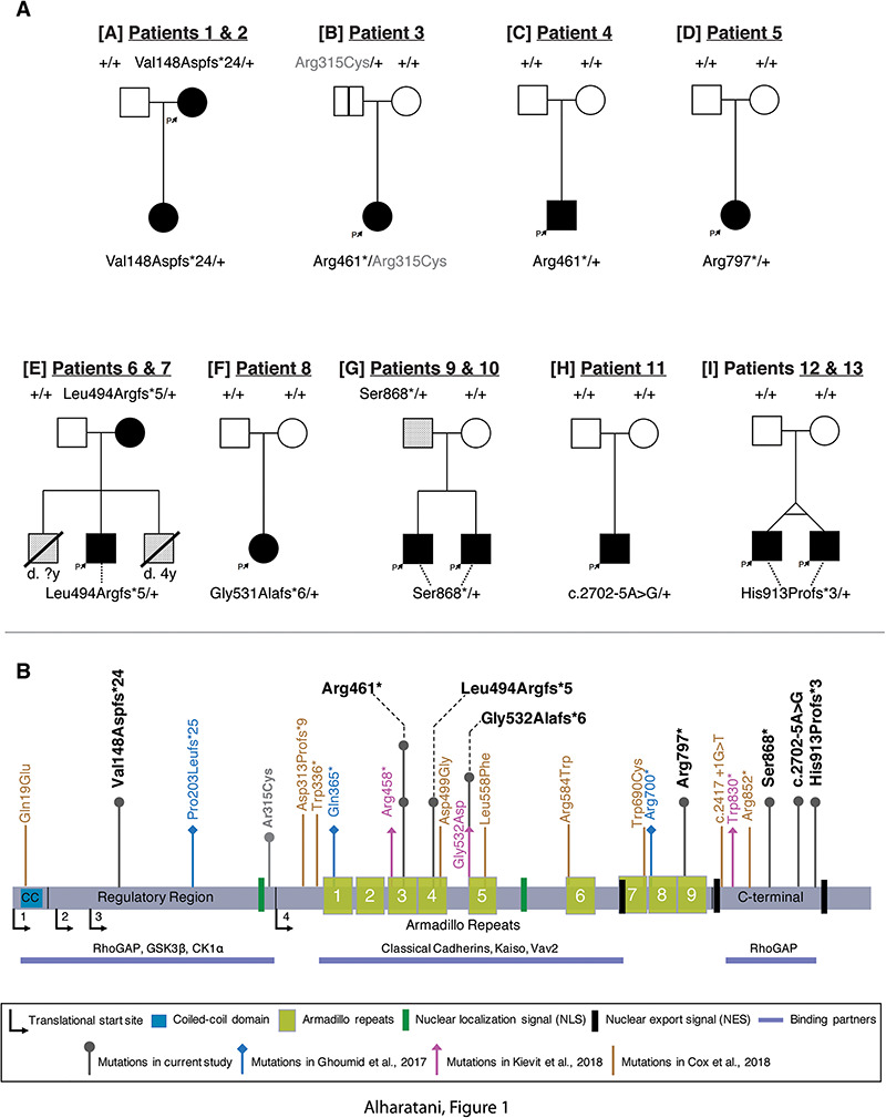
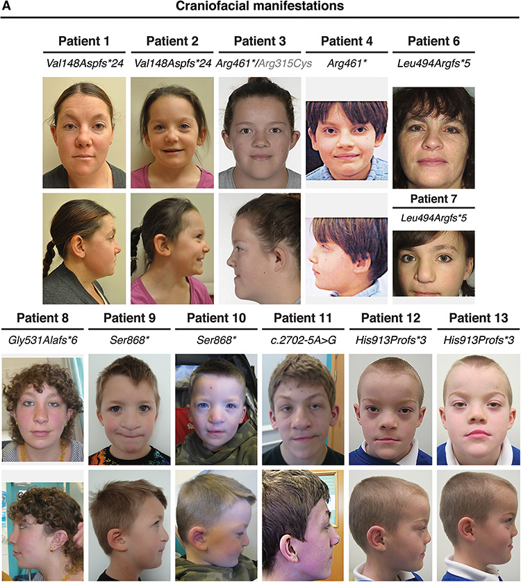
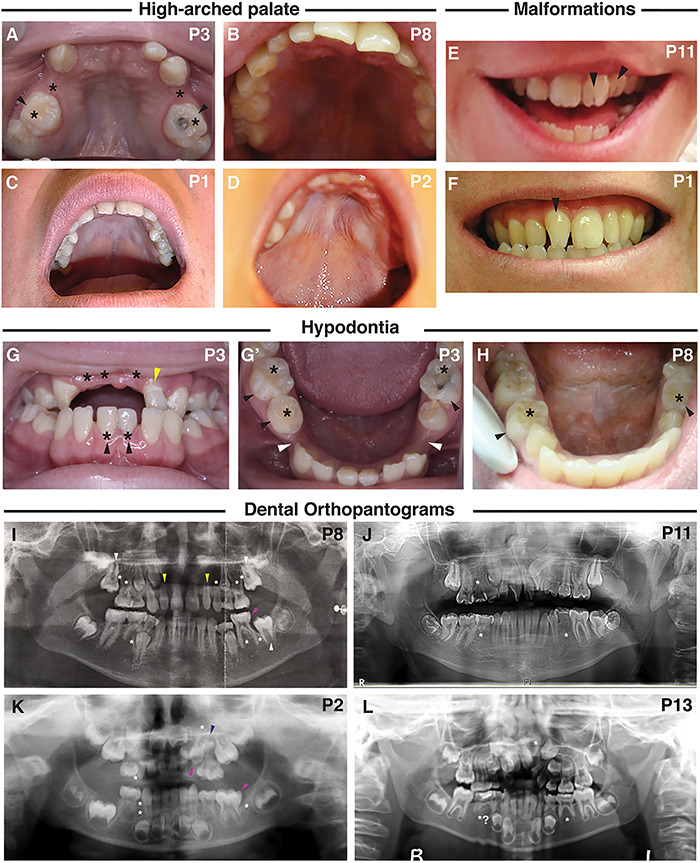
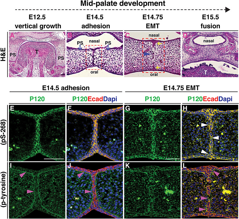
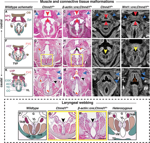
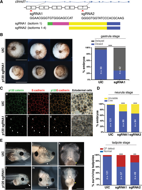
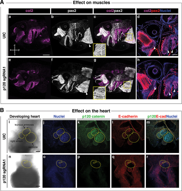
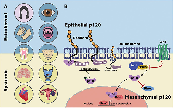
CTNND1 encodes the p120-catenin (p120) protein, which has a wide range of functions, including the maintenance of cell-cell junctions, regulation of the epithelial-mesenchymal transition and transcriptional signalling. Due to advances in next-generation sequencing, CTNND1 has been implicated in human diseases including cleft palate and blepharocheilodontic (BCD) syndrome albeit only recently. In this study, we identify eight novel protein-truncating variants, six de novo, in 13 participants from nine families presenting with craniofacial dysmorphisms including cleft palate and hypodontia, as well as congenital cardiac anomalies, limb dysmorphologies and neurodevelopmental disorders. Using conditional deletions in mice as well as CRISPR/Cas9 approaches to target CTNND1 in Xenopus, we identified a subset of phenotypes that can be linked to p120-catenin in epithelial integrity and turnover, and additional phenotypes that suggest mesenchymal roles of CTNND1. We propose that CTNND1 variants have a wider developmental role than previously described and that variations in this gene underlie not only cleft palate and BCD but may be expanded to a broader velocardiofacial-like syndrome.


Figures

Figure 1
Figure 1
Pedigrees and identification of CTNND1 variants. (A) Pedigrees of individuals with identified variants. Family identifications, in brackets [A-I], and patient identification numbers correlate with Table 1. Filled boxes indicate affected individuals demonstrating collective phenotypes described in our cohort. A blank box with a vertical black line indicates an asymptomatic carrier (clinically unaffected). A box with an oblique line indicates a deceased individual. Lightly shaded boxes indicate individuals affected with one or more of the conditions described. (B) Schematic representation of the human p120-catenin protein structure and its domains. The variants described in our cohort are shown above the protein with a dark gray arrow. The light gray arrow with the (p.Arg315Cys) variant indicates the other CTNND1 mutation found in Patient 3 which was inherited from the unaffected father [A]. Arrows in blue, pink and brown represent the variants and their locations reported in Ghoumid et al. (2), Kievit et al. (1) and Cox et al. (3), respectively.
Figure 2
Figure 2
Clinical presentation of individuals with a CTNND1 mutation. Facial photos (frontal and profile) show craniofacial features of patients. Note the narrow up-slanting palpebral fissures in Patients 3, 4 and 7–13; the hooded eyelids in Patients 3, 4 and 8–13; telecanthus in Patients 3, 4 and 9–13; the high arched eyebrows in Patients 1, 2, 6–8 and 11–13 and the thin lateral eyebrows in Patients 1 and 5–11. Patients 1 and 4 had missing eyelashes medially from the inner canthus; Patients 1, 2, 5 and 7 have distichiasis (double row of lashes), and mild ectropion of the lower eyelids was seen in Patients 1, 5 and 6. As evident, no patient shows signs of hair sparsity. Most patients had wide nasal bridges with broad nasal tips, while Patients 1, 2, 8 and 11 were also diagnosed with congenital choanal atresia. Patients 1, 2, 7–9, 11 and 12 showed thin upper lips, and while mid-face hypoplasia was observed, Patients 1, 3, 8, 11 and 13 also had mandibular prognathism. Scars from cleft lip operations are seen in Patients 7 and 9–13. Patient 3 was born with a sub-mucous cleft palate, a bifid uvula and VPI.
Figure 3
Figure 3
Dental manifestations and intra-oral phenotypes of patients with a CTNND1 mutation. (AD) A high-arched palate was seen, shown are palates of Patients 1, 2, 3 and 8. (E, F) Abnormalities in the morphology of the dentition included: fissured incisors in patient 11 (E, black arrowheads) and rotation of the incisors from the normal alignment shown in the non-cleft Patient 1 (F, black arrowhead). (G, H) Hypodontia (tooth agenesis) was a common phenotype, indicated by the black asterisk. Black arrowheads indicate retained primary teeth. Patient 3 also has a diminutive upper left lateral incisor (G, yellow arrowhead) and wide inter-dental spacing (G′, white arrowheads). (IL) Dental orthopantograms (OPGs); missing teeth are indicated by white asterisks; diminutive teeth by yellow, macrodont teeth by magenta and supernumerary teeth by blue arrowheads, respectively. (I) OPG of Patient 8 at age 11 shows eight missing permanent teeth (white asterisks) and shows the eruption of the second permanent molars (white arrowheads) in place of the missing first permanent molars. Also shown are diminutive upper right and left lateral incisors (peg-shaped) (yellow arrowheads) and a macrodont lower left second primary molar (magenta arrowhead). (J) OPG of Patient 11, at the age of 14, shows three missing permanent teeth (white asterisks), an ectopic maxillary left permanent canine and rotated maxillary centrals and left lateral incisors and dilacerated roots of the lower second permanent molars. (K) OPG of Patient 2, taken at 4 years, shows missing teeth including a missing lower left first permanent molar (white asterisks); a reported macrodont upper left primary canine (magenta arrowhead) with an underlying missing successor (white asterisk); a macrodont lower left second primary molar (magenta arrowhead) and a supernumerary tooth (blue arrowhead). (L) OPG for Patient 13, taken at 7.5 years, confirms the absence of the upper left permanent lateral incisor and possibly the lower second permanent premolars.
Figure 4
Figure 4
P120-catenin is expressed during relevant stages of human embryonic development. CTNND1 mRNA in situ hybridization at human Carnegie stages 13 (CS13) (AC) and 21 (DG). (A) Right lateral view of a CS13 human embryo, CTNND1 mRNA is strongly expressed in the head in all three pharyngeal arches (PA1, PA2 and PA3) and limb buds. Expression is specifically strong around the nasal placode and the maxillary and mandibular prominences. (B) Left lateral view, P120 is strongly expressed in the developing heart, frontonasal process, the trigeminal ganglion and the tenth cranial nerve. (C) P120 is ubiquitously expressed in the developing brain region in the rhombomeres, the forebrain and midbrain. (D–G) Coronal section through the head of a CS21 human embryo through a mid-palatal plane. (D) Strong expression is seen in the intrinsic muscles of the tongue: the superior longitudinal (magenta arrowhead), the transversal muscles of the tongue (black arrowhead) and the extrinsic genioglossus muscle (blue arrowhead). (E) CTNND1 mRNA is strongly expressed in the epithelium of the developing tooth bud. (F) CTNND1 is expressed on the dorsal epithelium of the palatal shelf (arrowhead) and in the epithelium of the tongue. (G) Expression is seen in the cardiomyocytes of the ventricular wall and the interventricular septum and in the cells of the endocardium (arrowhead). Scale bars = 100 μm. Abbreviations: PA1, first pharyngeal arch; PA2, second pharyngeal arch; PA3, third pharyngeal arch; Tg, trigeminal ganglion; Mx, maxillary process; Md, mandibular process; CN X, tenth cranial nerve; ULB, upper limb bud; S, somites; LLB, lower limb bud; NP, nasal placode; H, heart, FNP, frontonasal process; Tb, mandibular tooth bud; PS, palatal shelf; T, tongue; IVS, interventricular septum; VW, ventricular wall.
Figure 5
Figure 5
Expression of phosphorylated p120-catenin predicts fusion of the palatal seam. (AL) All images are coronal sections of CD1 wild-type murine embryos at consecutive stages of palatal development. (A–D) H&E staining illustrates successive stages of palatogenesis from embryonic day (E) 12.5 to E15.5. (B) At E14.5, following horizontal elevation, the opposing palatal shelves (blue arrows) meet and adhere to form the MES. (C) EMT occurs at E14.75 when the MES breaks down, forming epithelial islands (blue arrowhead); the nasal and oral epithelial triangles form (yellow arrows). (D) At E15.5 palatal shelves are fused. Red box in (B) marks the regions shown in (E, F, I and J). Red box in (C) marks the regions shown in (G, H, K and L). (E–L) Immunofluorescent staining for either pS-268 or p-tyrosine p120-catenin antibodies (green) shown independently in (E, G, I and K), or in a merge with E-cadherin antibody staining (red) and DNA/DAPI stain (blue) (F, H, J and L). (E, F, I and J) At E14.5, both forms of p120-catenin are expressed, with pS-268 strongly expressed in the periderm at the midline seam co-localizing with E-cadherin (E and F), while p-tyrosine clearly enriched in the area marking the border between the epithelial and mesenchymal populations (I, J, pink arrowheads). (G, H, K and L) At E14.75, pS-268 p120-catenin is strongly expressed in the epithelial islands and the oral and nasal epithelial triangles; this is co-localised with E-cadherin during EMT and endocytosis, while p120-catenin expression remains in some areas (H, white arrowheads). In contrast, p-tyrosine p120-catenin expression surrounds E-cadherin positive epithelial islands, while E-cadherin expression has disappeared in the intervening mesenchymal cells (L, pink arrowheads). Scale bars = 50 μm. Abbreviations: T, tongue; PS, palatal shelf.
Figure 6
Figure 6
Heterozygous loss of p120-catenin leads to structural changes in the laryngeal apparatus. (AO) Progression of the pharyngeal and laryngeal anomalies, (A, F and K) Schematics show the organization of the wild-type oropharynx from the more rostral (A) to caudal (K) planes. H&E staining of coronal sections through control (B, G, L: Ctnnd1fl/+) and heterozygous mutants (C, H, M: β-actin::cre/+; Ctnnd1fl/+) littermate at postnatal stage (P1). (B and C) The SPC (blue arrowhead) and PLP (red arrowhead) in mutants are disorganized with an increased thickness in the PLP cranio-caudally (C) as compared with the controls (B). (G and H) The FVC (vestibular folds) are well defined in the controls with abundant ligaments (G, red arrowhead). The FVC are fused in the mutant mice (H, black arrowhead) with ill-defined vestibular ligaments (H, red arrowhead). (L and M) The muscle attachments (blue arrowheads) superior to the FVC (black arrowhead) are well organized bilaterally in the controls surrounding the COC (L). Caudally, when the FVC separated in the mutants, it appeared hypoplastic (black arrowhead) as did the COC. The muscles (blue arrowheads) were ectopically fused to the LVP, producing an appearance of a ‘high-arched’ epiglottal area (M, orange hollow arrowhead). (D, E, I, J, N and O) Neural crest-specific mutants showed comparable laryngeal phenotype. μCT soft tissue scans of E16.5 control (D, I, N: Ctnnd1fl/+) or neural-crest-specific (E, J, O: Wnt1::cre/+; Ctnnd1fl/+) heterozygous mutant littermates. (D and E) Compare the PLP in control (D) to the very thick PLP muscle seen in mutant (E, red arrowheads). Compare the SPC in control (D) to the disorganized and hypoplastic SPC muscles seen in mutants (E, blue arrowheads). (I and J) Laryngeal webbing was observed in mutant TVF (J, yellow arrowhead) compared with parallel TVF in control littermate (I, yellow arrowhead). (N and O) Note aberrant muscle attachments (blue arrowheads) in (O) compared with control (N). Control (N) epiglottal region compared with the high-arched epiglottal area observed in mutant littermate (O, orange hollow arrowhead). (P–S) The laryngeal webbing phenotype. (P and S) Schematic representations of the wild-type (P) and mutant (S) anatomy at the vocal folds (TVF) from yellow-boxed insets in (G) and (H), respectively. (Q and R) H&E staining of coronal sections through control (Q: Ctnnd1fl/+) and heterozygous mutant (R: β-actin::cre/+;Ctnnd1fl/+) littermate at P1. (Q) In controls, well-defined VLs run parallel to the true vocal fold/cords (TVF). Underlying, the vocalis muscle (VM) and the thyroarytenoid muscle (TAM) are clearly attached and well-organised. (R) Laryngeal webbing is seen in the heterozygous mutant mice, where the VLs accumulate at a thin contact point (black arrowhead), thus perturbing the correct muscle attachments of the VM and TAM. Scale bars = 100 μm. Abbreviations: PLP, palatopharyngeus muscle; TAM, thyroarytenoid muscle; VM, vocalis muscle; HB, hyoid bone; Epi, epiglottis; OB, occipital bone; LVP, levator veli palatini muscle; AEF, aryepiglottic fold; FVC, false vocal cord; CC, cricoid cartilage; TC, thyroid cartilage; AC, arytenoid cartilage.
Figure 7
Figure 7
Ctnnd1 knockouts in Xenopus give rise to craniofacial and heart defects. (A) Embryos were injected at the one-cell stage with sgRNAs, sgRNA1 and sgRNA2 targeting exons 3 and 7, respectively. (B) Ventral view showing blastopores at stage 11. Embryos injected with sgRNA1 had delayed blastopore closure (bottom row) compared to un-injected controls (UIC) (top row). The bar chart shows quantitation. Scale bars = 100 μm. (C) Confocal sections through the apical surface of ectodermal cells at stage 11 of embryos injected with sgRNA1 (e–h) and UICs (a–d). (C) (a–d) p120-catenin (a, green) is expressed in puncta at the cell membranes. E-cadherin (b, red) is expressed more evenly through the cell membranes. Both are colocalized at the cell–cell interface (c, d). Endogenous levels of p120-catenin and E-cadherin are diminished at the cell–cell interface in the sgRNA1-injected embryos (e, f). Residual p120-catenin and E-cadherin are seen in a spot-like pattern, only at the tricellular junctions (e–h, white arrowheads). (D) p120-catenin depletion led to lethality in embryos by the neurula stage. (E) Stage 46 tadpoles. (E) (i, l) Lateral views show a flattened profile in p120 CRISPR tadpoles (l) compared with UICs (i). (E) (j, m) Frontal views showing a reduction in the size of mouth opening and a persistent cement gland (white arrowhead) in p120 CRISPR tadpoles (m) compared with UICs (j). (E) (k, n) Ventral views showing a reduction in the size of craniofacial cartilages, altered cardiac looping (black-dashed outline) and altered gut coiling (yellow arrowhead) in p120 CRISPR tadpoles (n) compared to UICs (k). Quantification of craniofacial defects in UIC and p120 depleted tadpoles. Scale bars = 100 μm. ****P < 0.0001; ***P < 0.001.
Figure 8
Figure 8
Ctnnd1 knockouts in Xenopus give rise to altered morphogenesis of the muscles and heart. (A) Immunofluorescent staining for collagen 2 (col2, magenta), muscle/pax2 (white) and nuclei (DAPI, blue); (a, anterior; p, posterior; d, dorsal; v, ventral). (A) (a, e) A lateral view of col2-positive branchial cartilages in UIC (a) and p120 CRISPR mutant (e) reveals the hypoplasia of mutant cartilages; however, cell morphology appears normal in p120 CRISPR mutants (h) (d and h, white arrowheads). (A) (b, c, f and g) Pax2-expressing muscles revealed a defect in the fibril organization of the rectus abdominus muscle in the p120 CRISPR tadpoles (f, white arrowhead) compared with the UIC muscles (b, white arrowhead); note insets in (c, g). (B) Ventral views of hearts of stage 46 tadpoles. Immunofluorescent staining for p120-catenin (green), E-cadherin (red) and DNA (blue). (B) (i–m) Controls; (n–r) p120 CRISPR mutant tadpoles. Morphologic defects are evident in the size of the heart and directionality of the loops (compare control heart (i) to mutant heart (n), yellow-dashed outlines). (B) (k, p) p120-catenin is strongly expressed in the heart of UIC tadpoles (k) but is lost in p120 CRISPR tadpoles (p). (B) (l, q) Note the absence of E-cadherin in the control and mutant hearts. Scale bars = 100 μm.
Figure 9
Figure 9
Model of CTNND1 function in systemic disease. (A) CTNND1 mutations are not only implicated in conditions that affect epithelial structures but also systemic conditions that originate from mesenchymal roles of p120-catenin. Structures in pink circles have been described in previous publications on CTNND1 (1,2); structures in blue circles have been implicated previously in CTNND1-related disorders (1,2) and in this study. Structures in yellow circles have been identified in this study. (B) BCD is primarily due to disturbances in E-cadherin/p120 interactions. The inclusion of other organ systems described here highlights the involvement of other known molecular functions of p120, such as its role in the WNT signalling pathway and its interactions with Rho-GTPases, demonstrating its mesenchymal roles in producing these systemic conditions.

References

    1. Kievit A., Tessadori F., Douben H., Jordens I., Maurice M., Hoogeboom J., Hennekam R., Nampoothiri S., Kayserili H., Castori M. et al. (2018) Variants in members of the cadherin-catenin complex, CDH1 and CTNND1, cause blepharocheilodontic syndrome. Eur. J. Hum. Genet., 26, 210–219. - PMC - PubMed
    1. Ghoumid J., Stichelbout M., Jourdain A.S., Frenois F., Lejeune-Dumoulin S., Alex-Cordier M.P., Lebrun M., Guerreschi P., Duquennoy-Martinot V., Vinchon M. et al. (2017) Blepharocheilodontic syndrome is a CDH1 pathway-related disorder due to mutations in CDH1 and CTNND1. Genet. Med., 19, 1013–1021. - PubMed
    1. Cox L.L., Cox T.C., Moreno Uribe L.M., Zhu Y., Richter C.T., Nidey N., Standley J.M., Deng M., Blue E., Chong J.X. et al. (2018) Mutations in the epithelial cadherin-p120-catenin complex cause Mendelian non-Syndromic cleft lip with or without cleft palate. Am. J. Hum. Genet., 102, 1143–1157. - PMC - PubMed
    1. Perez-Moreno M., Davis M.A., Wong E., Pasolli H.A., Reynolds A.B. and Fuchs E. (2006) p120-catenin mediates inflammatory responses in the skin. Cell, 124, 631–644. - PMC - PubMed
    1. Oas R.G., Xiao K., Summers S., Wittich K.B., Chiasson C.M., Martin W.D., Grossniklaus H.E., Vincent P.A., Reynolds A.B. and Kowalczyk A.P. (2010) p120-catenin is required for mouse vascular development. Circ. Res., 106, 941. - PMC - PubMed

Publication types

MeSH terms

Supplementary concepts