. 2020 Jul;161(7):1497-1517.
doi: 10.1097/j.pain.0000000000001866.

Pharmacological target-focused transcriptomic analysis of native vs cultured human and mouse dorsal root ganglia

Affiliations

Pharmacological target-focused transcriptomic analysis of native vs cultured human and mouse dorsal root ganglia

Andi Wangzhou et al. Pain. 2020 Jul.

Abstract

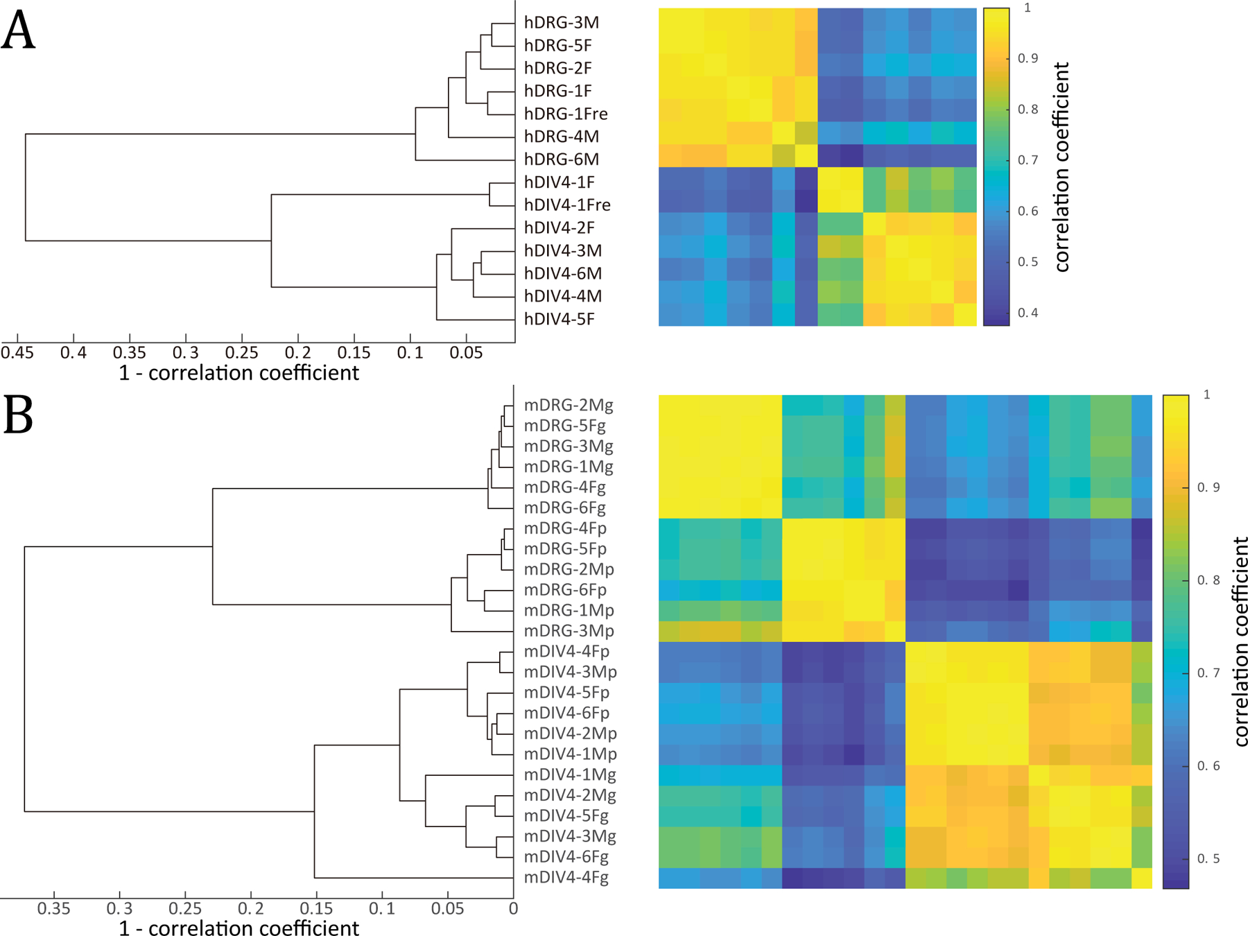
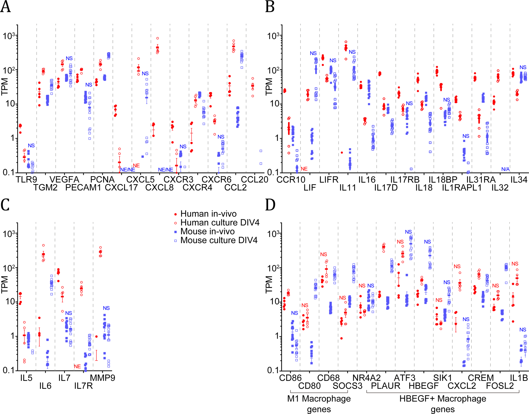
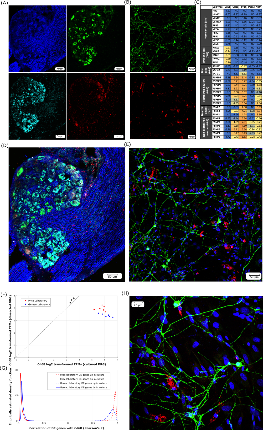
Dorsal root ganglion (DRG) neurons detect sensory inputs and are crucial for pain processing. They are often studied in vitro as dissociated cell cultures with the assumption that this reasonably represents in vivo conditions. However, to the best of our knowledge, no study has directly compared genome-wide transcriptomes of DRG tissue in vivo versus in vitro or between laboratories and culturing protocols. Comparing RNA sequencing-based transcriptomes of native to cultured (4 days in vitro) human or mouse DRG, we found that the overall expression levels of many ion channels and G-protein-coupled receptors specifically expressed in neurons are markedly lower although still expressed in culture. This suggests that most pharmacological targets expressed in vivo are present under the condition of dissociated cell culture, but with changes in expression levels. The reduced relative expression for neuronal genes in human DRG cultures is likely accounted for by increased expression of genes in fibroblast-like and other proliferating cells, consistent with their mitotic status in these cultures. We found that the expression of a subset of genes typically expressed in neurons increased in human and mouse DRG cultures relative to the intact ganglion, including genes associated with nerve injury or inflammation in preclinical models such as BDNF, MMP9, GAL, and ATF3. We also found a striking upregulation of a number of inflammation-associated genes in DRG cultures, although many were different between mouse and human. Our findings suggest an injury-like phenotype in DRG cultures that has important implications for the use of this model system for pain drug discovery.


Conflict of interest statement

The authors declare no conflicts of interest.

Figures

Figure 1.
Figure 1.
Hierarchical clustering of all human (A) and mouse (B) samples based on TPM-based whole genome gene abundances. A. Cultured and intact human DRG tissue samples are separated into two clusters. The outlier sample hDIV-1F and its paired dissected sample (hDRG-1F) were excluded from further analysis. B. Cultured and intact mouse DRG samples also segregate into separate clusters. Subclusters in the cultured DRG and dissected DRG clusters correspond to sample generated in Gereau and Price laboratories. The outlier sample mDIV4–4Fg shows moderate expression of neuronal genes, and clusters with other Gereau laboratory cultured samples when unrooted clustering is performed for cultured mouse DRG samples. (Sample id nomenclature -- Prefix: h - human; m - mouse; Infix: DRG - intact DRG samples; DIV4 – 4 days in vitro (4 DIV) DRG cultures; Suffix: M - male; F - female; p - Price laboratory; g - Gereau laboratory; re – repeated library preparation and sequencing.
Figure 2.
Figure 2.
Scatter plot and Venn diagrams showing a small amount of differential expression of GPCR genes (A), RK genes (B), and ion channel genes (C) in culture between the Price and Gereau laboratories. The number of genes consistently detected in RNA-sequencing assays for each laboratory are shown in Venn diagrams separated by gene families in (D). Expression levels of genes in all three families showed consistent correlation between the two laboratories: GPCR genes : Pearson’s R squared: 0.64, p < 0.01, RK genes : Pearson’s R squared: 0.81, p < 0.01, ion channel genes : Pearson’s R squared: 0.83, p < 0.01. Genes like Alk and Insrr are plotted on the diagonal, but marked as consistently detected only in Gereau laboratory samples. This is because they have comparable mean TPMs in samples from both laboratories, but are only consistently detected (in 5 or more samples out of 6) in the Gereau laboratory.
Figure 3.
Figure 3.
Empirical density distribution of log2 fold changes (ratio of means) for GPCRs, ion channels, RKs, and non-RK kinases in human (A) and mouse (B). RKs and kinases as a group are weakly de-enriched in human and weakly enriched in mouse cultures (in the context of mean expression). However, both GPCRs and ion channels are strongly de-enriched in both human and mouse cultures, likely because of the variety of these genes that are expressed in sensory neurons.
Figure 4.
Figure 4.
Expression levels in human and mouse intact vs. cultured DRGs. A wide diversity of genes involved in inflammation and proliferation, nerve and neuronal injury and repair, and immune signaling and response are profiled (A, B, and C). Key expressed genes for M1 macrophages and HBEGF+ macrophages are also shown (D). NS: | SSMD | <= 2, NE: not consistently detected for that condition, N/A: not applicable because orthologous gene not identified in that species.
Figure 5.
Figure 5.
Cd68 expression in intact vs. cultured mouse DRGs. RNAscope in situ hybridization imaging (20X) in pseudo-color for Cd68 (red) in combination with various neuronal markers including Calca (green), P2rx3 (cyan), and immunostained Nf200 (blue) in intact mouse DRG (A). RNAscope in situ hybridization imaging (20X) in pseudo-color for Cd68 (red) and immunostained Peripherin (green) and DAPI staining (blue) in cultured mouse DRG (B). log2 transformed expression levels of Cd68 and neuronal markers from mousebrain.org (C) in mouse DRG neuron and glial subtypes, and nervous system vascular and immune cells show Cd68 is detected only in macrophage-like cells (ND: Not Detectable). Overlay of images show that Cd68 mRNA is not expressed in neurons in either intact (D) or cultured (E) mouse DRG neurons. Expression levels (in log2-transformed TPMs) of Cd68 in intact versus cultured mouse DRGs are plotted (F) to show the consistent increase of Cd68 expression in cultures. Differentially expressed gene TPMs show strong correlation (or anti-correlation) to Cd68 abundance (in TPMs) (G), suggesting a consistent phenotype across samples and laboratories. Overlay of RNAscope in situ hybridization imaging (40X) for cultured mouse DRG (H) suggests Cd68+ cells have consistent shape and size with respect to DRG macrophages. Scale bar for 20X images equal to 50μm and 40x equal to 20μm.

References

    1. Abbadie C, Bhangoo S, De Koninck Y, Malcangio M, Melik-Parsadaniantz S, White FA. Chemokines and pain mechanisms. Brain research reviews 2009;60(1):125–134. - PMC - PubMed
    1. Amir R, Kocsis JD, Devor M. Multiple interacting sites of ectopic spike electrogenesis in primary sensory neurons. J Neurosci 2005;25(10):2576–2585. - PMC - PubMed
    1. Basbaum AI, Bautista DM, Scherrer G, Julius D. Cellular and molecular mechanisms of pain. Cell 2009;139(2):267–284. - PMC - PubMed
    1. Benjamini H, Hochberg Y. Controlling the false discovery rate: a practical and powerful approach to multiple testing. Journal of the royal statistical society: series B (Methodological) 1995;57(1):289–300.
    1. Berta T, Park CK, Xu ZZ, Xie RG, Liu T, Lu N, Liu YC, Ji RR. Extracellular caspase-6 drives murine inflammatory pain via microglial TNF-alpha secretion. J Clin Invest 2014;124(3):1173–1186. - PMC - PubMed