. 2020 May 15;201(10):1209-1217.
doi: 10.1164/rccm.201911-2105OC.

Localization of Macrophages in the Human Lung via Design-based Stereology

Affiliations

Localization of Macrophages in the Human Lung via Design-based Stereology

Patrick S Hume et al. Am J Respir Crit Care Med. .

Abstract

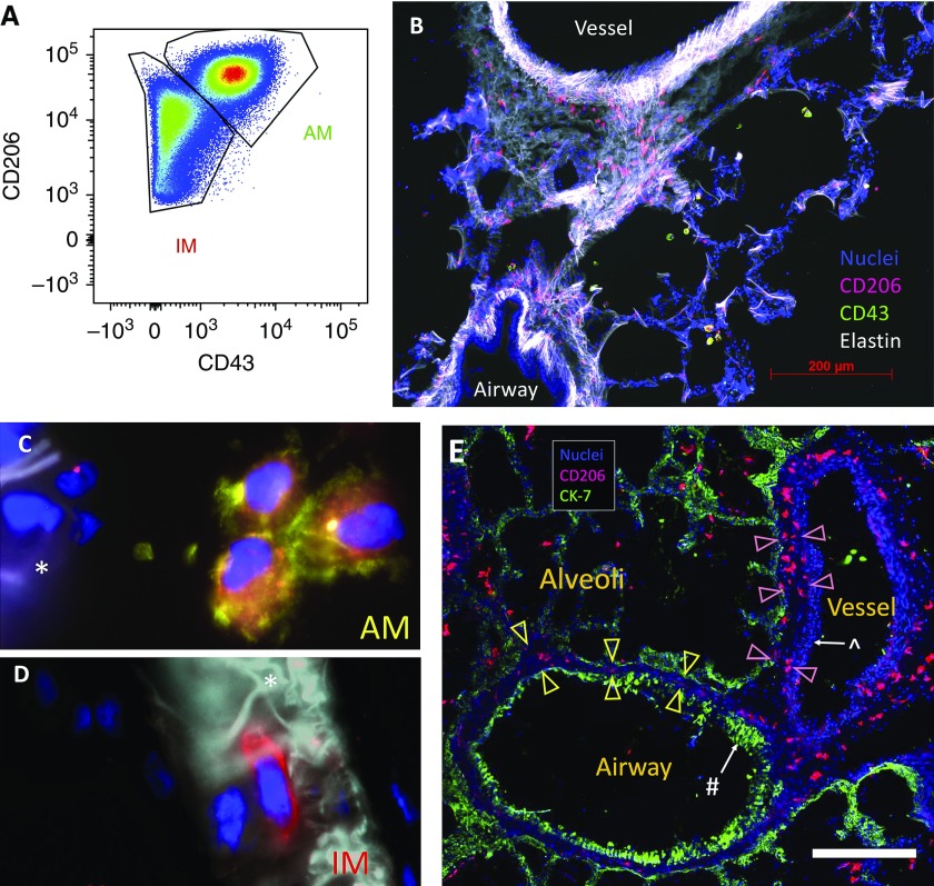
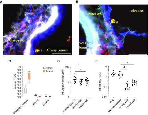
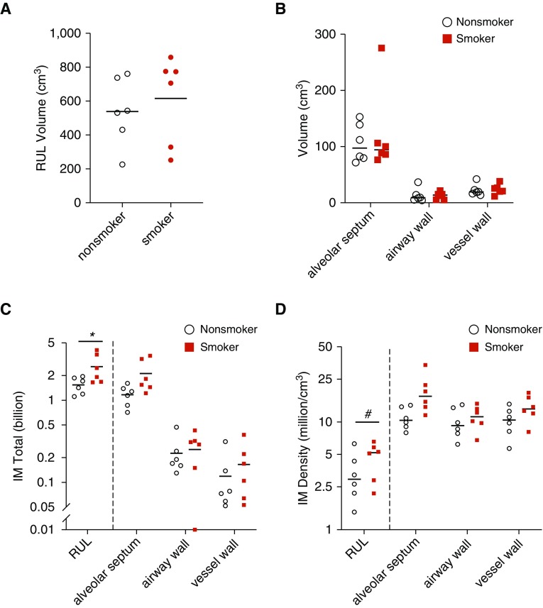
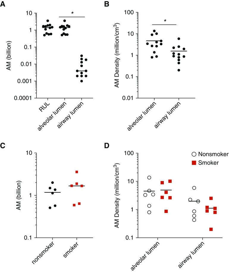
Rationale: Interstitial macrophages (IMs) and airspace macrophages (AMs) play critical roles in lung homeostasis and host defense, and are central to the pathogenesis of a number of lung diseases. However, the absolute numbers of macrophages and the precise anatomic locations they occupy in the healthy human lung have not been quantified.Objectives: To determine the precise number and anatomic location of human pulmonary macrophages in nondiseased lungs and to quantify how this is altered in chronic cigarette smokers.Methods: Whole right upper lobes from 12 human donors without pulmonary disease (6 smokers and 6 nonsmokers) were evaluated using design-based stereology. CD206 (cluster of differentiation 206)-positive/CD43+ AMs and CD206+/CD43- IMs were counted in five distinct anatomical locations using the optical disector probe.Measurements and Main Results: An average of 2.1 × 10⁹ IMs and 1.4 × 10⁹ AMs were estimated per right upper lobe. Of the AMs, 95% were contained in diffusing airspaces and 5% in airways. Of the IMs, 78% were located within the alveolar septa, 14% around small vessels, and 7% around the airways. The local density of IMs was greater in the alveolar septa than in the connective tissue surrounding the airways or vessels. The total number and density of IMs was 36% to 56% greater in the lungs of cigarette smokers versus nonsmokers.Conclusions: The precise locations occupied by pulmonary macrophages were defined in nondiseased human lungs from smokers and nonsmokers. IM density was greatest in the alveolar septa. Lungs from chronic smokers had increased IM numbers and overall density, supporting a role for IMs in smoking-related disease.

Keywords: alveolar; cigarette smokers; emphysema; interstitial; morphometry.


Figures

Figure 1.
Figure 1.
Identification of pulmonary macrophage populations. (A) Flow cytometry of CD45 (cluster of differentiation 45)-positive/CD3/CD19/CD15/DAPI myeloid cells from human lung digest illustrating that airspace macrophages (AMs) and interstitial macrophages (IMs) are CD206+ but only AMs are CD43+. (BE) Immunofluorescent staining of human lung tissue to localize AMs and IMs. (B) The luminal airway epithelial surface was elastin negative, whereas the vessel wall was elastin positive with a characteristic appearance, enabling the rapid, visual distinction between airways and vessels. CD206+/CD43+ AMs (red and green, respectively) are in the airspaces and CD206+/CD43 IMs (red) are easily identified in the interstitium. (C and D) High-power magnification shows CD206+/CD43+ AMs (red and green, respectively) located in the alveolus (C) and CD206+/CD43 (red) IMs located within the septum (D). The asterisks denote the alveolar septum. (E) Demonstration of airways and vessel wall compartments. CK-7 (green) highlights epithelium. CD206 (red) identifies macrophages. Nuclei are shown in blue. The # symbol denotes airway epithelium, whereas the ^ symbol denotes vessel endothelium. Airway-associated IMs are located within the subepithelial tissue (between yellow arrowheads), whereas vessel-associated IMs are within the subendothelial tissue (between pink arrowheads). Scale bar, 400 μm. CK = cytokeratin.
Figure 2.
Figure 2.
Quantification of interstitial macrophages (IMs) in lung subcompartments using stereology. (A) CD206 (cluster of differentiation 206)-positive IMs (red asterisks) are identified within the airway wall with CD206+/CD43+ airspace macrophages (red and green make yellow; #) located in the airway lumen. (B) Red CD206+ IMs visualized within the vessel wall and CD206+/CD43+ airspace macrophages (red and green make yellow; #) are visualized within the alveolus. Scale bars, 50 μm. (C) Volume fraction of each lung subcompartment was calculated via stereology. Diffusing airspaces consisted of alveoli, alveolar ducts, and respiratory bronchioles. (D) Density of IMs in lung subcompartments. *P = 0.02 and #P = 0.06. (E) Total number of IMs counted in human lung per subcompartment. &P < 0.0001 and +P < 0.003. RUL = right upper lobe.
Figure 3.
Figure 3.
Comparison of interstitial macrophage (IM) number and location in nonsmokers versus chronic cigarette smokers. (A and B) Lungs were structurally similar between smokers and nonsmokers with equivalent total volume (A) and volume of lung subcompartments (B). Plotted is mean ± SEM. (C) IM total number was increased overall in cigarette smokers. (D) IM density was increased overall in cigarette smokers with a trend toward increase in the alveolar septa. (C and D) *P = 0.03 and #P = 0.06. The y-axis is plotted as log10 scale. RUL = right upper lobe.
Figure 4.
Figure 4.
Quantification of airspace macrophages (AMs) in human lung. (A) The overwhelming majority of AMs were present in the diffusing airspaces rather than the airway lumens. (B) The density of AMs in the diffusing airspaces was significantly greater than that within the airway lumens (by t test). (C) Total AM number was equivalent in nonsmokers and smokers. (D) AM density in tissue subcompartments was not different in smokers versus nonsmokers. Diffusing airspace was defined to include alveoli, alveolar ducts, and respiratory bronchioles. *P < 0.05. The y-axis is plotted as log10 scale. RUL = right upper lobe.

Comment in

References

    1. Amit I, Winter DR, Jung S. The role of the local environment and epigenetics in shaping macrophage identity and their effect on tissue homeostasis. Nat Immunol. 2016;17:18–25. - PubMed
    1. Murray PJ, Wynn TA. Protective and pathogenic functions of macrophage subsets. Nat Rev Immunol. 2011;11:723–737. - PMC - PubMed
    1. Wynn TA, Vannella KM. Macrophages in tissue repair, regeneration, and fibrosis. Immunity. 2016;44:450–462. - PMC - PubMed
    1. Sabatel C, Radermecker C, Fievez L, Paulissen G, Chakarov S, Fernandes C, et al. Exposure to bacterial CpG DNA protects from airway allergic inflammation by expanding regulatory lung interstitial macrophages. Immunity. 2017;46:457–473. - PubMed
    1. Chakarov S, Lim HY, Tan L, Lim SY, See P, Lum J, et al. Two distinct interstitial macrophage populations coexist across tissues in specific subtissular niches. Science. 2019;363:eaau0964. - PubMed

Publication types

MeSH terms