

Electrostatic Environment of Proteorhodopsin Affects the pKa of Its Buried Primary Proton Acceptor
- PMID: 32197061
- PMCID: PMC7176579
- DOI: 10.1016/j.bpj.2020.02.027
Electrostatic Environment of Proteorhodopsin Affects the pKa of Its Buried Primary Proton Acceptor
Abstract
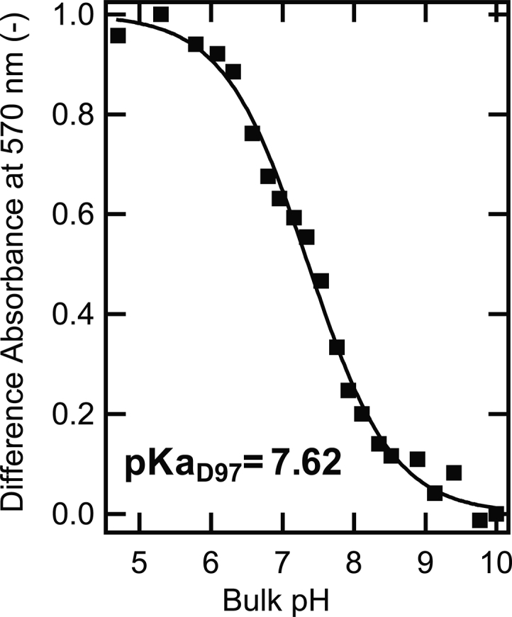
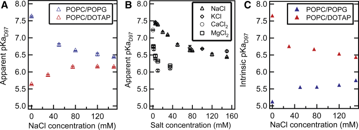
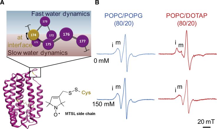
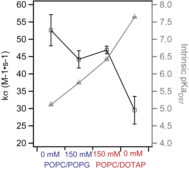
The protonation state of embedded charged residues in transmembrane proteins (TMPs) can control the onset of protein function. It is understood that interactions between an embedded charged residue and other charged or polar residues in the moiety would influence its pKa, but how the surrounding environment in which the TMP resides affects the pKa of these residues is unclear. Proteorhodopsin (PR), a light-responsive proton pump from marine bacteria, was used as a model to examine externally accessible factors that tune the pKa of its embedded charged residue, specifically its primary proton acceptor D97. The pKa of D97 was compared between PR reconstituted in liposomes with different net headgroup charges and equilibrated in buffer with different ion concentrations. For PR reconstituted in net positively charged compared to net negatively charged liposomes in low-salt buffer solutions, a drop of the apparent pKa from 7.6 to 5.6 was observed, whereas intrinsic pKa modeled with surface pH calculated from Gouy-Chapman predictions found an opposite trend for the pKa change, suggesting that surface pH does not account for the main changes observed in the apparent pKa. This difference in the pKa of D97 observed from PR reconstituted in oppositely charged liposome environments disappeared when the NaCl concentration was increased to 150 mM. We suggest that protein-intrinsic structural properties must play a role in adjusting the local microenvironment around D97 to affect its pKa, as corroborated with observations of changes in protein side-chain and hydration dynamics around the E-F loop of PR. Understanding the effect of externally controllable factors in tuning the pKa of TMP-embedded charged residues is important for bioengineering and biomedical applications relying on TMP systems, in which the onset of functions can be controlled by the protonation state of embedded residues.
Copyright © 2020. Published by Elsevier Inc.
Figures
References
-
- Luecke H., Richter H.-T., Lanyi J.K. Proton transfer pathways in bacteriorhodopsin at 2.3 angstrom resolution. Science. 1998;280:1934–1937. - PubMed
-
- Yoshikawa S., Shinzawa-Itoh K., Tsukihara T. Redox-coupled crystal structural changes in bovine heart cytochrome c oxidase. Science. 1998;280:1723–1729. - PubMed
-
- Churg A.K., Warshel A. Control of the redox potential of cytochrome c and microscopic dielectric effects in proteins. Biochemistry. 1986;25:1675–1681. - PubMed
-
- Warshel A., Sharma P.K., Parson W.W. Modeling electrostatic effects in proteins. Biochim. Biophys. Acta. 2006;1764:1647–1676. - PubMed
Publication types
MeSH terms
Substances
LinkOut - more resources
Full Text Sources
Research Materials
