. 2020 Mar 18;13(3):48.
doi: 10.3390/ph13030048.

Administration of Bacterial Lipopolysaccharide during Early Postnatal Ontogenesis Induces Transient Impairment of Long-Term Synaptic Plasticity Associated with Behavioral Abnormalities in Young Rats

Affiliations

Administration of Bacterial Lipopolysaccharide during Early Postnatal Ontogenesis Induces Transient Impairment of Long-Term Synaptic Plasticity Associated with Behavioral Abnormalities in Young Rats

Tatyana Y Postnikova et al. Pharmaceuticals (Basel). .

Abstract

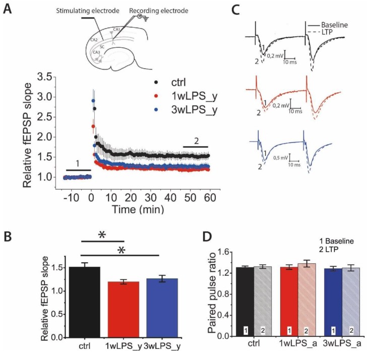
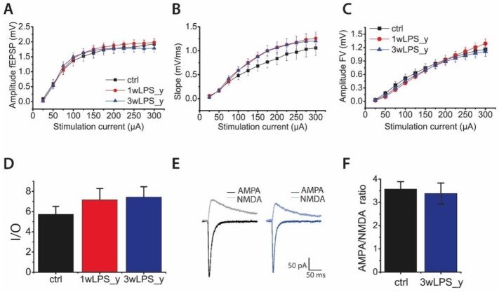
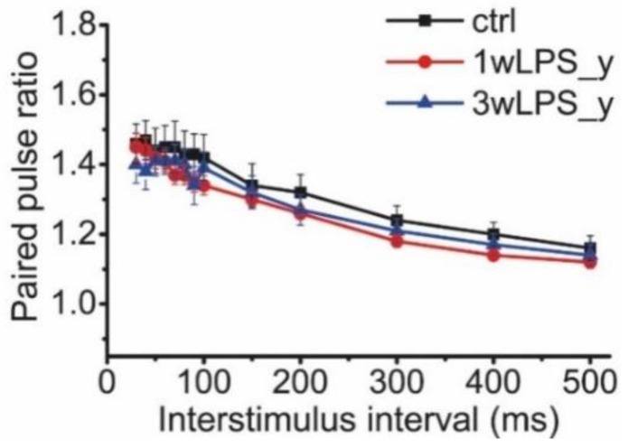
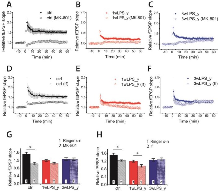
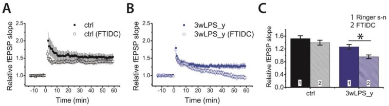
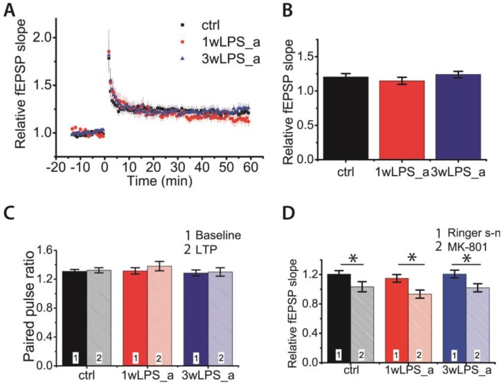
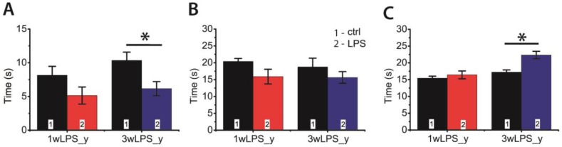
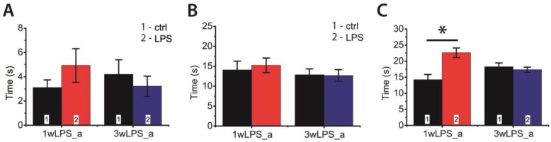
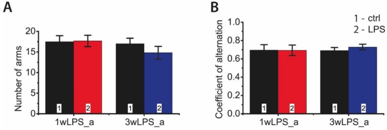
Infectious diseases in early postnatal ontogenesis often result in cognitive impairments, particularly learning and memory. The essential foundation of learning and memory is long-term synaptic plasticity, which depends on N-methyl-D-aspartate (NMDA) receptors. In the present study, bacterial infection was modeled by treating rat pups with bacterial lipopolysaccharide (LPS, 25 µg/kg) three times, during either the first or the third week of life. These time points are critical for the maturation of NMDA receptors. We assessed the effects of LPS treatments on the properties of long-term potentiation (LTP) in the CA1 hippocampus of young (21-23 days) and adolescent (51-55 days) rats. LTP magnitude was found to be significantly reduced in both groups of young rats, which also exhibited investigative and motor behavior disturbances in the open field test. No changes were observed in the main characteristics of synaptic transmission, although the LTP induction mechanism was disturbed. In rats treated with LPS during the third week, the NMDA-dependent form of LTP was completely suppressed, and LTP switched to the Type 1 metabotropic glutamate receptor (mGluR1)-dependent form. These impairments of synaptic plasticity and behavior were temporary. In adolescent rats, no difference was observed in LTP properties between the control and experimental groups. Lastly, the investigative and motor behavior parameters in both groups of adult rats were similar.

Keywords: bacterial lipopolysaccharide; early life; hippocampus; long-term potentiation; open field test.


Conflict of interest statement

The authors declare no conflict of interest. The funders had no role in the design of the study; in the collection, analyses, or interpretation of data; in the writing of the manuscript, or in the decision to publish the results.

Figures

Figure 1
Figure 1
Hippocampal long-term potentiation (LTP) in young rats is weakened after the administration of lipopolysaccharide (LPS) in early postnatal ontogenesis. (A) The normalized field excitatory postsynaptic potentials (fEPSP) slope after theta-burst stimulation (TBS) in control (ctrl) and experimental animals injected with LPS during the first (1wLPS_y) or third (3wLPS_y) postnatal week. The insert (above) shows the positions of electrodes in the hippocampus. (B) Diagram illustrating the difference in LTP values between control and experimental animals (one-way ANOVA: F2,48 = 3.30; p < 0.05; the significant difference with the control group: * p < 0.05). (C) Representative examples of paired fEPSP responses before (1) and after (2) TBS in control and experimental animals. (D) The paired-pulse ratio (PPR) of the fEPSP amplitudes before and after the induction protocol did not change in either control or experimental rats. Repeated measures ANOVA: F2,49 = 2.53; p = 0.09 (control, n = 14; 1wLPS_y, n = 18; 3wLPS_y, n = 20).
Figure 2
Figure 2
The main features of excitatory synaptic transmission in hippocampal pyramidal neurons are not altered after LPS treatment. The relationships between stimulation current and fEPSP amplitudes (A), slopes (B), and presynaptic fiber volley (FV) amplitude (C) recorded from the hippocampal CA1 area. (D) The diagram shows the maximum slope of the input/output (I/O) curve in different groups. (E) Representative examples of evoked EPSCs recorded at −80 mV and +40 mV from control rats and rats treated with LPS. The AMPA component was obtained by measuring the excitatory postsynaptic current (EPSC) peak amplitude at −80 mV in the presence of the GABAAR blockers bicuculline (10 μM) and picrotoxin (50 μM). The N-methyl-D-aspartate (NMDA) component was obtained by measuring the EPSC peak amplitude at +40 mV in the presence of gamma-aminobutyric acid type A receptor (GABAa) blockers and the AMPAR blocker, CNQX (20 μM). (F) The diagram illustrates the AMPA/NMDA ratio in the control and 3wLPS_y groups. Error bars indicate standard errors SE.
Figure 3
Figure 3
The PPRs of the fEPSPs evoked at different interstimulus intervals in the range from 30 to 500 ms in the control and experimental groups. Repeated measures ANOVA: F24,336 = 1.23; p = 0.22. Ctrl (n = 9), 1wLPS_y (n = 12), 3wLPS_y (n = 10).
Figure 4
Figure 4
The NMDA receptor (NMDAR)-dependent mechanism of LTP induction is disrupted in the CA1 hippocampus of juvenile animals treated with LPS during the third postnatal week. (AC) The normalized fEPSP slope in the control and experimental groups in the presence of the NMDAR blocker Dizocilpine (MK-801) (10 μM) before and after TBS. (DF) The relative fEPSP slope in the control and experimental groups in the presence of ifenprodil (If, 3 μM), a selective GluN2B-containing NMDAR antagonist, before and after TBS. (GH) Diagrams illustrating the magnitude of plasticity in the control and experimental groups in the presence of MK-801 or ifenprodil. Two-way ANOVA following Tukey post hoc tests were used. * p < 0.05.
Figure 5
Figure 5
The effect of ifenprodil on eEPSCs in CA1 pyramidal neurons. (A) Representative examples of evoked NMDAR-mediated EPSCs recorded at +40 mV in the presence of bicuculline (10 μM), picrotoxin (50 μM), and CNQX (20 μM) in the control and 3wLPS_y groups. (B) The same traces as in (A) normalized by amplitude. Note that ifenprodil decreases tau decay. (C,D) The effect of ifenprodil on amplitude and area under the curve of NMDAR-mediated eEPSC. Dot points represent data from individual cells. Error bars indicate SE.
Figure 6
Figure 6
Metabotropic glutamate receptor (mGluR)-dependent form of LTP presents in the hippocampal CA1 area of juvenile rats administered with LPS during the third postnatal week. Diagrams showing the normalized slope of fEPSP in the control (A) and experimental (B) groups. Note that FTIDC (4-[1-(2-fluoropyridin-3-yl)-5-methyltriazol-4-yl]-N-methylN-propan-2-yl-3,6dihydro-2H-pyridine-1-carboxamide) (5 μM) affected LTP only in the 3wLPS_y group. (C) Diagram illustrating the effect of FTIDS on LTP values in different groups of rats, respectively * p < 0.05 (t-test).
Figure 7
Figure 7
LTP properties are not disturbed in adolescent rats administered with LPS during the first postnatal week (1wLPS_a) or third postnatal week (3wLPS_a). (A) Diagram showing the normalized slope of fEPSP in the control (ctrl) and experimental groups (1wLPS_a and 3wLPS_a) before and after TBS. (B) Diagram illustrating average LTP values in these groups. One-way ANOVA: F2;48 = 0.27; p = 0.77. (C) Diagram showing the PPRs of fEPSPs before (baseline) and after (LTP) TBS in different groups. (D) Diagram illustrating the effect of MK-801 (10 μM) on LTP magnitude in the control (ctrl) and experimental (1wLPS_a; 3wLPS_a) groups * p < 0.05.
Figure 8
Figure 8
The behavior of P23-25 rats in the open field-test, which were treated with LPS during the first (1wLPS_y) or third (3wLPS_y) weeks. (A) Total time of hole exploration (effect of treatment: F1,50 = 11.2; p = 0.002); (B) Time of locomotion (effect of treatment: F1,54 = 4.01; p = 0.05); (C) Duration of movement on the spot (interaction time × treatment: F1,51 = 5.3; p = 0.03; time: F1,51 = 17.9; p < 0.001; treatment: F1,51 = 11.1; p = 0.002). * p < 0.05 (Tukey post hoc test).
Figure 9
Figure 9
The behavior of P90 rats in the open field-test, which were treated with LPS during the first (1wLPS_a) or third (3wLPS_a) weeks. (A) Total time of hole exploration (interaction time × treatment: F1,45 = 1.77; p = 0.19; time: F1,45 = 0.09; p = 0.77; treatment: F1,45 = 0.16; p = 0.69); (B) Time of locomotion (interaction time × treatment: F1,45 = 0.14; p = 0.71; time: F1,45 = 1.14; p = 0.29; treatment: F1,45 = 0.08; p = 0.78); (C) Duration of movement on place (interaction time × treatment: F1,45 = 13.21; p < 0.01; time: F1,45 = 0.25; p = 0.62; treatment: F1,45 = 8.41; p < 0.01). * p < 0.05 (Tukey post hoc test).
Figure 10
Figure 10
The behavior of P90 rats in the Y-maze test, which were treated with LPS in early postnatal ontogenesis. (A) Number of visited arms (interaction time × treatment: F1,38 = 0.66; p = 0.42; time: F1,38 = 1.34; p = 0.25; treatment: F1,38 = 0.49; p = 0.49). (B) Coefficient of alternation (interaction time × treatment: F1,38 = 0.19; p = 0.67; time: F1,38 = 0.11; p = 0.74; treatment: F1,38 = 0.16; p = 0.70).

Similar articles

Cited by

References

    1. Carter J.A., Neville B.G., Newton C.R. Neuro-cognitive impairment following acquired central nervous system infections in childhood: A systematic review. Brain Res. Brain Res. Rev. 2003;43:57–69. doi: 10.1016/S0165-0173(03)00192-9. - DOI - PubMed
    1. Harre E.M., Galic M.A., Mouihate A., Noorbakhsh F., Pittman Q.J. Neonatal inflammation produces selective behavioural deficits and alters N-methyl-D-aspartate receptor subunit mRNA in the adult rat brain. Eur. J. Neurosci. 2008;27:644–653. doi: 10.1111/j.1460-9568.2008.06031.x. - DOI - PMC - PubMed
    1. Wang X., Rousset C.I., Hagberg H., Mallard C. Lipopolysaccharide-induced inflammation and perinatal brain injury. Semin. Fetal Neonatal Med. 2006;11:343–353. doi: 10.1016/j.siny.2006.04.002. - DOI - PubMed
    1. Golia M.T., Poggini S., Alboni S., Garofalo S., Albanese N.C., Viglione A., Ajmone-Cat M.A., St-Pierre A., Brunello N., Limatola C., et al. Interplay between inflammation and neural plasticity: Both immune activation and suppression impair LTP and BDNF expression. Brain Behav. Immun. 2019;81:484–494. doi: 10.1016/j.bbi.2019.07.003. - DOI - PubMed
    1. Kubera M., Curzytek K., Duda W., Leskiewicz M., Basta-Kaim A., Budziszewska B., Roman A., Zajicova A., Holan V., Szczesny E., et al. A new animal model of (chronic) depression induced by repeated and intermittent lipopolysaccharide administration for 4 months. Brain Behav. Immun. 2013;31:96–104. doi: 10.1016/j.bbi.2013.01.001. - DOI - PubMed

LinkOut - more resources