. 2020 Mar 18;11(3):192.
doi: 10.3390/insects11030192.

Identification, Characterization and Expression Analysis of TRP Channel Genes in the Vegetable Pest, Pieris rapae

Affiliations

Identification, Characterization and Expression Analysis of TRP Channel Genes in the Vegetable Pest, Pieris rapae

Fen Mao et al. Insects. .

Abstract

Transient receptor potential (TRP) channels are critical for insects to detect environmental stimuli and regulate homeostasis. Moreover, this superfamily has become potential molecular targets for insecticides or repellents. Pieris rapae is one of the most common and widely spread pests of Brassicaceae plants. Therefore, it is necessary to study TRP channels (TRPs) in P. rapae. In this study, we identified 14 TRPs in P. rapae, including two Water witch (Wtrw) genes. By contrast, only one Wtrw gene exists in Drosophila and functions in hygrosensation. We also found splice isoforms of Pyrexia (Pyx), TRPgamma (TRPγ) and TRP-Melastatin (TRPM). These three genes are related to temperature and gravity sensation, fine motor control, homeostasis regulation of Mg2+ and Zn2+ in Drosophila, respectively. Evolutionary analysis showed that the TRPs of P. rapae were well clustered into their own subfamilies. Real-time quantitative PCR (qPCR) showed that PrTRPs were widely distributed in the external sensory organs, including antennae, mouthparts, legs, wings and in the internal physiological organs, including brains, fat bodies, guts, Malpighian tubules, ovaries, as well as testis. Our study established a solid foundation for functional studies of TRP channels in P. rapae, and would be benefit to developing new approaches to control P. rapae targeting these important ion channels.

Keywords: Pieris rapae; gene duplication; splice isoform; transient receptor potential channel.


Conflict of interest statement

The authors declare no conflict of interest.

Figures
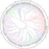
Figure 1
Figure 1
Phylogenetic analysis of TRP channels in P. rapae, D. plexippus, P. Xuthus, M. sexta, B. mori and other organisms. Amino acid sequences of P. rapae (Pr), D. plexippus (Dp), P. Xuthus (Px), M. sexta (Ms), B. mori (Bm), B. germanica (Bg), Z. nevadensis (Zn), N. lugens (Nl), Apis mellifera (Am), D. melanogaster (Dm), A. gambiae (Ag), Tribolium castaneum (Tc), Caenorhabditis elegans (Ce), Homo sapiens (Hs), Danio rerio (Dr) and Mus musculus (Mm) TRP channels were aligned through MAFFT software v7.123b with the default parameters [18]. Under the LG + G4 nucleotide substitution model, IQ-TREE was applied for the maximum likelihood analyses with 1000 bootstrap replicates [19]. The bootstrap value is indicated next to each branch. The accession number of TRP channels identified in this study are listed in an Additional file (Table S3). Other accession numbers of identified TRP channels originated from previous studies [26,27].
Figure 2
Figure 2
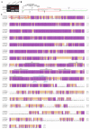
Detection and amino acid sequence alignment of the two PrPyx splice forms. (A) Two molecular isoforms of PrPyx mRNAs identified by PCR. (B) Two-dimensional gene structure map of PrPyx. The boxes represented the exons of PrPyx, the black solid lines connecting the boxes represented the introns, and the black dotted lines on both sides represent other genome sequences. (C) Multiple sequence alignment deduced for PrPyx(A), PrPyx(B) and DmPyx. The red box indicates the different amino acid residues of the two isoforms. Ankyrin repeats were indicated with dotted lines and marked as ANK1–9. Transmembrane regions were indicated with solid lines and marked as TM1–6. The protein sequences used for multiple sequence alignment were in the attached fasta file.
Figure 3
Figure 3
Detection and amino acid sequence alignment of the two PrTRPγ splice forms. (A) Two molecular isoforms of PrTRPγ mRNAs identified by PCR. (B) Two-dimensional gene structure map of PrTRPγ. The boxes represented the exons of PrTRPγ, the black solid lines connecting the boxes represented the introns and the black dotted lines on both sides represent other genome sequences. The deletion of 9 nucleotides at the end of exon 14 produced by splicing were showed in the black box. (C) Multiple sequence alignment deduced for PrTRPγ(A), PrTRPγ(B) and DmTRPγ. The red box indicates the different amino acid residues of the two isoforms. TRP boxes and TRP domain were indicated with blue lines. Ankyrin repeats were indicated with dotted lines and marked as ANK1–2. Transmembrane regions were indicated with solid lines and marked as TM1–6. The protein sequences used for multiple sequence alignment were in the attached fasta file.
Figure 4
Figure 4
Detection and amino acid sequence alignment of the two PrPRPM splice forms. (A) Two molecular isoforms of PrTRPM mRNAs identified by PCR. The bands of PrTRPM mRNAs were indicated with open red box, and the bands below it were primer dimers or none-specific bands. (B) Two-dimensional gene structure map of PrTRPM. The boxes represented the exons of PrTRPM, the black solid lines connecting the boxes represented the introns and the black dotted lines on both sides represent other genome sequences. (C) Multiple sequence alignment deduced for PrTRPM(A), PrTRPM(B) and DmTRPM. The red box indicates the different amino acid residues of the two isoforms. TRP boxes and TRP domain were indicated with blue lines. Ankyrin repeats were indicated with dotted lines and marked as ANK1–2. Transmembrane regions were indicated with solid lines and marked as TM1–6. The protein sequences used for multiple sequence alignment were in the attached fasta file.
Figure 5
Figure 5
Sequence analysis of two PrWtrw genes. (A) Duplication diagram of the PrWtrw gene. Length of two coding sequences (CDSes) and interval length of two genes were shown as the figure. Arrows showed the direction of gene transcription. (B) Multiple sequence alignment deduced for PrWtrw-1, PrWtrw-2 and DmWtrw. Ankyrin repeats were indicated with dotted lines and marked as ANK1–2. Transmembrane regions were indicated with solid lines and marked as TM1–6. The protein sequences used for multiple sequence alignment were in the attached fasta file.
Figure 6
Figure 6
Relative transcriptional levels of two isoforms of PrPyx (A), PrWtrw (B), PrTRPM (C), PrTRP (D), PrTRPL (E), PrTRPγ (F), PrTRPA1 (G), PrTRPA5 (H), PrPain (I), PrNompC (J), PrIav (K), PrNan (L) and PrTRPML (M) in various tissues of 3–5 days old adult P. rapae. “MT” represented malpighian tubules. Asterisks showed the significant difference in expression levels of two isoforms and gene duplications in the same samples. Data represent means  ±  SEM.

References

    1. Venkatachalam K., Montell C. TRP channels. Annu. Rev. Biochem. 2007;76:387–417. doi: 10.1146/annurev.biochem.75.103004.142819. - DOI - PMC - PubMed
    1. Fowler M.A., Montell C. Drosophila TRP channels and animal behavior. Life Sci. 2013;92:394–403. doi: 10.1016/j.lfs.2012.07.029. - DOI - PMC - PubMed
    1. Cosens D.J., Manning A. Abnormal electroretinogram from a Drosophila mutant. Nature. 1969;224:285–287. doi: 10.1038/224285a0. - DOI - PubMed
    1. Montell C., Birnbaumer L., Flockerzi V. The TRP channels, a remarkably functional family. Cell. 2002;108:595–598. doi: 10.1016/S0092-8674(02)00670-0. - DOI - PubMed
    1. Montell C. Drosophila visual transduction. Trends Neurosci. 2012;35:356–363. doi: 10.1016/j.tins.2012.03.004. - DOI - PMC - PubMed

LinkOut - more resources