

Generation and functional assessment of nonhuman primate regulatory dendritic cells and their therapeutic efficacy in renal transplantation
- PMID: 32197811
- PMCID: PMC7197030
- DOI: 10.1016/j.cellimm.2020.104087
Generation and functional assessment of nonhuman primate regulatory dendritic cells and their therapeutic efficacy in renal transplantation
Abstract
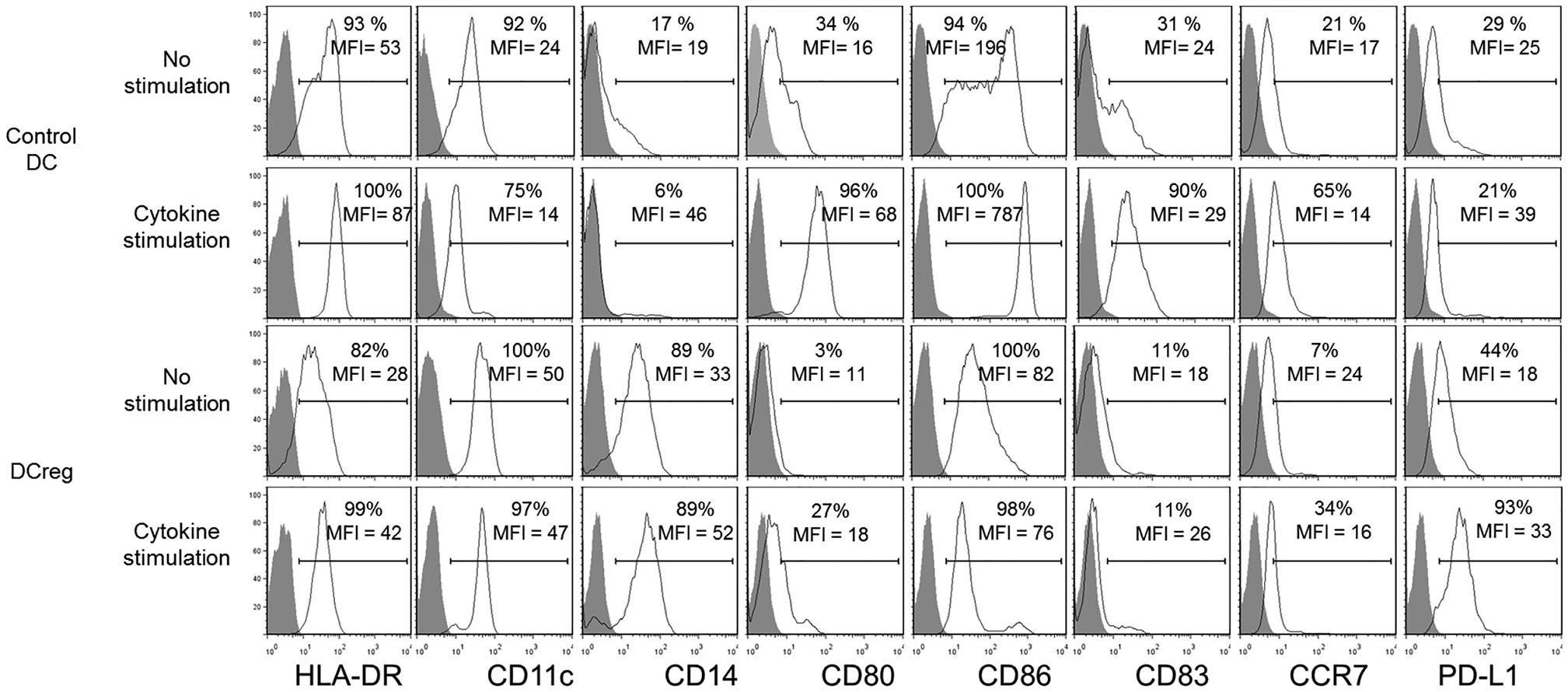
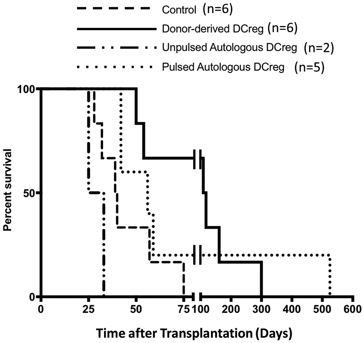
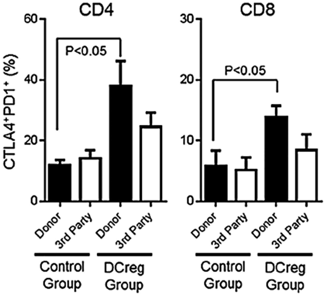
Nonhuman primates (NHP) are important pre-clinical models for evaluation of the safety and efficacy of the most promising potential therapeutic advances in organ transplantation based on rodent studies. Although rare, dendritic cells (DC) play important roles in preservation of self tolerance and DC with immunoregulatory properties (regulatory DC; DCreg) can promote transplant tolerance in rodents when adoptively transferred to allograft recipients. NHP DCreg can be generated ex vivo from bone marrow precursors or blood monocytes of cynomolgus or rhesus macaques or baboons. NHP DCreg generated in the presence of anti-inflammatory factors that confer stability and resistance to maturation, subvert alloreactive T cell responses. When infused into rhesus renal allograft recipients before transplant, they safely prolong MHC mis-matched graft survival, associated with attenuation of anti-donor immune reactivity. In this concise review we describe the properties of NHP DCreg and discuss their influence on T cell responses, alloimmunity and organ transplant survival.
Keywords: Dendritic cells; Immune regulation; Nonhuman primates; Tolerance; Transplantation.
Copyright © 2020 Elsevier Inc. All rights reserved.
Figures
Similar articles
-
Renal Allograft Survival in Nonhuman Primates Infused With Donor Antigen-Pulsed Autologous Regulatory Dendritic Cells.Am J Transplant. 2017 Jun;17(6):1476-1489. doi: 10.1111/ajt.14182. Epub 2017 Feb 2. Am J Transplant. 2017. PMID: 28009481 Free PMC article.
-
Regulatory dendritic cell infusion prolongs kidney allograft survival in nonhuman primates.Am J Transplant. 2013 Aug;13(8):1989-2005. doi: 10.1111/ajt.12310. Epub 2013 Jun 11. Am J Transplant. 2013. PMID: 23758811 Free PMC article.
-
Regulatory dendritic cells for promotion of liver transplant operational tolerance: Rationale for a clinical trial and accompanying mechanistic studies.Hum Immunol. 2018 May;79(5):314-321. doi: 10.1016/j.humimm.2017.10.017. Epub 2017 Oct 31. Hum Immunol. 2018. PMID: 29100944 Free PMC article. Review.
-
Donor-Derived Regulatory Dendritic Cell Infusion Maintains Donor-Reactive CD4+CTLA4hi T Cells in Non-Human Primate Renal Allograft Recipients Treated with CD28 Co-Stimulation Blockade.Front Immunol. 2018 Feb 19;9:250. doi: 10.3389/fimmu.2018.00250. eCollection 2018. Front Immunol. 2018. PMID: 29520267 Free PMC article.
-
Dendritic Cell-Mediated Regulation of Liver Ischemia-Reperfusion Injury and Liver Transplant Rejection.Front Immunol. 2021 Jun 28;12:705465. doi: 10.3389/fimmu.2021.705465. eCollection 2021. Front Immunol. 2021. PMID: 34262574 Free PMC article. Review.
Cited by
-
The Role of Innate Immune Cells in the Prediction of Early Renal Allograft Injury Following Kidney Transplantation.J Clin Med. 2022 Oct 18;11(20):6148. doi: 10.3390/jcm11206148. J Clin Med. 2022. PMID: 36294469 Free PMC article.
-
The emerging role of regulatory cell-based therapy in autoimmune disease.Front Immunol. 2022 Dec 14;13:1075813. doi: 10.3389/fimmu.2022.1075813. eCollection 2022. Front Immunol. 2022. PMID: 36591309 Free PMC article. Review.
-
Regulatory Dendritic Cells, T Cell Tolerance, and Dendritic Cell Therapy for Immunologic Disease.Front Immunol. 2021 Mar 10;12:633436. doi: 10.3389/fimmu.2021.633436. eCollection 2021. Front Immunol. 2021. PMID: 33777019 Free PMC article. Review.
-
Ultrasonographic study of hemodynamics and contrast-enhanced ultrasound in the rhesus monkey kidney.Exp Anim. 2022 May 20;71(2):116-122. doi: 10.1538/expanim.20-0194. Epub 2021 Nov 19. Exp Anim. 2022. PMID: 34803125 Free PMC article.
References
Publication types
MeSH terms
Grants and funding
LinkOut - more resources
Full Text Sources
Medical
Research Materials
