

Aggressive Posterior Retinopathy of Prematurity: Clinical and Quantitative Imaging Features in a Large North American Cohort
- PMID: 32197913
- PMCID: PMC7384953
- DOI: 10.1016/j.ophtha.2020.01.052
Aggressive Posterior Retinopathy of Prematurity: Clinical and Quantitative Imaging Features in a Large North American Cohort
Abstract
Purpose: Aggressive posterior retinopathy of prematurity (AP-ROP) is a vision-threatening disease with a significant rate of progression to retinal detachment. The purpose of this study was to characterize AP-ROP quantitatively by demographics, rate of disease progression, and a deep learning-based vascular severity score.
Design: Retrospective analysis.
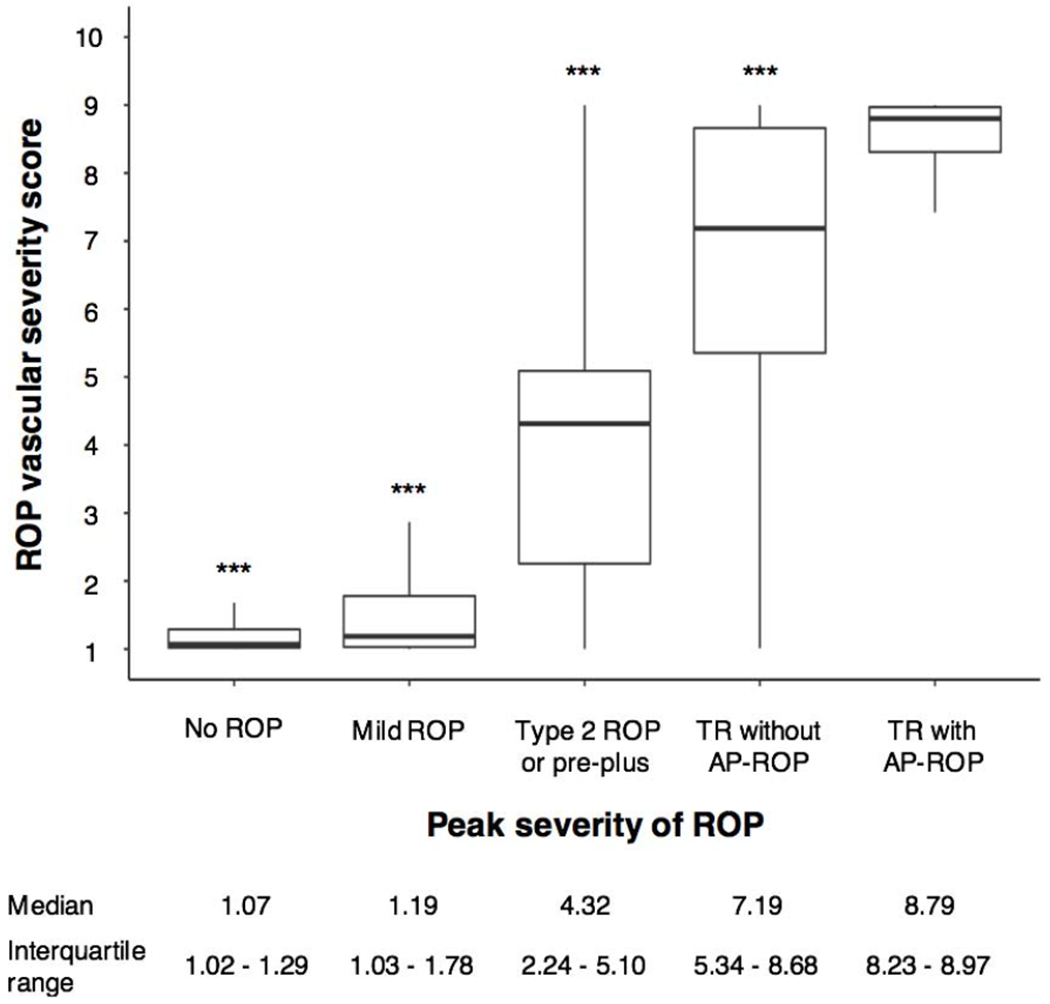
Participants: The Imaging and Informatics in ROP cohort from 8 North American centers, consisting of 947 patients and 5945 clinical eye examinations with fundus images, was used. Pretreatment eyes were categorized by disease severity: none, mild, type 2 or pre-plus, treatment-requiring (TR) without AP-ROP, TR with AP-ROP. Analyses compared TR with AP-ROP and TR without AP-ROP to investigate differences between AP-ROP and other TR disease.
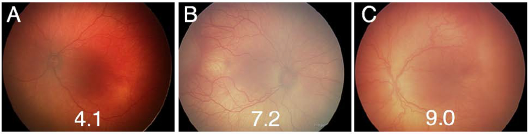
Methods: A reference standard diagnosis was generated for each eye examination using previously published methods combining 3 independent image-based gradings and 1 ophthalmoscopic grading. All fundus images were analyzed using a previously published deep learning system and were assigned a score from 1 through 9.
Main outcome measures: Birth weight, gestational age, postmenstrual age, and vascular severity score.
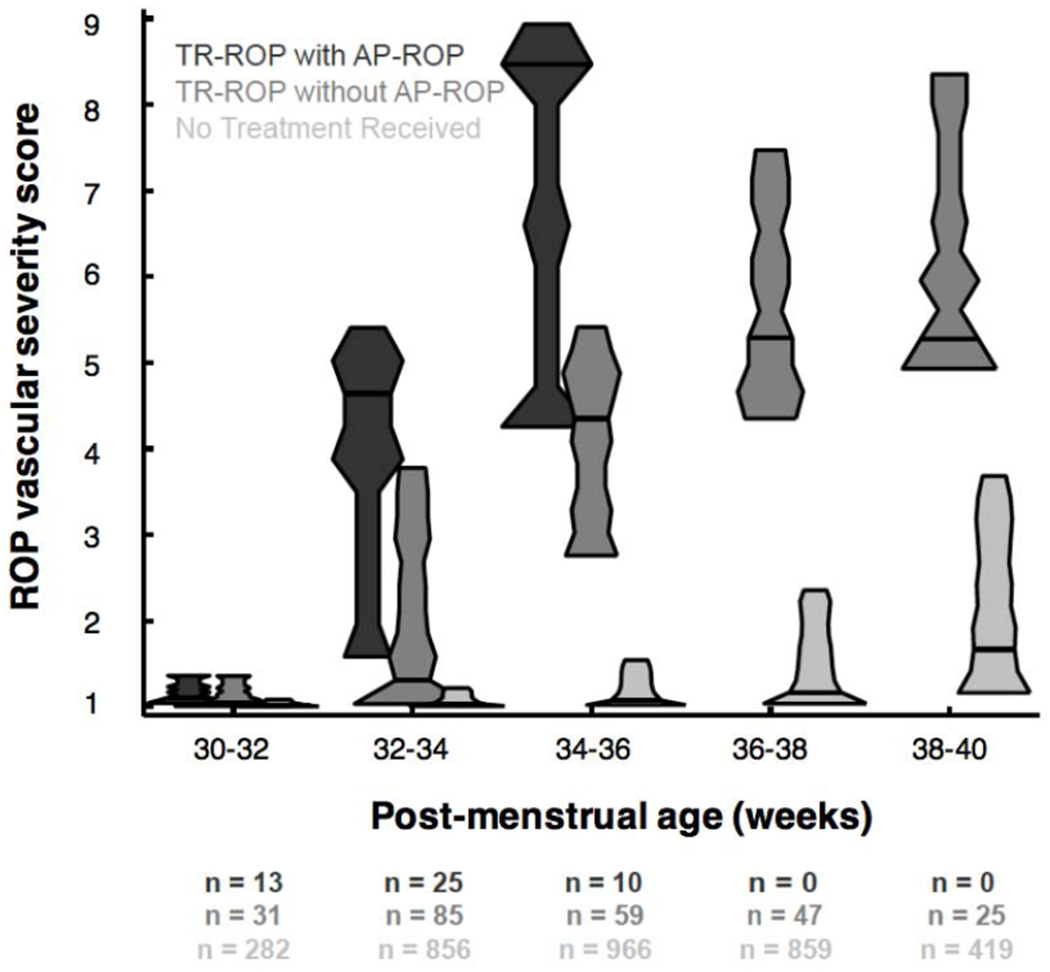
Results: Infants who demonstrated AP-ROP were more premature by birth weight (617 g vs. 679 g; P = 0.01) and gestational age (24.3 weeks vs. 25.0 weeks; P < 0.01) and reached peak severity at an earlier postmenstrual age (34.7 weeks vs. 36.9 weeks; P < 0.001) compared with infants with TR without AP-ROP. The mean vascular severity score was greatest in TR with AP-ROP infants compared with TR without AP-ROP infants (8.79 vs. 7.19; P < 0.001). Analyzing the severity score over time, the rate of progression was fastest in infants with AP-ROP (P < 0.002 at 30-32 weeks).
Conclusions: Premature infants in North America with AP-ROP are born younger and demonstrate disease earlier than infants with less severe ROP. Disease severity is quantifiable with a deep learning-based score, which correlates with clinically identified categories of disease, including AP-ROP. The rate of progression to peak disease is greatest in eyes that demonstrate AP-ROP compared with other treatment-requiring eyes. Analysis of quantitative characteristics of AP-ROP may help improve diagnosis and treatment of an aggressive, vision-threatening form of ROP.
Copyright © 2020 American Academy of Ophthalmology. Published by Elsevier Inc. All rights reserved.
Figures
References
-
- International Committee for the Classification of Retinopathy of Prematurity. The International Classification of Retinopathy of Prematurity revisited. Arch Ophthalmol 2005;123:991–999. - PubMed
-
- Shapiro MJ, Blair MP, Garcia-Gonzalez JM. Experts contradict established classification. Graefes Arch Clin Exp Ophthalmol 2016;254:199. - PubMed
-
- Patel SN, Singh Ranjodh, Jonas Karen E, et al. Inconsistencies in the Diagnosis of Aggressive Posterior Retinopathy of Prematurity. Journal of Vitreo Retinal Diseases 2017;1:181–186.
-
- Fielder AR, Wallace DK, Stahl A, et al. Describing Retinopathy of Prematurity: Current Limitations and New Challenges. Ophthalmology 2019;126:652–654. - PubMed
Publication types
MeSH terms
Grants and funding
LinkOut - more resources
Full Text Sources
Medical
