

First European Haplotype of Echinococcus multilocularis Identified in the United States: An Emerging Disease?
- PMID: 32198510
- PMCID: PMC8028098
- DOI: 10.1093/cid/ciaa245
First European Haplotype of Echinococcus multilocularis Identified in the United States: An Emerging Disease?
Abstract
Background: Echinococcus multilocularis is one of the most severe and lethal parasitic diseases of humans, most often reported in Europe and Asia. Only 1 previous case has been documented in the contiguous United States from Minnesota in 1977. European haplotypes have been identified in carnivores and domestic dogs as well as recently in patients in western and central Canada.
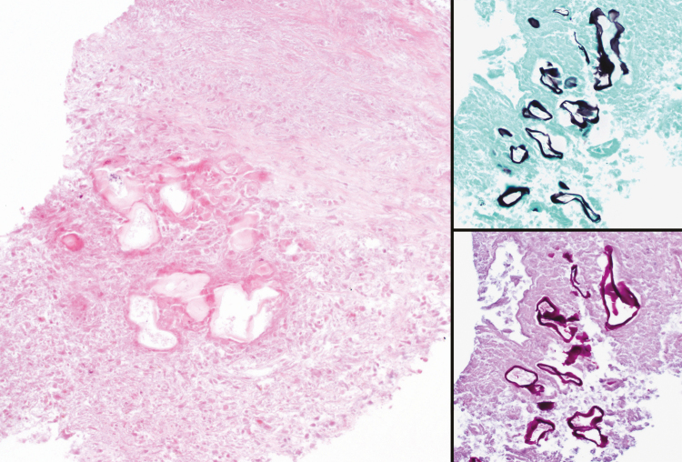
Methods: We used immunohistochemical testing with the monoclonal antibody Em2G11 and a species-specific enzyme-linked immunosorbent assay affinity-purified antigen Em2, as well as COX1 gene sequencing.
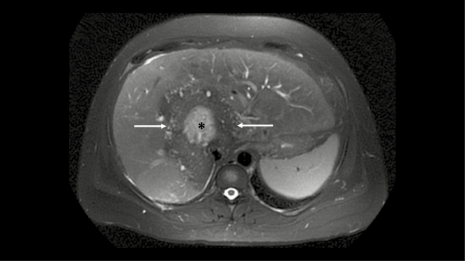
Results: Using pathology, immunohistochemical staining, specific immunodiagnostic testing, and COX1 gene sequencing, we were able to definitively identify E. multilocularis as the causative agent of our patient's liver and lung lesions, which clustered most closely with the European haplotype.
Conclusions: We have identified the first case of a European haplotype E. multilocularis in the United States and the first case of this parasitic infection east of the Mississippi River. Given the identification of this haplotype in Canada, this appears to be an emerging infectious disease in North America.
Keywords: Echinococcus multilocularis; Echinococcus multilocularis COX gene; alveolar echinococcus spectrum of disease; liver masses; parasitic infection.
© The Author(s) 2020. Published by Oxford University Press for the Infectious Diseases Society of America. All rights reserved. For permissions, e-mail: journals.permissions@oup.com.
Figures


Comment in
-
Advanced Alveolar Echinococcosis in a New Geographic Area.Clin Infect Dis. 2021 Apr 8;72(7):1124-1126. doi: 10.1093/cid/ciaa257. Clin Infect Dis. 2021. PMID: 32198509 No abstract available.
References
-
- Thompson RC. Biology and systematics of Echinococcus. Adv Parasitol 2017; 95:65–109. - PubMed
-
- Brunetti E, Kern P, Vuitton DA; Writing Panel for the WHO-IWGE . Expert consensus for the diagnosis and treatment of cystic and alveolar echinococcosis in humans. Acta Trop 2010; 114:1–16. - PubMed
-
- Deplazes P, Rinaldi L, Alvarez Rojas CA, et al. Global distribution of alveolar and cystic echinococcosis. Adv Parasitol 2017; 95:315–493. - PubMed
-
- Wilson JF, Rausch RL. Alveolar hydatid disease. A review of clinical features of 33 indigenous cases of Echinococcus multilocularis infection in Alaskan Eskimos. Am J Trop Med Hyg 1980; 29:1340–55. - PubMed
