Review
. 2020 Mar;41(3):503-513.
doi: 10.1007/s00246-020-02301-6. Epub 2020 Mar 20.

MRI Catheterization: Ready for Broad Adoption

Affiliations
Review

MRI Catheterization: Ready for Broad Adoption

Stephen J Nageotte et al. Pediatr Cardiol. 2020 Mar.

Abstract

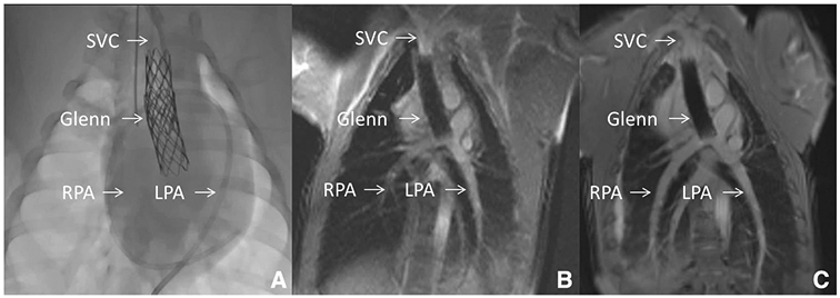
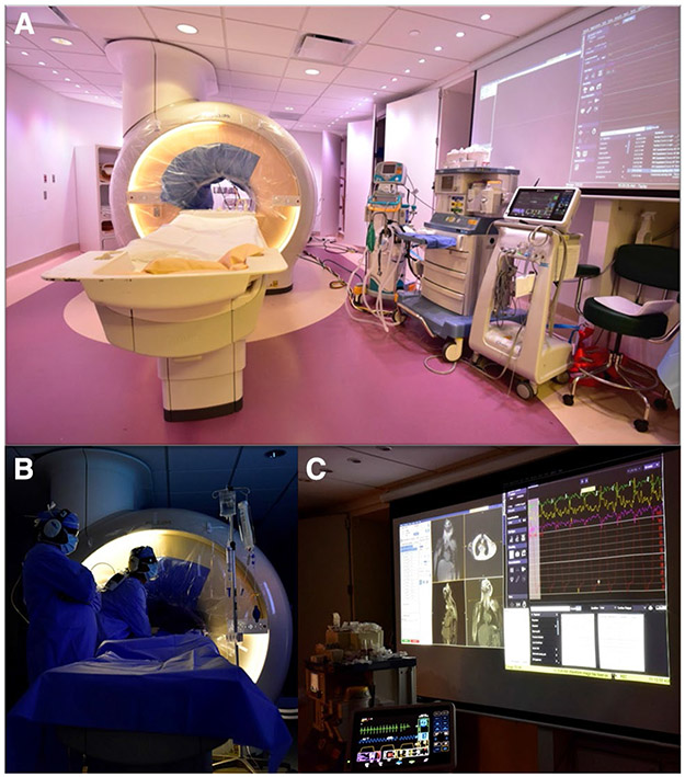
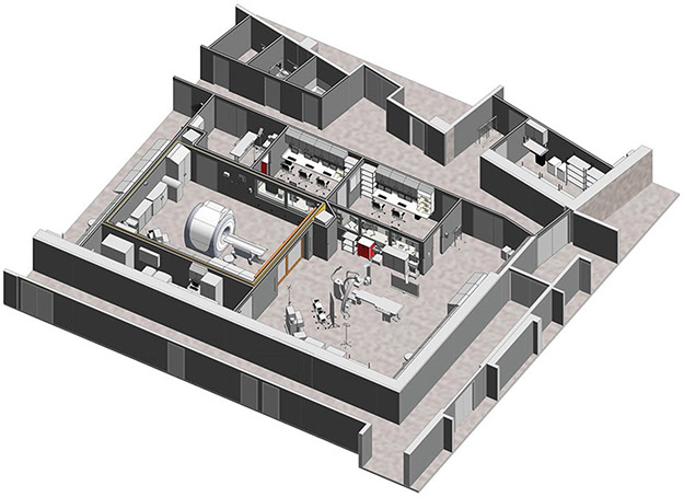
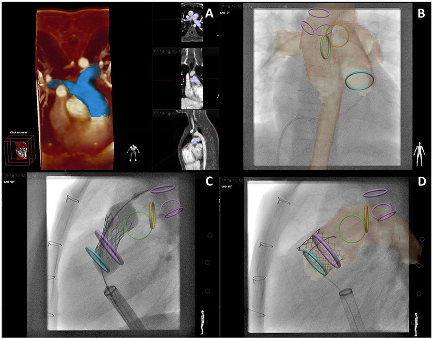
In recent years, interventional cardiac magnetic resonance imaging (iCMR) has evolved from attractive theory to clinical routine at several centers. Real-time cardiac magnetic resonance imaging (CMR fluoroscopy) adds value by combining soft-tissue visualization, concurrent hemodynamic measurement, and freedom from radiation. Clinical iCMR applications are expanding because of advances in catheter devices and imaging. In the near future, iCMR promises novel procedures otherwise unsafe under standalone X-Ray guidance.

Keywords: Congenital heart disease; Interventional MRI; MR fluoroscopy; Magnetic resonance imaging (MRI).


Conflict of interest statement

Conflict of interest The authors declare that they have no conflict of interest.

Figures

Fig. 1
Fig. 1
X-Ray Fluoroscopy versus Interventional Cardiac MRI (iCMR). This figure demonstrates a novel percutaneous intervention: transcatheter cavopulmonary anastomosis [58]. a Shows traditional X-Ray fluoroscopy of the endograft based cavopulmonary anastomosis (Glenn) in situ. Superior Vena Cava (SVC), Right and Left Pulmonary Artery (RPA, LPA) expected locations are shown. b Shows real-time CMR fluoroscopy with superior anatomic visualization. SVC, RPA, LPA and entire thoracic context are clearly and continuously displayed. c Shows higher quality diagnostic CMR (steady-state free precession “white” blood imaging) of the Glenn. During iCMR cases, intermittent higher quality non-real-time CMR can be obtained at any time
Fig. 2
Fig. 2
Interventional cardiac MRI (iCMR) room, a Demonstrates the iCMR room at Children’s Medical Center of Dallas. The CMR bore has a sterile drape. CMR compatible ventilator, nitric oxide setup, and hemodynamic monitoring system are visible. b The interventional team is performing clinical CMR catheterization. c Shows a large projected image of real-time CMR as catheterization is being performed and live high fidelity hemodynamic recording. Images are courtesy Dr. Surendranath Veeram Reddy and Children’s Medical Center of Dallas team
Fig. 3
Fig. 3
Interventional Cardiac MRI (iCMR) suite (co-localized, adjacent CMR and X-Ray cardiac catheterization rooms). A 3-D rendering of future iCMR suite at Rady Children’s Hospital—San Diego show co-localized, adjacent CMR and X-Ray cardiac catheterization rooms separated by sliding radiofrequency shielded doors. Image is courtesy Rady Children’s Hospital—San Diego
Fig. 4
Fig. 4
X-Ray Fused with MRI (XFM) Guidance of Percutaneous Pulmonary Valve Implantation. a Demonstrates three-dimensional (3D) reconstruction of the right ventricular outflow tract and proximal branch pulmonary arteries (blue). Multiple imaging slices of that region of interest are shown in the vertical panel. bd demonstrate critical structures depicted by CMR with overlay on live X-Ray fluoroscopy. Three purple rings (most posterior in c, d) show main trachea and right, left bronchus. Blue ring shows proximal right ventricular outflow tract. Green and yellow rings show proximal right and left pulmonary artery, respectively. Large purple circle in c, d highlights intended landing zone. b Shows soft-tissue CMR contour of aorta and pulmonary artery circulation as well as a venous catheter advanced to the right pulmonary artery. c Shows implantation of a percutaneous pulmonary valve. Rigid guidewires displace anatomy making XFM less accurate (see wire in left pulmonary artery above yellow or left pulmonary artery ring). d Shows percutaneous pulmonary valve after implantation and removal of rigid guidewire restoring XFM accuracy. Co-registration was performed with trachea, bronchus, and spine markers. Images are courtesy Dr. Sebastian Goreczny [Polish Mother’s Memorial Hospital, Research Institute (Lodz, Poland) and Children’s Hospital Colorado (Aurora, CO)] [60]
Fig. 5
Fig. 5
CMR-Guided Endomyocardial Biopsy. a Demonstrates real-time MR-guided endomyocardial biopsy within the left ventricle using an “active” visualization device (green). The arrow indicates the jaws of the bioptome. b demonstrates the late-gadolinium enhancement of biopsy targets (arrows) [5]
Fig. 6
Fig. 6
CMR-Guided Chemoablation for Ventricular Arrhythmia Therapy. a Late-gadolinium enhancement highlights two areas of infarct (green arrows) with an isthmus of normal myocardium (black arrow). b Shows real-time MR–guided chemoablation with an “active” visualization injection needle in green. c Post-chemoablation the areas of infarct continue to be highlighted with green arrows and the red arrow demonstrates a transmural chemoablation lesion eliminating the gap between infarct zones [55]

References

    1. Razavi R, Hill DL, Keevil SF et al. (2003) Cardiac catheterisation guided by MRI in children and adults with congenital heart disease. The Lancet 362:1877–1882. 10.1016/S0140-6736(03)14956-2 - DOI - PubMed
    1. Rogers T, Ratnayaka K, Khan JM et al. (2017) CMR fluoroscopy right heart catheterization for cardiac output and pulmonary vascular resistance: results in 102 patients. J Cardiovasc Magn Reson. 10.1186/s12968-017-0366-2 - DOI - PMC - PubMed
    1. Ratnayaka K, Kanter JP, Faranesh AZ et al. (2017) Radiation-free CMR diagnostic heart catheterization in children. J Cardiovasc Magn Reson. 10.1186/s12968-017-0374-2 - DOI - PMC - PubMed
    1. Pushparajah K, Tzifa A, Bell A et al. (2015) Cardiovascular Magnetic Resonance catheterization derived pulmonary vascular resistance and medium-term outcomes in congenital heart disease. J Cardiovasc Magn Reson 17:28 10.1186/s12968-015-0130-4 - DOI - PMC - PubMed
    1. Rogers T, Ratnayaka K, Karmarkar P et al. (2016) Real-time magnetic resonance imaging guidance improves the diagnostic yield of endomyocardial biopsy. JACC Basic Transl Sci 1:376–383. 10.1016/j.jacbts.2016.05.007 - DOI - PMC - PubMed

MeSH terms

LinkOut - more resources