

CD133+Exosome Treatment Improves Cardiac Function after Stroke in Type 2 Diabetic Mice
- PMID: 32198711
- PMCID: PMC7502550
- DOI: 10.1007/s12975-020-00807-y
CD133+Exosome Treatment Improves Cardiac Function after Stroke in Type 2 Diabetic Mice
Abstract
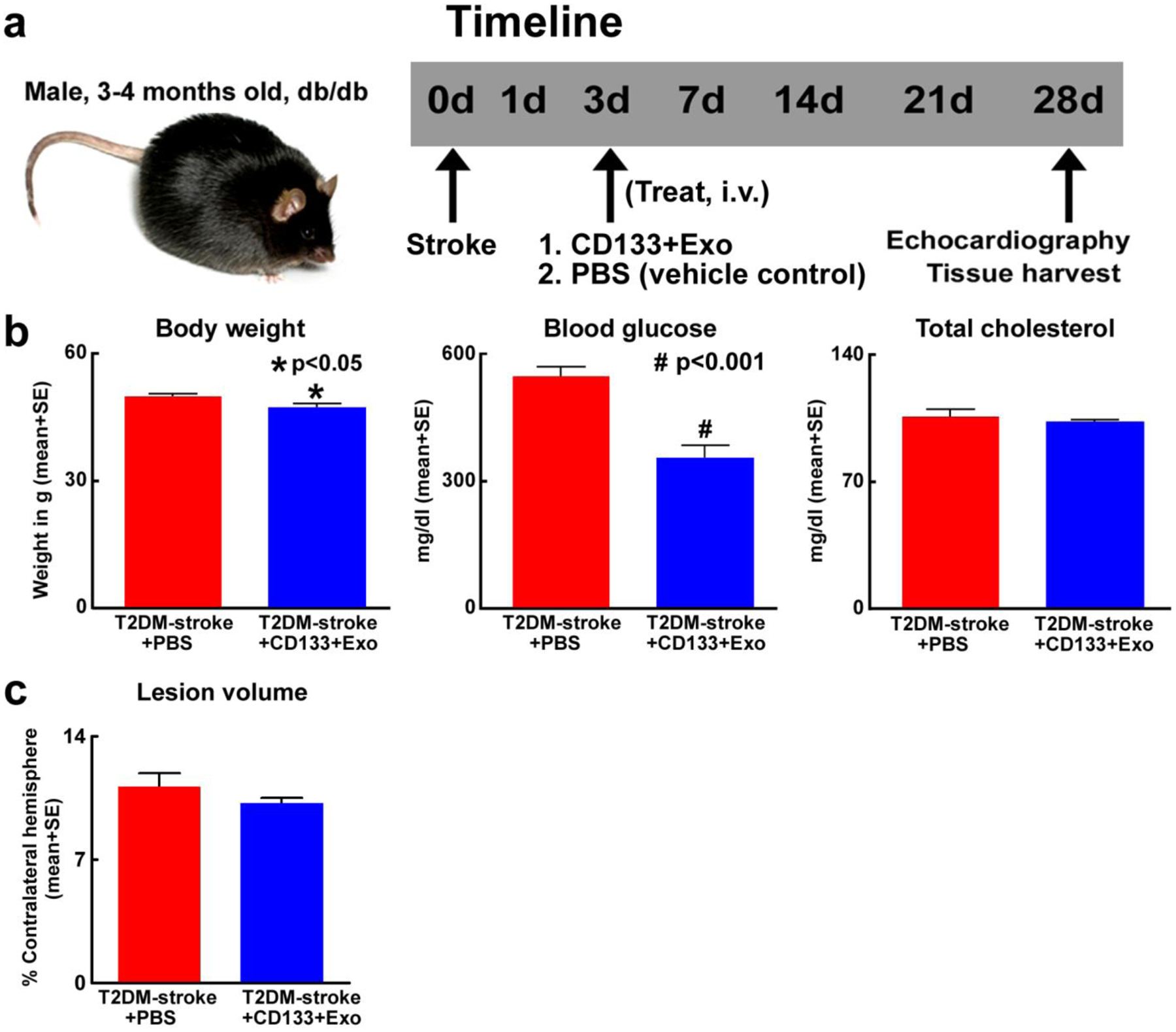
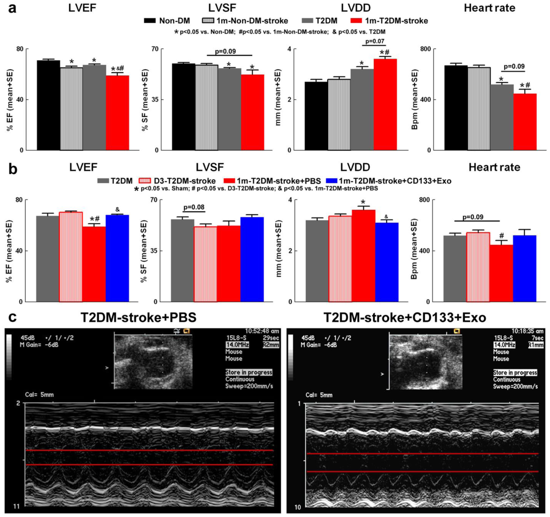
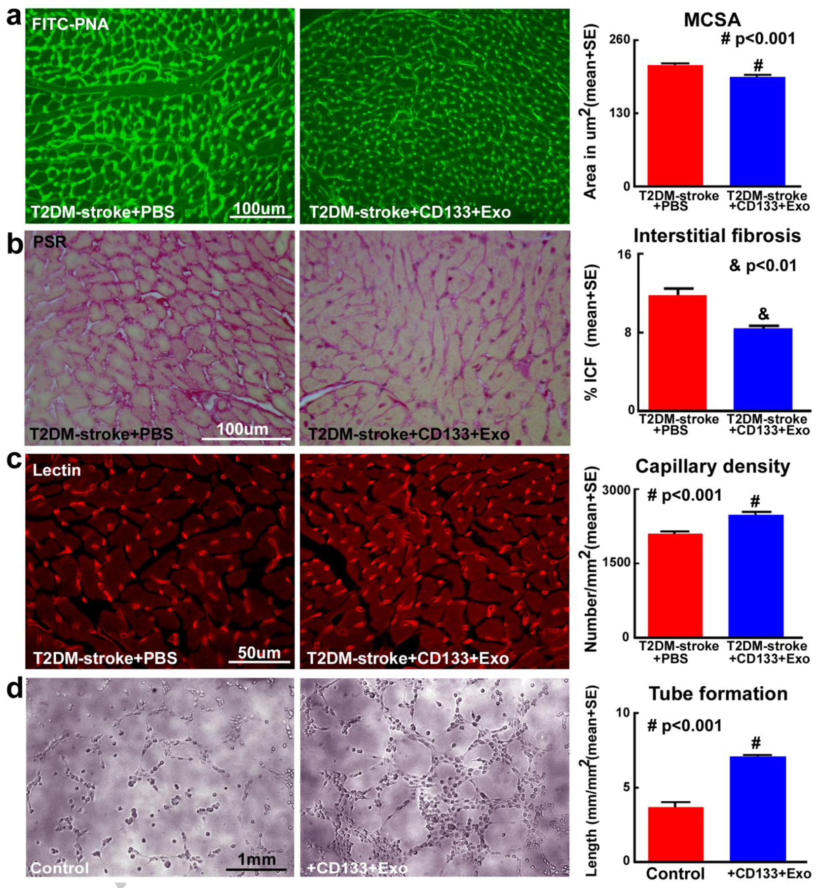
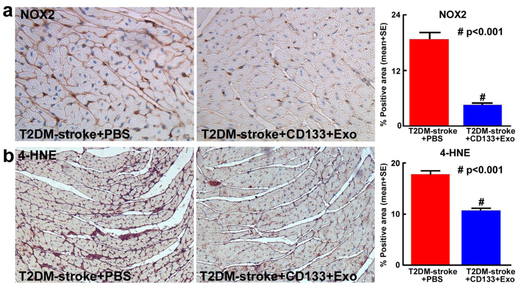
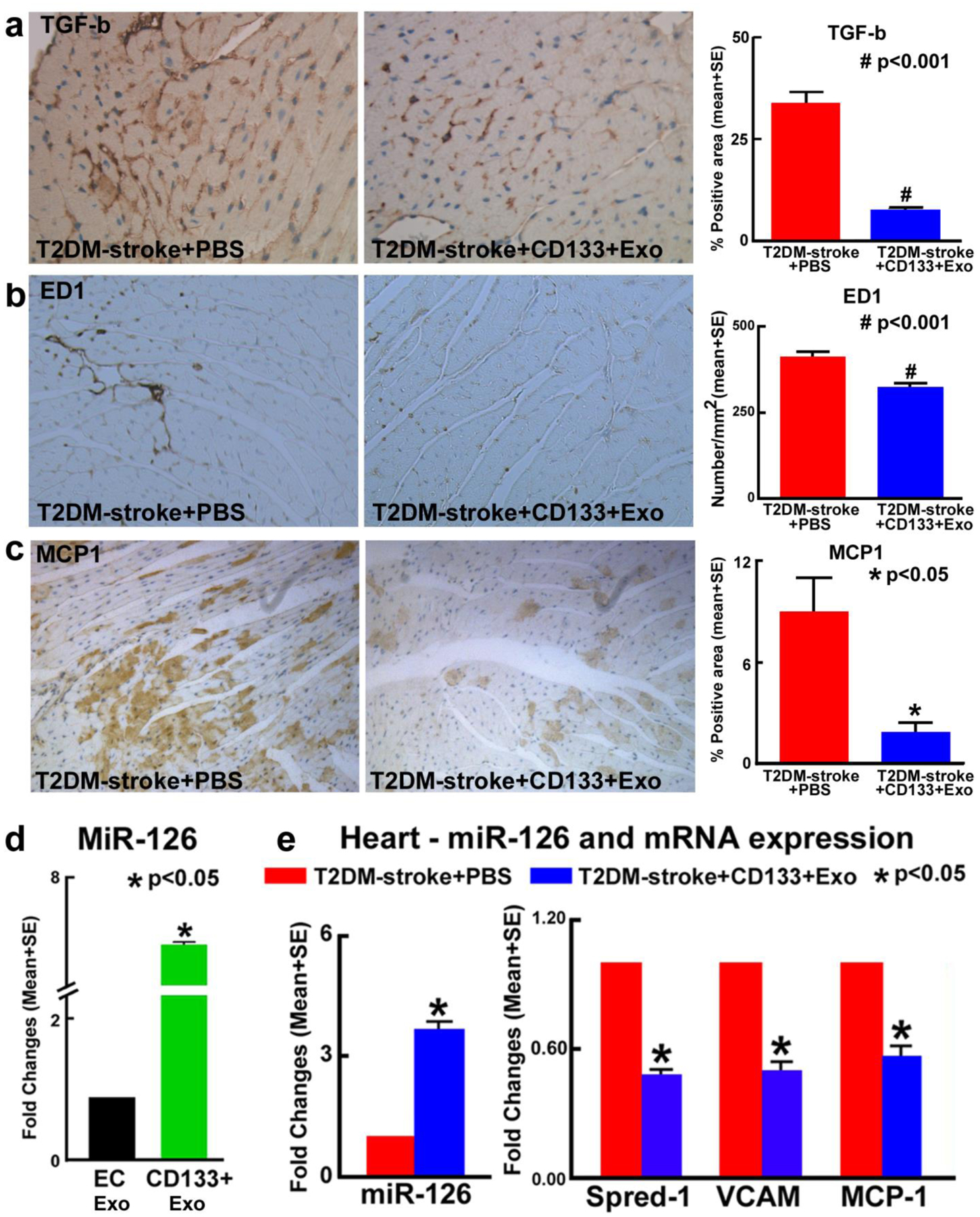
Cardiac complications post-stroke are common, and diabetes exacerbates post-stroke cardiac injury. In this study, we tested whether treatment with exosomes harvested from human umbilical cord blood derived CD133+ cells (CD133+Exo) improves cardiac function in type 2 diabetes mellitus (T2DM) stroke mice. Adult (3-4 m), male, BKS.Cg-m+/+Leprdb/J (db/db, T2DM) and non-DM (db+) mice were randomized to sham or photothrombotic stroke groups. T2DM-stroke mice were treated with phosphate-buffered saline (PBS) or CD133+Exo (20 μg, i.v.) at 3 days after stroke. T2DM sham and T2DM+CD133+Exo treatment groups were included as controls. Echocardiography was performed, and mice were sacrificed at 28 days after stroke. Cardiomyocyte hypertrophy, myocardial capillary density, interstitial fibrosis, and inflammatory factor expression were measured in the heart. MicroRNA-126 expression and its target gene expression were measured in the heart. T2DM mice exhibit significant cardiac deficits such as decreased left ventricular ejection fraction (LVEF) and shortening fraction (LVSF), increased left ventricular diastolic dimension (LVDD), and reduced heart rate compared to non-DM mice. Stroke in non-DM and T2DM mice significantly decreases LVEF compared to non-DM and T2DM-sham, respectively. Cardiac dysfunction is worse in T2DM-stroke mice compared to non-DM-stroke mice. CD133+Exo treatment of T2DM-stroke mice significantly improves cardiac function identified by increased LVEF and decreased LVDD compared to PBS treated T2DM-stroke mice. In addition, CD133+Exo treatment significantly decreases body weight and blood glucose but does not decrease lesion volume in T2DM-stroke mice. CD133+Exo treatment of T2DM mice significantly decreases body weight and blood glucose but does not improve cardiac function. CD133+Exo treatment in T2DM-stroke mice significantly decreases myocardial cross-sectional area, interstitial fibrosis, transforming growth factor beta (TGF-β), numbers of M1 macrophages, and oxidative stress markers 4-HNE (4-hydroxynonenal) and NADPH oxidase 2 (NOX2) in heart tissue. CD133+Exo treatment increases myocardial capillary density in T2DM-stroke mice as well as upregulates endothelial cell capillary tube formation in vitro. MiR-126 is highly expressed in CD133+Exo compared to exosomes derived from endothelial cells. Compared to PBS treatment, CD133+Exo treatment significantly increases miR-126 expression in the heart and decreases its target gene expression such as Sprouty-related, EVH1 domain-containing protein 1 (Spred-1), vascular cell adhesion protein (VCAM), and monocyte chemoattractant protein 1 (MCP1) in the heart of T2DM-stroke mice. CD133+Exo treatment significantly improves cardiac function in T2DM-stroke mice. The cardio-protective effects of CD133+Exo in T2DM-stroke mice may be attributed at least in part to increasing miR-126 expression and decreasing its target protein expression in the heart, increased myocardial capillary density and decreased cardiac inflammatory factor expression.
Keywords: CD133; Cardiac function; Echocardiography; Exosomes; Stroke; Type 2 diabetes mellitus.
Conflict of interest statement
Figures

Similar articles
-
MiR-126 Mediates Brain Endothelial Cell Exosome Treatment-Induced Neurorestorative Effects After Stroke in Type 2 Diabetes Mellitus Mice.Stroke. 2019 Oct;50(10):2865-2874. doi: 10.1161/STROKEAHA.119.025371. Epub 2019 Aug 9. Stroke. 2019. PMID: 31394992 Free PMC article.
-
Exosomes derived from bone marrow mesenchymal stem cells harvested from type two diabetes rats promotes neurorestorative effects after stroke in type two diabetes rats.Exp Neurol. 2020 Dec;334:113456. doi: 10.1016/j.expneurol.2020.113456. Epub 2020 Sep 2. Exp Neurol. 2020. PMID: 32889008
-
Astragaloside IV increases PDHA1 in mesenchymal stem cell exosomes to treat myocardial infarction.Sci Rep. 2025 Jul 15;15(1):25461. doi: 10.1038/s41598-025-08628-5. Sci Rep. 2025. PMID: 40659732 Free PMC article.
-
Dipeptidyl-peptidase (DPP)-4 inhibitors and glucagon-like peptide (GLP)-1 analogues for prevention or delay of type 2 diabetes mellitus and its associated complications in people at increased risk for the development of type 2 diabetes mellitus.Cochrane Database Syst Rev. 2017 May 10;5(5):CD012204. doi: 10.1002/14651858.CD012204.pub2. Cochrane Database Syst Rev. 2017. PMID: 28489279 Free PMC article.
-
Insulin secretagogues for prevention or delay of type 2 diabetes mellitus and its associated complications in persons at increased risk for the development of type 2 diabetes mellitus.Cochrane Database Syst Rev. 2016 Oct 17;10(10):CD012151. doi: 10.1002/14651858.CD012151.pub2. Cochrane Database Syst Rev. 2016. PMID: 27749986 Free PMC article.
Cited by
-
Anti-inflammatory Prowess of endothelial progenitor cells in the realm of biology and medicine.NPJ Regen Med. 2024 Sep 30;9(1):27. doi: 10.1038/s41536-024-00365-z. NPJ Regen Med. 2024. PMID: 39349482 Free PMC article. Review.
-
Therapeutic effects of CD133 + Exosomes on liver function after stroke in type 2 diabetic mice.Front Neurosci. 2023 Mar 9;17:1061485. doi: 10.3389/fnins.2023.1061485. eCollection 2023. Front Neurosci. 2023. PMID: 36968490 Free PMC article.
-
Exosome treatment for stroke with diabetic comorbidity.Neural Regen Res. 2022 Feb;17(2):315-317. doi: 10.4103/1673-5374.319190. Neural Regen Res. 2022. PMID: 34269198 Free PMC article. No abstract available.
-
Therapeutic Potentials of Extracellular Vesicles for the Treatment of Diabetes and Diabetic Complications.Int J Mol Sci. 2020 Jul 21;21(14):5163. doi: 10.3390/ijms21145163. Int J Mol Sci. 2020. PMID: 32708290 Free PMC article. Review.
-
Role of Extracellular Vesicles in Placental Inflammation and Local Immune Balance.Mediators Inflamm. 2021 Jun 18;2021:5558048. doi: 10.1155/2021/5558048. eCollection 2021. Mediators Inflamm. 2021. PMID: 34239366 Free PMC article. Review.
References
-
- Oppenheimer SM. Neurogenic cardiac effects of cerebrovascular disease. Curr Opin Neurol. 1994;7(1):20–4. - PubMed
Publication types
MeSH terms
Substances
Grants and funding
LinkOut - more resources
Full Text Sources
Medical
Research Materials
Miscellaneous
