. 2020 May;111(5):1596-1606.
doi: 10.1111/cas.14391. Epub 2020 Apr 13.

Helicobacter pylori CagA oncoprotein interacts with SHIP2 to increase its delivery into gastric epithelial cells

Affiliations

Helicobacter pylori CagA oncoprotein interacts with SHIP2 to increase its delivery into gastric epithelial cells

Yumiko Fujii et al. Cancer Sci. 2020 May.

Abstract

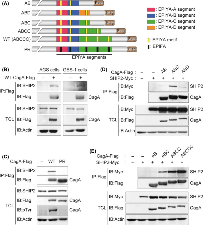
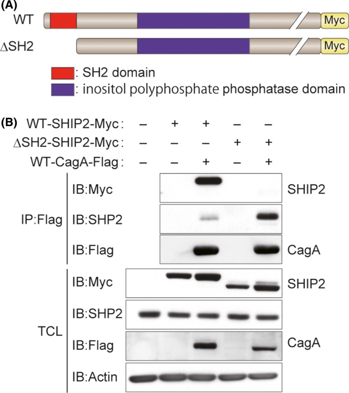
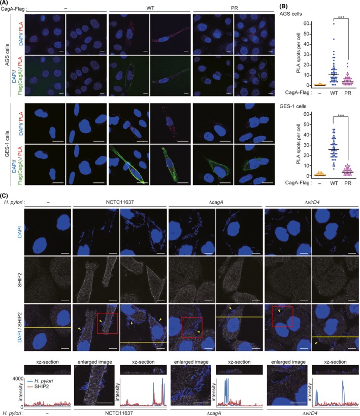
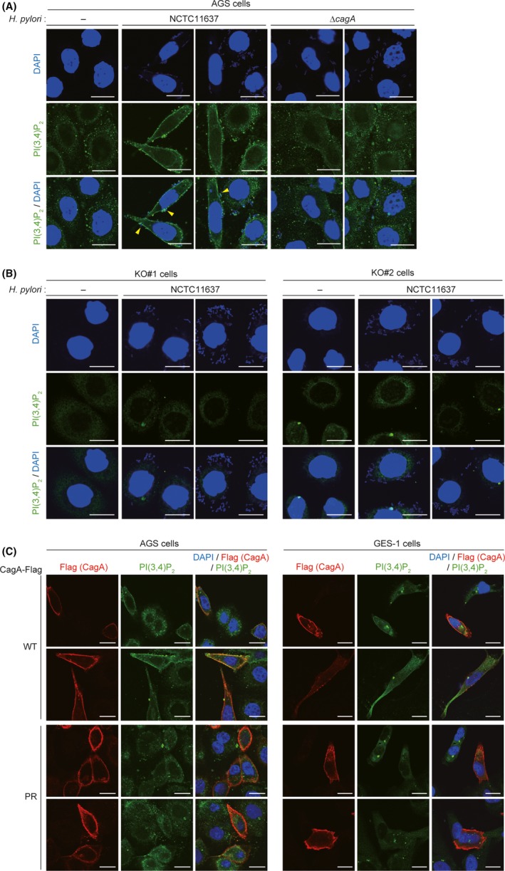
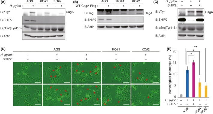
Chronic infection with Helicobacter pylori cagA-positive strains is causally associated with the development of gastric diseases, most notably gastric cancer. The cagA-encoded CagA protein, which is injected into gastric epithelial cells by bacterial type IV secretion, undergoes tyrosine phosphorylation at the Glu-Pro-Ile-Tyr-Ala (EPIYA) segments (EPIYA-A, EPIYA-B, EPIYA-C, and EPIYA-D), which are present in various numbers and combinations in its C-terminal polymorphic region, thereby enabling CagA to promiscuously interact with SH2 domain-containing host cell proteins, including the prooncogenic SH2 domain-containing protein tyrosine phosphatase 2 (SHP2). Perturbation of host protein functions by aberrant complex formation with CagA has been considered to contribute to the development of gastric cancer. Here we show that SHIP2, an SH2 domain-containing phosphatidylinositol 5'-phosphatase, is a hitherto undiscovered CagA-binding host protein. Similar to SHP2, SHIP2 binds to the Western CagA-specific EPIYA-C segment or East Asian CagA-specific EPIYA-D segment through the SH2 domain in a tyrosine phosphorylation-dependent manner. In contrast to the case of SHP2, however, SHIP2 binds more strongly to EPIYA-C than to EPIYA-D. Interaction with CagA tethers SHIP2 to the plasma membrane, where it mediates production of phosphatidylinositol 3,4-diphosphate [PI(3,4)P2 ]. The CagA-SHIP2 interaction also potentiates the morphogenetic activity of CagA, which is caused by CagA-deregulated SHP2. This study indicates that initially delivered CagA interacts with SHIP2 and thereby strengthens H. pylori-host cell attachment by altering membrane phosphatidylinositol compositions, which potentiates subsequent delivery of CagA that binds to and thereby deregulates the prooncogenic phosphatase SHP2.

Keywords: Helicobacter pylori; CagA; PI(3,4)P2; SHIP2; gastric cancer.


Conflict of interest statement

The authors have no conflict of interest to declare.

Figures

Figure 1
Figure 1
CagA interacts with SH2 domain‐containing phosphatidylinositol 5′‐phosphatase 2 (SHIP2) in a tyrosine phosphorylation‐dependent manner. A, Schematic diagram of Flag‐tagged CagA constructs. B‐E, AGS cells (B, C) or GES‐1 cells (B) were transiently transfected with a Flag‐tagged WT‐CagA or phosphorylation‐resistant (PR)‐CagA vector. COS‐7 cells (D, E) were transiently transfected with the indicated Flag‐tagged CagA vector together with a Myc‐tagged SHIP2 or control empty vector. Total cell lysates (TCLs) were immunoprecipitated (IP) with an anti‐Flag Ab and were subjected to immunoblotting (IB) with the respective Abs. EPIYA, Glu‐Pro‐Ile‐Tyr‐Ala motif
Figure 2
Figure 2
SH2 domain of SH2 domain‐containing phosphatidylinositol 5′‐phosphatase 2 (SHIP2) is essential for binding to CagA. A, Schematic diagram of Myc‐tagged SHIP2 constructs. B, COS‐7 cells were transiently transfected with a Myc‐tagged WT‐SHIP2 or mutant SHPI2 lacking the SH2 domain (ΔSH2‐SHIP2) vector together with a Flag‐tagged WT‐CagA or control empty vector. Total cell lysates (TCLs) were immunoprecipitated (IP) with an anti‐Flag Ab and subjected to immunoblotting (IB) with the respective Abs
Figure 3
Figure 3
CagA induces translocalization of SH2 domain‐containing phosphatidylinositol 5′‐phosphatase 2 (SHIP2) to the plasma membrane. A, B, AGS (upper) or GES‐1 (lower) cells were transiently transfected with a Flag‐tagged WT‐ or phosphorylation‐resistant (PR)‐CagA vector. Physical interactions between endogenous SHIP2 and CagA in cells were analyzed by the proximity ligation assay (PLA) with an anti‐Flag Ab and an anti‐SHIP2 Ab. A, Red PLA spots indicate CagA‐SHIP2 interaction. Cellular nuclei and CagA‐expressing cells were visualized in blue and green, respectively. Upper images of AGS cells show maximum intensity projections (2.5 μm thick) and others are confocal images. Scale bar, 20 μm. B, Dots show the number of PLA spots per CagA‐expressing cell in (A). Bars indicate median. n = 50 cells. ***P < .001 (Mann‐Whitney U test). C, AGS cells were infected with the Helicobacter pylori NCTC11637 strain or its isogenic mutants (ΔcagA or ΔvirD4) at an MOI of 100 for 6 h, and were then immunofluorescence stained with an anti‐SHIP2 Ab. SHIP2 was visualized in gray. Cellular nuclei and H. pylori were visualized in blue by DAPI staining. Arrowheads indicate H. pylori dense area. Scale bar, 10 μm. Enlarged images of red boxes indicated at the third row panels are shown in the bottom row of panels (panels 2, 4, and 6 from left). Vertical xz‐sections (upper) and their scanning fluorescence intensities (lower) along the yellow lines indicated at the third row panels are also shown in the bottom row of panels (panels 1, 3, 5, and 7 from left). In the scanning data, intensities of H. pylori and SHIP2 are shown in blue and red lines, respectively
Figure 4
Figure 4
Phosphatidylinositol 3,4‐diphosphate (PI(3,4)P2) accumulates in the plasma membrane of CagA‐injected cells. A, B, AGS cells (A) or AGS‐derived SHIP2‐KO KO#1 and KO#2 cells (B) were infected with the Helicobacter pylori NCTC11637 strain or its isogenic ΔcagA strain at an MOI of 100 for 6 h, and subjected to PI(3,4)P2 staining. PI(3,4)P2 was visualized in green. Cellular nuclei and H. pylori were visualized in blue by DAPI staining. Arrowheads indicate CagA‐injected cells. Scale bar, 20 μm. C, AGS or GES‐1 cells were transiently transfected with a Flag‐tagged WT‐ or PR‐CagA vector, and subjected to PI(3,4)P2 staining. PI(3,4)P2, CagA‐Flag, and cellular nuclei were visualized in green, red, and blue, respectively. Scale bar, 20 μm
Figure 5
Figure 5
CagA‐SH2 domain‐containing phosphatidylinositol 5′‐phosphatase 2 (SHIP2) interaction potentiated delivery of CagA from Helicobacter pylori to gastric epithelial cells. A, AGS, AGS‐derived SHIP2‐KO KO#1, or AGS‐derived SHIP2‐KO KO#2 cells were infected with the H. pylori NCTC11637 strain at an MOI of 100 for 9 h. Total cell lysates were then immunoblotted (IB) with the respective Abs. B, AGS, AGS‐derived SHIP2‐KO KO#1, or AGS‐derived SHIP2‐KO KO#2 cells were transiently transfected with a WT‐CagA vector or a control empty vector for 24 h. Total cell lysates were IB with the respective Abs. C, AGS cells were transfected with a SHIP2 vector or control empty vector. At 24 h after transfection, cells were infected with the H. pylori NCTC11637 strain at an MOI of 100 for an additional 9 h. Total cell lysates were subjected to IB with the respective Abs. D, AGS, AGS‐derived SHIP2‐KO KO#1, or AGS‐derived SHIP2‐KO KO#2 cells were transfected with a SHIP2 vector or control empty vector. At 24 h after transfection, cells were infected with the H. pylori NCTC11637 strain at an MOI of 100 for an additional 9 h before microscopic analysis. Arrowheads indicate the hummingbird phenotype. Scale bar, 100 μm. E, Percentage of cells with the hummingbird phenotype shown in (D). Error bars, ±SD; n = 3. *P < .05, **P < .01 (Student’s t test)

References

    1. Blaser MJ, Perez‐Perez GI, Kleanthous H, et al. Infection with Helicobacter pylori strains possessing cagA is associated with an increased risk of developing adenocarcinoma of the stomach. Cancer Res. 1995;55:2111‐2115. - PubMed
    1. Parsonnet J, Friedman GD, Orentreich N, Vogelman H. Risk for gastric cancer in people with CagA positive or CagA negative Helicobacter pylori infection. Gut. 1997;40:297‐301. - PMC - PubMed
    1. Covacci A, Rappuoli R. Tyrosine‐phosphorylated bacterial proteins: Trojan horses for the host cell. J Exp Med. 2000;191:587‐592. - PMC - PubMed
    1. Higashi H, Tsutsumi R, Muto S, et al. SHP‐2 tyrosine phosphatase as an intracellular target of Helicobacter pylori CagA protein. Science. 2002;295:683‐686. - PubMed
    1. Hatakeyama M. Oncogenic mechanisms of the Helicobacter pylori CagA protein. Nat Rev Cancer. 2004;4:688‐694. - PubMed

MeSH terms

Substances