. 2020 May:32:101491.
doi: 10.1016/j.redox.2020.101491. Epub 2020 Mar 3.

Comprehensive chemical proteomics for target deconvolution of the redox active drug auranofin

Affiliations

Comprehensive chemical proteomics for target deconvolution of the redox active drug auranofin

Amir Ata Saei et al. Redox Biol. 2020 May.

Abstract

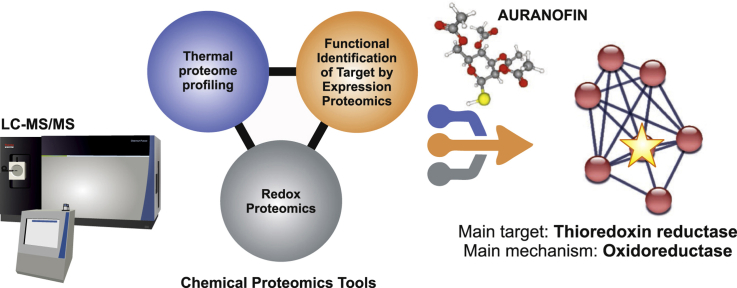
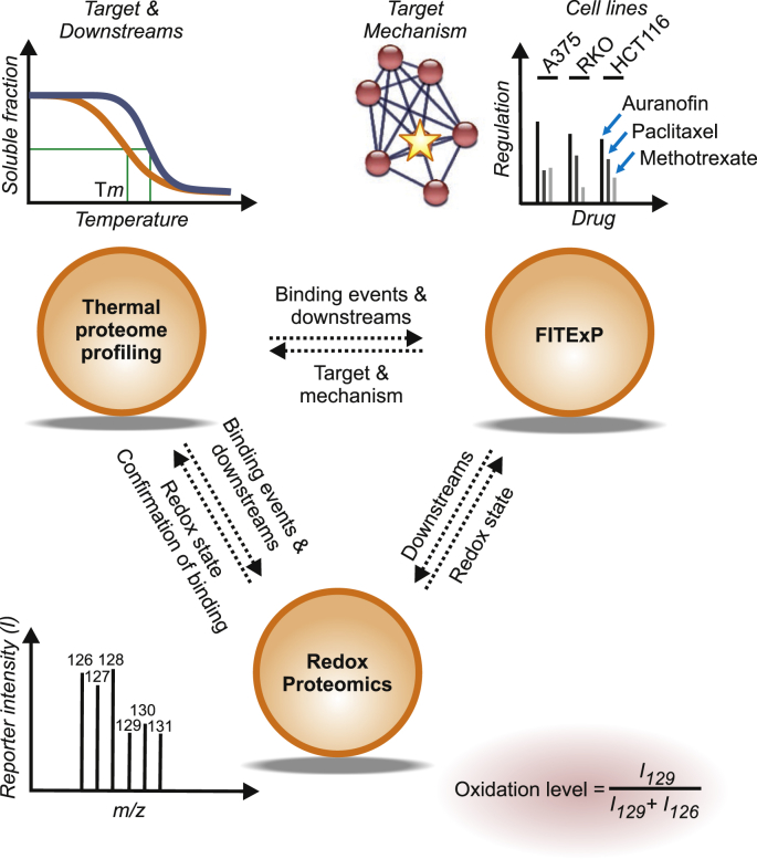
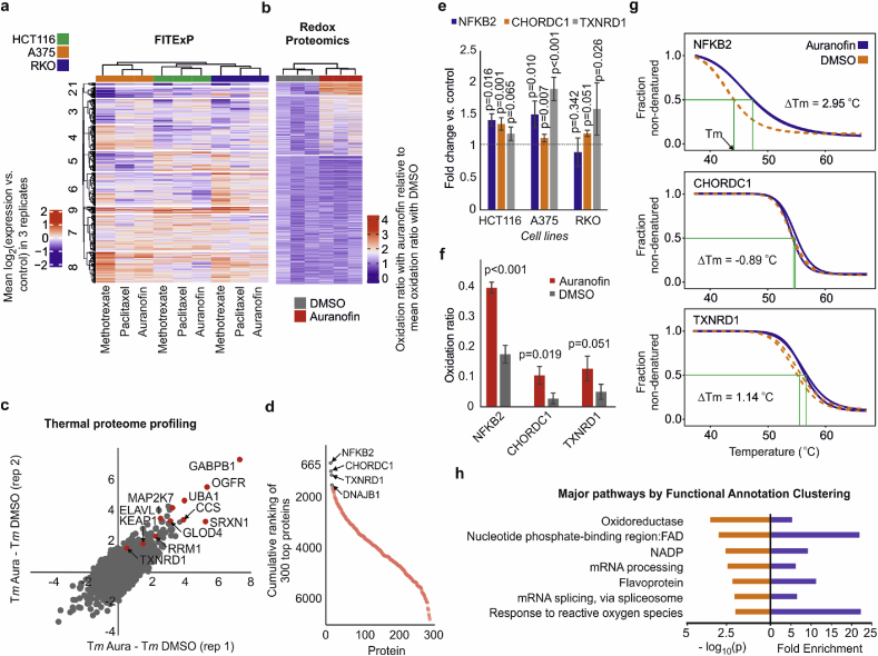
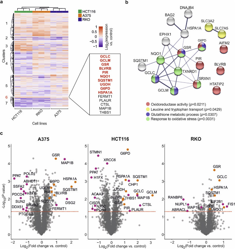
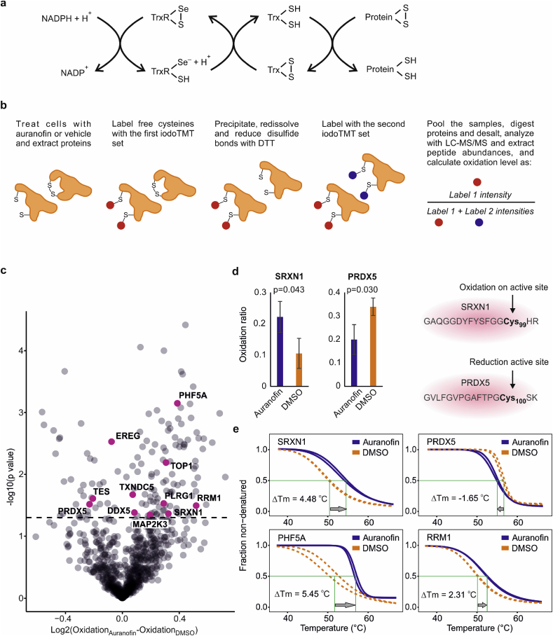
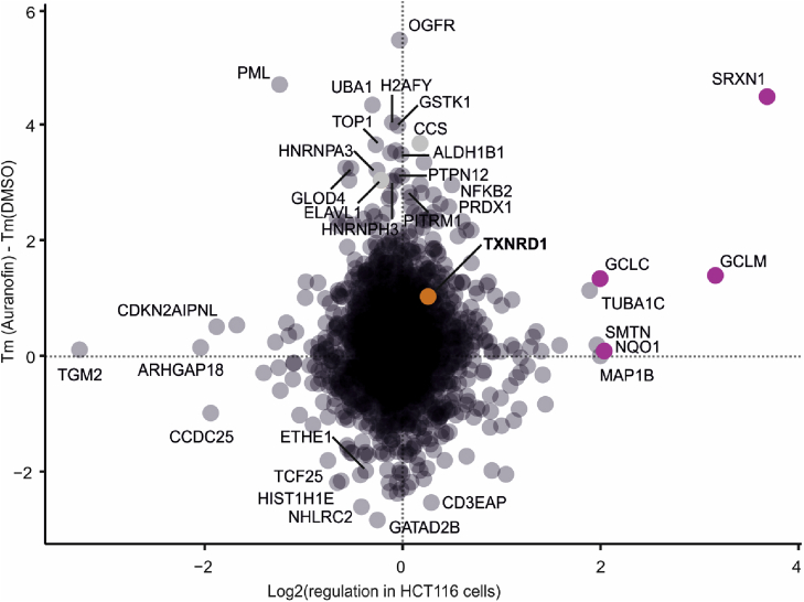
Chemical proteomics encompasses novel drug target deconvolution methods in which compound modification is not required. Herein we use Thermal Proteome Profiling, Functional Identification of Target by Expression Proteomics and multiplexed redox proteomics for deconvolution of auranofin targets to aid elucidation of its mechanisms of action. Auranofin (Ridaura®) was approved for treatment of rheumatoid arthritis in 1985. Because several clinical trials are currently ongoing to repurpose auranofin for cancer therapy, comprehensive characterization of its targets and effects in cancer cells is important. Together, our chemical proteomics tools confirmed thioredoxin reductase 1 (TXNRD1, EC:1.8.1.9) as a main auranofin target, with perturbation of oxidoreductase pathways as the top mechanism of drug action. Additional indirect targets included NFKB2 and CHORDC1. Our comprehensive data can be used as a proteomic signature resource for further analyses of the effects of auranofin. Here we also assessed the orthogonality and complementarity of different chemical proteomics methods that can furnish invaluable mechanistic information and thus the approach can facilitate drug discovery efforts in general.

Keywords: Ligand; Mechanism of action; Melting temperature; Protein expression; Target.


Conflict of interest statement

Declaration of competing interest Katarina Johansson is currently an employee of Pfizer Innovations AB. The remaining authors declare no conflicts of interest.

Figures

Image 1
Graphical abstract
Fig. 1
Fig. 1
Complementary chemical proteomics approaches for characterization of auranofin targets and mechanism space. While TPP provides information related to stability changes in drug targets and downstream proteins, FITExP reveals proteins with affected abundances, which include both targets and mechanistic proteins. Redox proteomics reveals the changes in the oxidation states of cysteines at the peptide level, which can furthermore hypothetically correlate with protein stability changes in TPP.
Fig. 2
Fig. 2
Unbiased de novo prediction of auranofin targets and mechanism space by chemical proteomics tools. a, Combining the FITExP data from three cell lines and three drugs, which was followed by identification of compound-specific proteome signatures by hierarchical clustering. Data in the heatmap is presented as mean log2 (fold change vs. control) in 3 replicates. b, Redox proteomics revealed an increase in the oxidation level of many peptide in response to auranofin, as expected. Data is plotted as oxidation ratio upon auranofin treatment relative to the mean oxidation ratio upon DMSO treatment. c, TPP data for proteins with ≤1 °C difference between the two replicates in 3 μM auranofin treatment of HCT116 cells shows stabilization of several proteins. Some of these proteins, including GABPB1 [30], RRM1 [31,32] and SRXN1 [33] are known to be regulated by the thioredoxin system. d, The cumulative sum of individual target rankings in four different types of analysis (FITExP, TR-TPP in cells and lysate, as well as deep redox proteomics). Proteins with the lowest overall sum are top candidate targets (TXNRD1 is the known cognate target). e-g, Changes in 3 top target proteins: (e) – specific expression in three cells lines in FITExP, (f) – oxidation level of top peptide (f), and (g) – melting curves in TPP experiments in cells (none of these proteins changed their stability in cell lysate). h, Top 15 proteins from each method were combined (60 proteins in total from 4 methods) and analyzed with Functional Annotation Clustering tool in DAVID. Top enriched biological pathways with minimal redundancy (fold enrichment > 5 and p < 0.01), representing the dominant mechanisms for auranofin, are shown (n = 3 biological replicates for all experiments, TPP experiment was performed in 2 replicates. P values were calculated with two-sided student t-test, mean ± SD).
Fig. 3
Fig. 3
Perturbation of “oxidoreductase” pathway as the dominant auranofin mechanism. a, Clustering of FITExP data for auranofin in three cell lines in 3 replicates. Cluster 6 contains a group of tightly and consistently upregulated proteins. Most of these proteins are Nrf2 targets (bold red) [44,[51], [52], [53]]. The other 5 proteins might be putative Nrf2 targets. b, The enriched pathways for 30 most consistently upregulated proteins in three cell lines in FITExP (disconnected proteins have been removed). c, The consistent upregulation of Nrf2 target proteins from cluster 6 in (a) in different cell lines (in orange). Other significantly regulated proteins are shown in purple. (For interpretation of the references to color in this figure legend, the reader is referred to the Web version of this article.)
Fig. 4
Fig. 4
Peptide oxidation state linked to protein stability. a, Schematics of the role of TXNRD1 (TrxR) and the thioredoxin system in the cell and. b, redox experiment design with sequential multiplexed iodoTMT labeling to measure oxidation levels in the peptide level. c, Peptides from proteins with stability changes in TPP are highlighted in purple. d, Oxidation of SRXN1 and reduction of PRDX5 on the active sites. e, Change in the stability of SRXN1 and PRDX5 in opposite direction. Other representative proteins PHF5A and RRM1 had significantly oxidized peptides and were more stable upon exposure to auranofin. (n = 3 biological replicates for redox proteomics, TPP experiment was performed in 2 replicates. P values were calculated using two-tailed Student's t-test, mean ± SD). (For interpretation of the references to color in this figure legend, the reader is referred to the Web version of this article.)
Fig. 5
Fig. 5
Comparison of protein expression vs. thermal stability for HCT116 cells shows the orthogonality of FITExP and TPP. Some Nrf2 target genes are shown in purple. (For interpretation of the references to color in this figure legend, the reader is referred to the Web version of this article.)

References

    1. Savitski M.M. Tracking cancer drugs in living cells by thermal profiling of the proteome. Science. 2014;346(6205):1255784. - PubMed
    1. Chernobrovkin A. Functional Identification of Target by Expression Proteomics (FITExP) reveals protein targets and highlights mechanisms of action of small molecule drugs. Sci. Rep. 2015;5 - PMC - PubMed
    1. Koshland D. Application of a theory of enzyme specificity to protein synthesis. Proc. Natl. Acad. Sci. U. S. A. 1958;44(2):98–104. - PMC - PubMed
    1. Molina D.M. Monitoring drug target engagement in cells and tissues using the cellular thermal shift assay. science. 2013;341(6141):84–87. - PubMed
    1. Franken H. Thermal proteome profiling for unbiased identification of direct and indirect drug targets using multiplexed quantitative mass spectrometry. Nat. Protoc. 2015;10(10):1567. - PubMed

Publication types