. 2020 May;131(5):1087-1098.
doi: 10.1016/j.clinph.2020.02.014. Epub 2020 Mar 4.

Effect of interictal epileptiform discharges on EEG-based functional connectivity networks

Affiliations

Effect of interictal epileptiform discharges on EEG-based functional connectivity networks

Derek K Hu et al. Clin Neurophysiol. 2020 May.

Abstract

Objective: Functional connectivity networks (FCNs) based on interictal electroencephalography (EEG) can identify pathological brain networks associated with epilepsy. FCNs are altered by interictal epileptiform discharges (IEDs), but it is unknown whether this is due to the morphology of the IED or the underlying pathological activity. Therefore, we characterized the impact of IEDs on the FCN through simulations and EEG analysis.

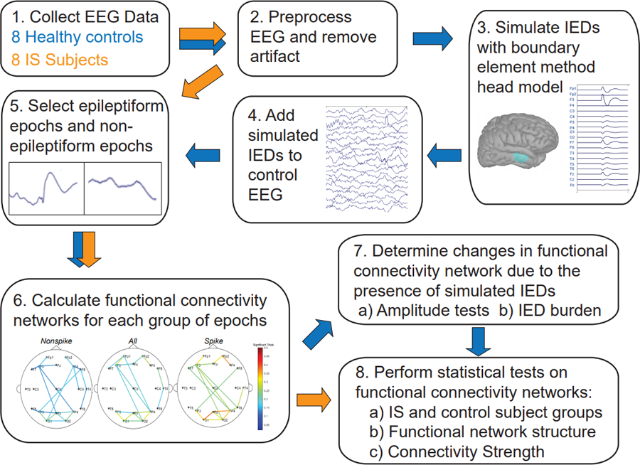
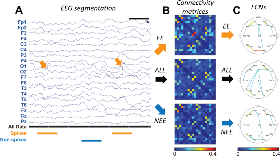
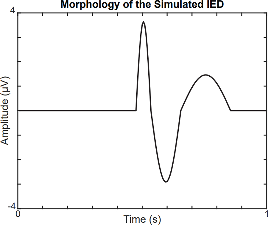
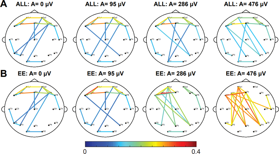
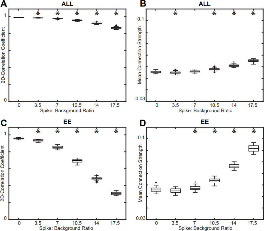
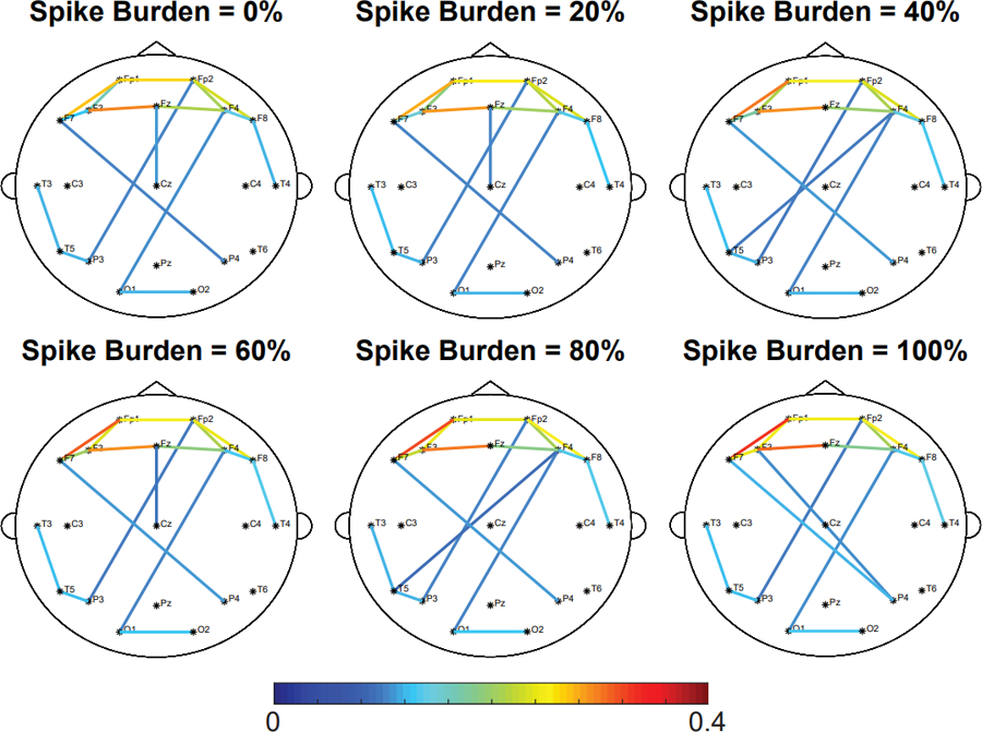
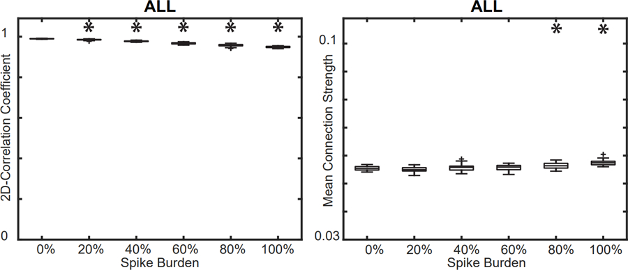
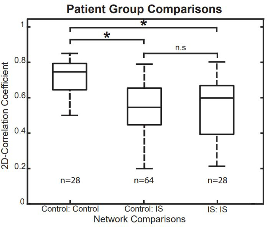
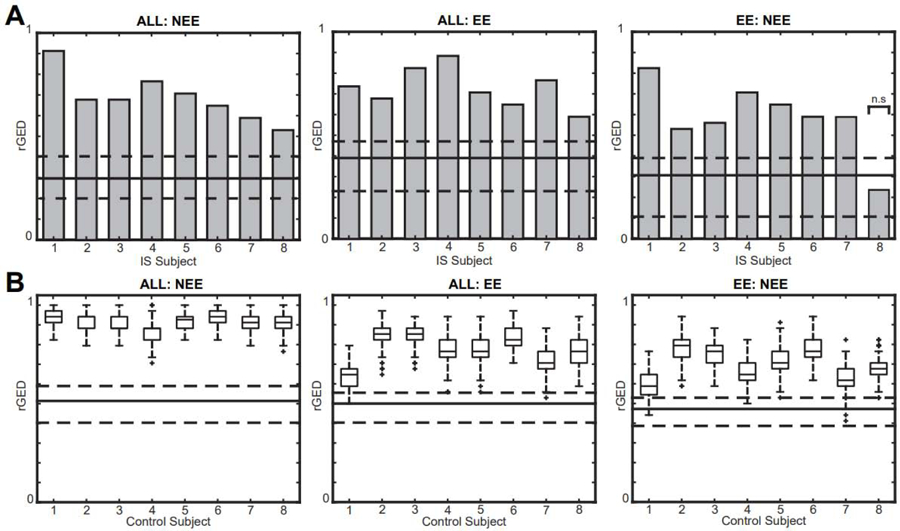
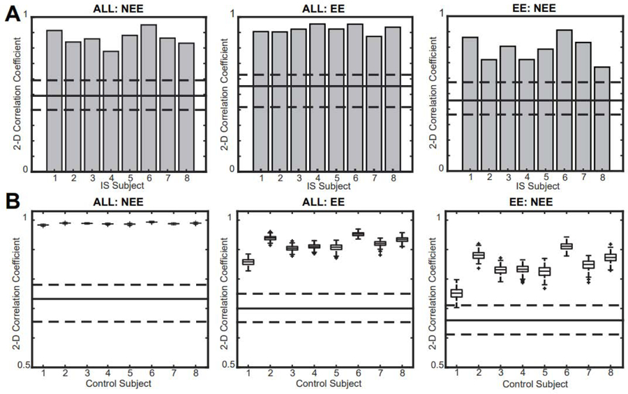
Methods: We introduced simulated IEDs to sleep EEG recordings of eight healthy controls and analyzed the effect of IED amplitude and rate on the FCN. We then generated FCNs based on epochs with and without IEDs and compared them to the analogous FCNs from eight subjects with infantile spasms (IS), based on 1340 visually marked IEDs. Differences in network structure and strength were assessed.

Results: IEDs in IS subjects caused increased connectivity strength but no change in network structure. In controls, simulated IEDs with physiological amplitudes and rates did not alter network strength or structure.

Conclusions: Increases in connectivity strength in IS subjects are not artifacts caused by the interictal spike waveform and may be related to the underlying pathophysiology of IS.

Significance: Dynamic changes in EEG-based FCNs during IEDs may be valuable for identification of pathological networks associated with epilepsy.

Keywords: Brain mapping; Electroencephalography; Epilepsy; Functional connectivity; Infantile spasms; Interictal epileptiform discharges.


Conflict of interest statement

Conflict of Interest Statement

None of the authors have potential conflicts of interest to be disclosed.

Figures

Figure 1.
Figure 1.
Summary of the functional connectivity analysis. Blue arrows indicate the data analysis procedure for the control subject EEG, while orange arrows indicate the procedure for analysis of EEG data from subjects with epilepsy.
Figure 2.
Figure 2.
(A) Sample of EEG data segmentation. Orange segments are 1s epochs containing an IED (termed EE), black segments are all 1s epochs (ALL), and blue segments are 1s epochs with non-spike activity (NEE). (B) Connectivity matrices for spikes (orange arrow), all data (black arrow), and non-spikes (blue arrow), represented as a percentage of significant connections. (C) FCNs for EE (orange arrow), ALL (black arrow), and NEE (blue arrow). For clarity, only the strongest 10% of all connections are shown. IED: interictal epileptiform discharge; EE: epileptiform epochs; NEE: non-epileptiform epochs; FCN: functional connectivity network.
Figure 3.
Figure 3.
Morphology of the simulated IED at the focal electrode F3. The amplitude of the IED is scaled to match the spike:background ratio of IS subjects. IED: interictal epileptiform discharge; IS: infantile spasms.
Figure 4.
Figure 4.
Increasing the amplitude of the IED has a small effect on the strength and structure of the ALL network and a large effect on the EE network. Representative example showing the effect of adding simulated focal IEDs at F3 with varying amplitudes on the (A) ALL FCN and the (B) EE FCN of control subject 6. The strongest 10% of connections are shown. Results for all other control subjects are shown in Supplementary Figures S1–S7. IED: interictal epileptiform discharge; EE: epileptiform epochs; FCN: functional connectivity network; ALL: all 1s epochs.
Figure 5.
Figure 5.
Effect of varying spike amplitude on the (A) 2D correlation and (B) mean connection strength of the ALL FCN compared to the CONTROL FCN. Effect of varying spike amplitude on the (C) 2D correlation and (D) mean connection strength of the EE FCN compared to the CONTROL FCN. The mean spike:background ratio for IS subjects was 2.62, and changes in network strength and structure occur well above this value. Results for all control subjects were similar (Supplementary Figures S1–S7); a representative example from control subject 6 is shown. Significance tests compared each result to the base CONTROL FCN (spike burden = 0%). EE: epileptiform epochs; FCN: functional connectivity network; ALL: all 1s epochs; IS: infantile spasms.
Figure 6.
Figure 6.
Increasing the spike burden does not affect the network strength and structure of the ALL network. Representative example of the effect of spike burden on the ALL FCN of control subject 6. Simulated focal IEDs are added at electrode F3. The strongest 10% of connections are shown. Results for all other control subjects are shown in Supplementary Figures S1–S7. IED: interictal epileptiform discharge; ALL: all 1s epochs.
Figure 7.
Figure 7.
(A) Effect of spike burden on the 2D correlation between the ALL FCN and CONTROL FCN. (B) Effect of spike burden on the mean connection strength in the ALL FCN. Results for all control subjects were similar (Supplementary Figures S1–S7); a representative example from control subject 6 is shown. Significance tests compared each result to the base CONTROL FCN (spike burden = 0%). FCN: functional connectivity network; ALL: all 1s epochs.
Figure 8.
Figure 8.
Rank-sum tests for 2D correlation coefficients across different subject groups. The correlation coefficients in control-control comparisons were significantly higher than in control-IS and IS-IS comparisons, indicating that control subjects have more consistent, stereotyped FCNs. IS: infantile spasms; FCN: functional connectivity network.
Figure 9.
Figure 9.
Relative graph edit distance (rGED) values for intra-subject network comparisons are significantly higher than inter-subject comparisons in (A) IS subjects and (B) control subjects with simulated IEDs. Gray bars represent intra-subject comparisons for IS subjects, and box plots represent intra-subject values from 200 simulations in controls. The solid lines represent the median of the inter-subject rGED values and the dashed lines represent the 25th and 75th percentiles. All tests are significant except EE:NEE in IS subject 8. IED: interictal epileptiform discharge; EE: epileptiform epochs; NEE: non-epileptiform epochs; IS: infantile spasms.
Figure 10.
Figure 10.
2D correlation coefficients for all intra-subject network comparisons are significantly higher than inter-subject comparisons in (A) IS subjects and (B) controls. Gray bars represent intra-subject comparisons for IS subjects, and box plots represent intra-subject values from 200 simulations in controls. The solid line represents the median of the inter-subject correlation values and the dashed lines represent the 25th and 75th percentiles. IS: infantile spasms.

References

    1. Adebimpe A, Aarabi A, Bourel-Ponchel E, Mahmoudzadeh M, Wallois F. EEG resting state analysis of cortical sources in patients with benign epilepsy with centrotemporal spikes. NeuroImage Clin 2015;9:275–82. - PMC - PubMed
    1. Blinowska KJ. Review of the methods of determination of directed connectivity from multichannel data. Med Biol Eng Comput 2011;49(5):521–9. - PMC - PubMed
    1. Blinowska KJ, Kaminski M. Functional brain networks: random, “small world” or deterministic? PLoS One. 2013;8(10):e78763. - PMC - PubMed
    1. Chu-Shore CJ, Kramer MA, Pathmanathan J, Bianchi MT, Westover MB, Wizon L, et al. Emergence of stable functional networks in long-term human EEG. NIH Public Access. 2012;32(8):2703–13. - PMC - PubMed
    1. Clemens B, Puskás S, Spisák T, Lajtos I, Opposits G, Besenyei M, et al. Increased resting-state EEG functional connectivity in benign childhood epilepsy with centro-temporal spikes. Seizure 2016;35:50–5. - PubMed

Publication types