

The characteristics and clinical value of chest CT images of novel coronavirus pneumonia
- PMID: 32199619
- PMCID: PMC7156121
- DOI: 10.1016/j.crad.2020.03.002
The characteristics and clinical value of chest CT images of novel coronavirus pneumonia
Abstract
Aim: To investigate the characteristics and clinical value of chest computed tomography (CT) images of novel coronavirus pneumonia (NCP).
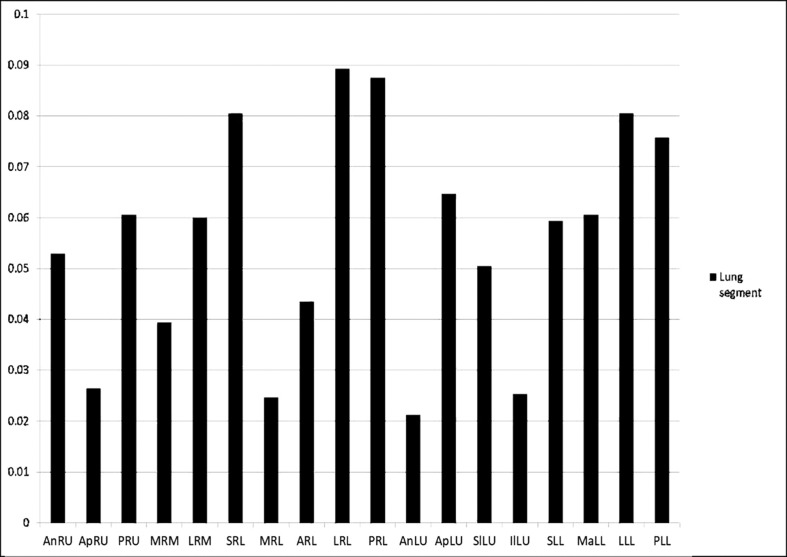
Materials and methods: Clinical data and CT images of 80 cases of NCP were collected. The clinical manifestations and laboratory test results of the patients were analysed. The lesions in each lung segment of the patient's chest CT images were characterised. Lesions were scored according to length and diffusivity.
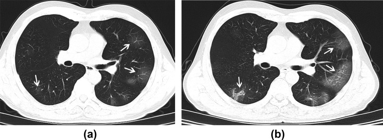
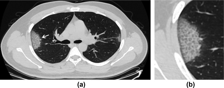
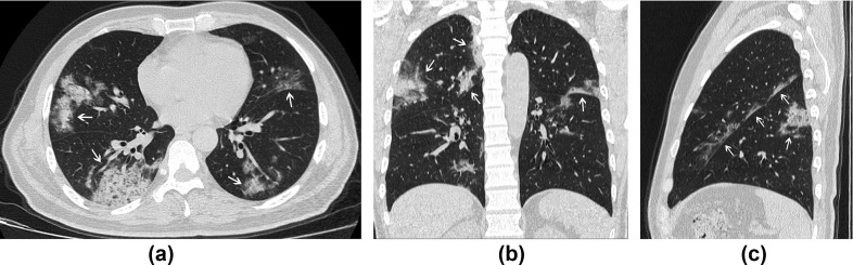
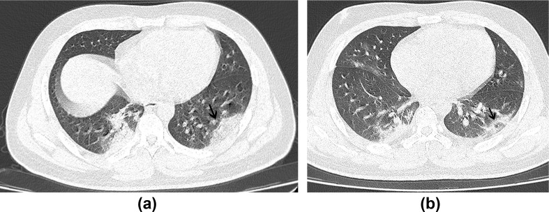
Results: The main clinical manifestations were fever, dry cough, fatigue, a little white sputum, or diarrhoea. A total of 1,702 scored lesions were found in the first chest CT images of 80 patients. The lesions were located mainly in the subpleural area of the lungs (92.4%). Most of the lesions were ground-glass opacity, and subsequent fusions could increase in range and spread mainly in the subpleural area. Pulmonary consolidation accounted for 44.1% of all of the lesions. Of the 80 cases, 76 patients (95%) had bilateral lung disease, four (5%) patients had unilateral lung disease, and eight (10%) patients had cord shadow.
Conclusion: The chest CT of NCP patients is characterised by the onset of bilateral ground-glass lesions located in the subpleural area of the lung, and progressive lesions that result in consolidation with no migratory lesions. Pleural effusions and mediastinal lymphadenopathy are rare. As patients can have inflammatory changes in the lungs alongside a negative early nucleic acid test, chest CT, in combination with epidemiological and laboratory tests, is a useful examination to evaluate the disease and curative effect.
Copyright © 2020 The Royal College of Radiologists. Published by Elsevier Ltd. All rights reserved.
Figures
Similar articles
-
Imaging and clinical features of patients with 2019 novel coronavirus SARS-CoV-2.Eur J Nucl Med Mol Imaging. 2020 May;47(5):1275-1280. doi: 10.1007/s00259-020-04735-9. Epub 2020 Feb 28. Eur J Nucl Med Mol Imaging. 2020. PMID: 32107577 Free PMC article.
-
Preliminary CT findings of coronavirus disease 2019 (COVID-19).Clin Imaging. 2020 Sep;65:124-132. doi: 10.1016/j.clinimag.2020.04.042. Epub 2020 May 12. Clin Imaging. 2020. PMID: 32464579 Free PMC article.
-
Clinical and computed tomographic imaging features of novel coronavirus pneumonia caused by SARS-CoV-2.J Infect. 2020 Apr;80(4):394-400. doi: 10.1016/j.jinf.2020.02.017. Epub 2020 Feb 25. J Infect. 2020. PMID: 32109443 Free PMC article.
-
Similarities and Differences of Early Pulmonary CT Features of Pneumonia Caused by SARS-CoV-2, SARS-CoV and MERS-CoV: Comparison Based on a Systemic Review.Chin Med Sci J. 2020 Sep 30;35(3):254-261. doi: 10.24920/003727. Chin Med Sci J. 2020. PMID: 32972503 Free PMC article.
-
Clinical and radiological features of novel coronavirus pneumonia.J Xray Sci Technol. 2020;28(3):391-404. doi: 10.3233/XST-200687. J Xray Sci Technol. 2020. PMID: 32538893 Free PMC article. Review.
Cited by
-
Comorbidities, clinical signs and symptoms, laboratory findings, imaging features, treatment strategies, and outcomes in adult and pediatric patients with COVID-19: A systematic review and meta-analysis.Travel Med Infect Dis. 2020 Sep-Oct;37:101825. doi: 10.1016/j.tmaid.2020.101825. Epub 2020 Aug 4. Travel Med Infect Dis. 2020. PMID: 32763496 Free PMC article.
-
Which criteria were used to describe patients with COVID-19? A systematic review and meta analysis of clinical, laboratory, and imaging features.Med J Islam Repub Iran. 2021 Sep 2;35:112. doi: 10.47176/mjiri.35.112. eCollection 2021. Med J Islam Repub Iran. 2021. PMID: 34956958 Free PMC article. Review.
-
HQDCNet: Hybrid Quantum Dilated Convolution Neural Network for detecting covid-19 in the context of Big Data Analytics.Multimed Tools Appl. 2023 May 12:1-27. doi: 10.1007/s11042-023-15515-6. Online ahead of print. Multimed Tools Appl. 2023. PMID: 37362720 Free PMC article.
-
The Limited Sensitivity of Chest Computed Tomography Relative to Reverse Transcription Polymerase Chain Reaction for Severe Acute Respiratory Syndrome Coronavirus-2 Infection: A Systematic Review on COVID-19 Diagnostics.Invest Radiol. 2020 Dec;55(12):754-761. doi: 10.1097/RLI.0000000000000700. Invest Radiol. 2020. PMID: 32554983 Free PMC article.
-
Clinical and CT features of the COVID-19 infection: comparison among four different age groups.Eur Geriatr Med. 2020 Oct;11(5):843-850. doi: 10.1007/s41999-020-00356-5. Epub 2020 Jul 13. Eur Geriatr Med. 2020. PMID: 32662041 Free PMC article.
References
-
- World Health Organization WHO handbook for guideline development. 2014. https://apps.who.int/iris/handle/10665/145714 2nd edn.
-
- General Office of National Health Committee Office of State Administration of Traditional Chinese Medicine. Notice on the issuance of a programme for the diagnosis and treatment of novel coronavirus (2019-nCoV) infected pneumonia (trial 4th edn) (2020-0128) http://bgs.satcm.gov.cn/zhengcewenjian/2020-01-28/12576.html
Publication types
MeSH terms
LinkOut - more resources
Full Text Sources
Research Materials
