

Prognostic Value of Myocardial Extracellular Volume Fraction and T2-mapping in Heart Transplant Patients
- PMID: 32199848
- PMCID: PMC8809107
- DOI: 10.1016/j.jcmg.2020.01.014
Prognostic Value of Myocardial Extracellular Volume Fraction and T2-mapping in Heart Transplant Patients
Abstract
Objectives: The purpose of this study was to examine prognostic value of T1- and T2-mapping techniques in heart transplant patients.
Background: Myocardial characterization using T2 mapping (evaluation of edema/inflammation) and pre- and post-gadolinium contrast T1 mapping (calculation of extracellular volume fraction [ECV] for assessment of interstitial expansion/fibrosis) are emerging modalities that have been investigated in various cardiomyopathies.
Methods: A total of 99 heart transplant patients underwent the magnetic resonance imaging (MRI) scans including T1- (n = 90) and T2-mapping (n = 79) techniques. Relevant clinical characteristics, MRI parameters including late gadolinium enhancement (LGE), and invasive hemodynamics were collected. Median clinical follow-up duration after the baseline scan was 2.4 to 3.5 years. Clinical outcomes include cardiac events (cardiac death, myocardial infarction, coronary revascularization, and heart failure hospitalization), noncardiac death and noncardiac hospitalization.
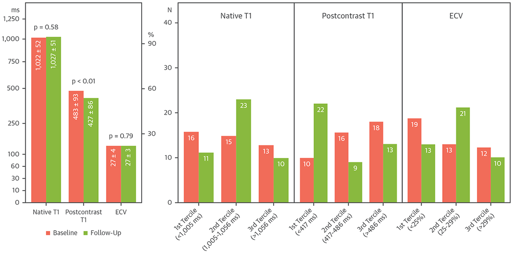
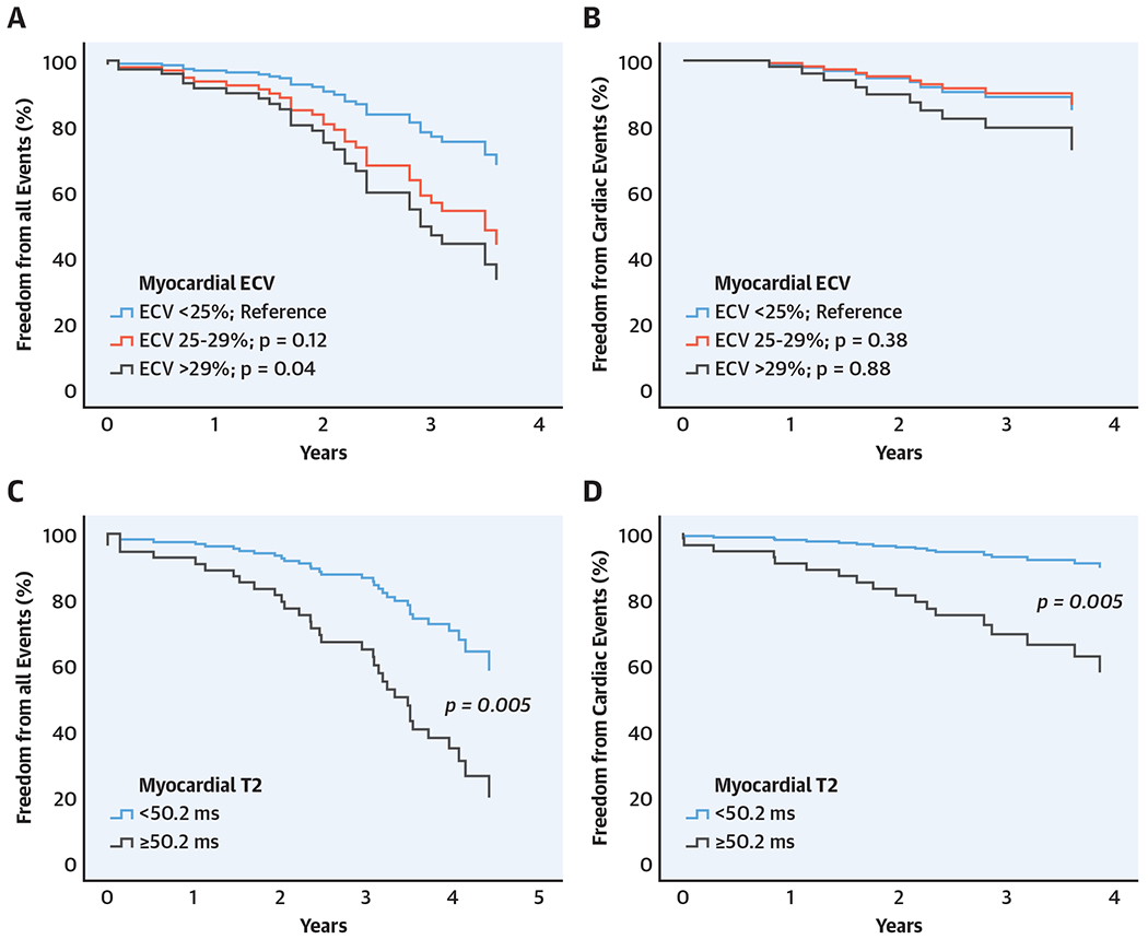
Results: Overall, the global native T1, postcontrast T1, ECV, and T2 were 1,030 ± 56 ms, 458 ± 84 ms, 27 ± 4% and 50 ± 4 ms, respectively. Top-tercile-range ECV (ECV >29%) independently predicted adverse clinical outcomes compared with bottom-tercile-range ECV (ECV <25%) (hazard ratio [HR]: 2.87; 95% confidence interval [CI]: 1.07 to 7.68; p = 0.04) in a multivariable model with left ventricular end-systolic volume and LGE. Higher T2 (T2 ≥50.2 ms) independently predicted adverse clinical outcomes (HR: 3.01; 95% CI: 1.39 to 6.54; p = 0.005) after adjustment for left ventricular ejection fraction, left ventricular end-systolic volume, and LGE. Additionally, higher T2 (T2 ≥50.2 ms) also independently predicted cardiac events (HR: 4.92; CI: 1.60 to 15.14; p = 0.005) in a multivariable model with left ventricular ejection fraction.
Conclusions: MRI-derived myocardial ECV and T2 mapping in heart transplant patients were independently associated with cardiac and noncardiac outcomes. Our findings highlight the need for larger prospective studies.
Keywords: T1 mapping; T2 mapping; extracellular volume fraction; heart transplantation; magnetic resonance imaging; natural history; prognosis.
Copyright © 2020 American College of Cardiology Foundation. Published by Elsevier Inc. All rights reserved.
Figures
Comment in
-
Cardiac Magnetic Resonance Parametric Mapping Following Heart Transplantation: Moving Beyond Acute Rejection and Coronary Allograft Vasculopathy Assessment.JACC Cardiovasc Imaging. 2020 Jul;13(7):1531-1533. doi: 10.1016/j.jcmg.2020.02.015. Epub 2020 Apr 15. JACC Cardiovasc Imaging. 2020. PMID: 32305482 No abstract available.
References
-
- Di Marco A, Anguera I, Schmitt M, et al. Late gadolinium enhancement and the risk for ventricular arrhythmias or sudden death in dilated cardiomyopathy: systematic review and meta-analysis. J Am Coll Cardiol HF 2017;5:28–38. - PubMed
-
- Green JJ, Berger JS, Kramer CM, Salerno M. Prognostic value of late gadolinium enhancement in clinical outcomes for hypertrophic cardiomyopathy. J Am Coll Cardiol Cardiol Img 2012;5: 370–7. - PubMed
-
- Kim RJ, Wu E, Rafael A, et al. The use of contrast-enhanced magnetic resonance imaging to identify reversible myocardial dysfunction. N Engl J Med 2000;343:1445–53. - PubMed
Publication types
MeSH terms
Substances
Grants and funding
LinkOut - more resources
Full Text Sources
Medical
