Review
. 2020 Jun;158(8):2072-2081.
doi: 10.1053/j.gastro.2020.02.059. Epub 2020 Mar 19.

Carcinogenesis of Pancreatic Ductal Adenocarcinoma

Affiliations
Review

Carcinogenesis of Pancreatic Ductal Adenocarcinoma

Peter Storz et al. Gastroenterology. 2020 Jun.

Abstract

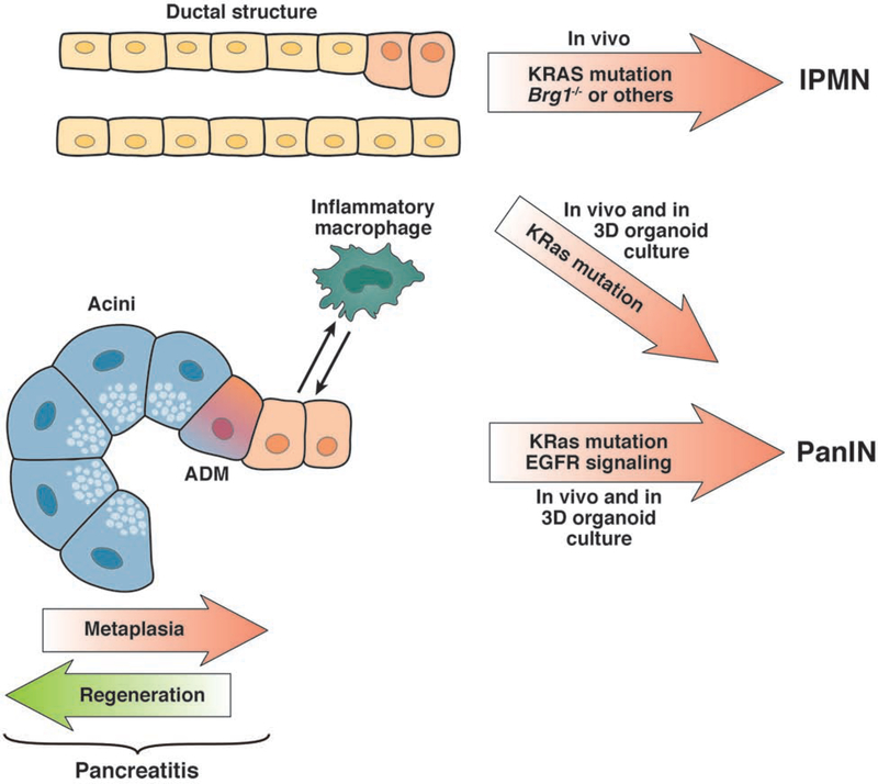
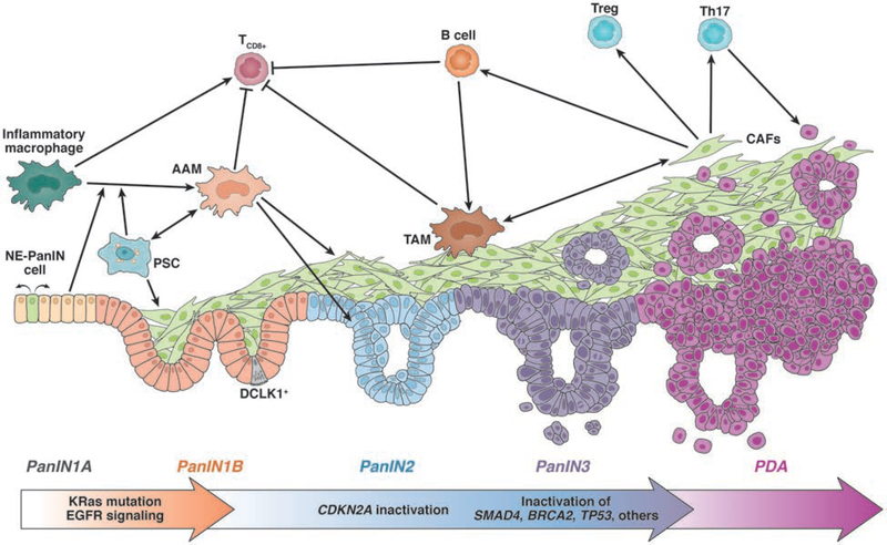
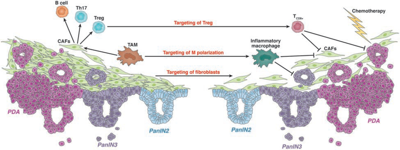
Although the estimated time for development of pancreatic ductal adenocarcinoma (PDA) is more than 20 years, PDAs are usually detected at late, metastatic stages. PDAs develop from duct-like cells through a multistep carcinogenesis process, from low-grade dysplastic lesions to carcinoma in situ and eventually to metastatic disease. This process involves gradual acquisition of mutations in oncogenes and tumor suppressor genes, as well as changes in the pancreatic environment from a pro-inflammatory microenvironment that favors the development of early lesions, to a desmoplastic tumor microenvironment that is highly fibrotic and immune suppressive. This review discusses our current understanding of how PDA originates.

Keywords: Carcinogenesis; Desmoplastic Reaction; Microenvironment; Pancreatic Ductal Adenocarcinoma; Pancreatic Intraepithelial Neoplasia.


Conflict of interest statement

Conflicts of Interest:

The authors certify that they have no affiliations with or involvement in any organization or entity with any financial interest or non-financial interest in the subject matter discussed in this manuscript.

Figures

Figure 1.
Figure 1.. Cells of origin for PDA.
There is evidence that PDAs develop from acinar and/or duct cells. In 3-dimensional organoid culture, duct cells that express oncogenic KRAS can develop into PanIN-like cells; when these are transplanted into mice they form low-grade dysplasias. After additional disruption of Brg1, duct cells with a KRAS mutation can progress to IPMN. Acinar cells are highly plastic and, in crosstalk with inflammatory macrophages, can transdifferentiate to a ductal phenotype (acinar to ductal metaplasia). With an oncogenic KRAS mutation these ADM cells stay locked in a ductal stage, show increased EGFR signaling, and progress to PanIN and PDA.
Figure 2.
Figure 2.. Progression of PDA and the associated microenvironment.
PDA develops through different stages of low-grade dysplasia (PanIN1A/B, PanIN2). While PanIN1 lesions develop after acquisition of an oncogenic Kras mutation and upregulated EGFR signaling, further progression to PanIN2 requires additional gene mutations in tumor suppressors such as CDKN2A. NE-PanIN cells respond to neuropeptides and promote expansion of lesions. Increased fibrogenesis accompanies low grade dysplasia formation. This is mediated by AAM, which are attracted by PanIN cells and PSCs. AAM generate a tumor-promoting and immunosuppressive microenvironment by inducing fibrogenesis, inhibiting CD8+ T-cells, and by releasing factors that promote expansion of lesions. A strong desmoplastic reaction can be observed at PanIN3 lesions (carcinoma in situ) and PDA, with different types of cancer-associated fibroblasts (CAFs) that in part originate from activated PSC. CAFs stimulate TAMs and contribute to the presence of CD4+ T cells (such as Treg cells and Th17 cells) and B cells. At this stage, inactivating mutations in tumor suppressor genes (e.g. SMAD4, BRCA2, TP53) are observed.
Figure 3.
Figure 3.. Strategies for Therapeutic Targeting of PDA.
Chemotherapy alone is not very effective against PDAs, so strategies have been developed to target cells that support tumor growth and progression. These include targeting CD4+ T cells and strategies to shift macrophage populations to an inflammatory phenotype, to generate an inflammatory microenvironment. Strategies to decrease fibrosis include depletion of AAMs or TAMs, or direct targeting of fibroblasts.

References

    1. Petersen GM. Familial Pancreatic Adenocarcinoma. Hematol Oncol Clin North Am 2015;29:641–53. - PMC - PubMed
    1. Shi C, Hong SM, Lim P, et al. KRAS2 mutations in human pancreatic acinar-ductal metaplastic lesions are limited to those with PanIN: implications for the human pancreatic cancer cell of origin. Mol Cancer Res 2009;7:230–6. - PMC - PubMed
    1. Mueller S, Engleitner T, Maresch R, et al. Evolutionary routes and KRAS dosage define pancreatic cancer phenotypes. Nature 2018;554:62–68. - PMC - PubMed
    1. Eibl G, Cruz-Monserrate Z, Korc M, et al. Diabetes Mellitus and Obesity as Risk Factors for Pancreatic Cancer. J Acad Nutr Diet 2018;118:555–567. - PMC - PubMed
    1. Korc M, Jeon CY, Edderkaoui M, et al. Tobacco and alcohol as risk factors for pancreatic cancer. Best Pract Res Clin Gastroenterol 2017;31:529–536. - PMC - PubMed

Publication types

MeSH terms

Substances