. 2020 Jul 15;37(14):1645-1655.
doi: 10.1089/neu.2019.6932. Epub 2020 Apr 17.

Neuroinflammation Mediated by Glia Maturation Factor Exacerbates Neuronal Injury in an in vitro Model of Traumatic Brain Injury

Affiliations

Neuroinflammation Mediated by Glia Maturation Factor Exacerbates Neuronal Injury in an in vitro Model of Traumatic Brain Injury

Mohammad Ejaz Ahmed et al. J Neurotrauma. .

Abstract

Traumatic brain injury (TBI) is the primary cause of death and disability affecting over 10 million people in the industrialized world. TBI causes a wide spectrum of secondary molecular and cellular complications in the brain. However, the pathological events are still not yet fully understood. Previously, we have shown that the glia maturation factor (GMF) is a mediator of neuroinflammation in neurodegenerative diseases. To identify the potential molecular pathways accompanying TBI, we used an in vitro cell culture model of TBI. A standardized injury was induced by scalpel cut through a mixed primary cell culture of astrocytes, microglia and neurons obtained from both wild type (WT) and GMF-deficient (GMF-KO) mice. Cell culture medium and whole-cell lysates were collected at 24, 48, and 72 h after the scalpel cuts injury and probed for oxidative stress using immunofluorescence analysis. Results showed that oxidative stress markers such as glutathione and glutathione peroxidase were significantly reduced, while release of cytosolic enzyme lactate dehydrogenase along with nitric oxide and prostaglandin E2 were significantly increased in injured WT cells compared with injured GMF-KO cells. In addition, injured WT cells showed increased levels of oxidation product 4-hydroxynonenal and 8-oxo-2'-deoxyguanosine compared with injured GMF-KO cells. Further, we found that injured WT cells showed a significantly increased expression of glial fibrillary acidic protein, ionized calcium binding adaptor molecule 1, and phosphorylated ezrin/radixin/moesin proteins, and reduced microtubule associated protein expression compared with injured GMF-KO cells after injury. Collectively, our results demonstrate that GMF exacerbates the oxidative stress-mediated neuroinflammation that could be brought about by TBI-induced astroglial activation.

Keywords: ezrin/radixin/moesin proteins; glia maturation factor; neurodegeneration; neuroinflammation; traumatic brain injury.


Conflict of interest statement

No competing financial interests exist.

Figures
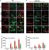
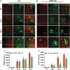
FIG. 1.
FIG. 1.
The absence of glia maturation factor (GMF) attenuates in vitro injury-induced reactive gliosis. Representative immunofluorescence staining of glial fibrillary acidic protein (GFAP; green) and ionized calcium binding adaptor molecule 1 (IBA1; red) in mixed primary neurons and glial cells shows over the different time course effects (0, 24, 48 and 72 h) following post scratch induced injury. GMF-deficient (GMF-KO) mixed primary neurons and glial cells staining showed a gradually huge reduction in expression and co-localization of GFAP and compared with wild type primary mixed neuronal cells showed in (A, B). The immunoactivity associated with GFAP and Iba1 were quantified and shown as percentage changes versus 0 h control group (C, D). Scale bar = 50-μm and dash line represent the injury site. Data are presented as mean ± standard error of the mean (n = 3). *p < 0.05 versus uninjured control. Color image is available online.
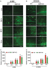
FIG. 2.
FIG. 2.
The absence of glia maturation factor (GMF) promotes regeneration of neurons following neuronal injury in primary neuronal cultures. We performed immunofluorescence staining using neuronal-specific marker microtubule associated protein (MAP2), a potent and sensitive indicator of neuronal morphology and determined of neuronal pathology following injury. Subsequent to the injury to neuronal cultures obtained from wild type mice, the primary mixed neuronal cells shows poor neurons morphology with less growth cone towards the cut line over the time course 0 to 72 h (A) compared with primary mixed neuronal cells from GMF-deficient (GMF-KO) mice (B). The GMF-KO group neurons start to generate new and bigger growth cones and shows widespread MAP2 staining towards the cut line. (C) Quantitative analysis of MAP2 intensity towards inury line. Scale bar = 50 μm and dash lines represents the injury sites. Color image is available online.
FIG. 3.
FIG. 3.
The absence of glia maturation factor (GMF) reduces the expression of Ezrin/Radixin/Moesin (ERM) proteins in astrocytes following cell injury to primary neurons and glial cultures. Representative immunofluorescence staining of mixed neurons and glial cells cultures for a marker of ERM (red) with glial fibrillary acidic protein (GFAP; green) over the different time course study (0, 24, 48 and 72 h following injury). Mixed primary neurons and glial cells from GMF-deficient (GMF-KO) mice shows gradually decreased expression of ERM and GFAP compared with wild type mixed primary neurons and glial cells (A, B). Scale bar = 50 μm. (C, D) Fluorescent intensity was quantified using ImageJ analysis software and expressed as percentage change versus respective 0 h control group. The dash line represents the injury site. Data are presented as mean ± standard error of the mean (n = 3). *p < 0.05 versus 0 h control group. Color image is available online.
FIG. 4.
FIG. 4.
The absence of glia maturation factor (GMF) greatly reduced the phosphorylation of Ezrin/Radixin/Moesin proteins after injury in mice primary mixed neurons and glial cells. Representative immunofluorescence staining of phosphorylated Ezrin/Radixin/Moesin (pERM; red) with glial fibrillary acidic protein (GFAP; green) in primary mixed neurons and glial cells from wild type (WT) and GMF-deficient (GMF-KO) mice subjected to scratch injury. There is a gradual reduction in the expression of pERM and GFAP in the GMF-KO group compared with the WT group (A, B). Dash line represent injury site, scale bar = 50 μm. Fluorescent intensity was quantified using ImageJ software and expressed as a percentage change in intensity versus 0 h control group (C, D). Data are represented as mean ± standard error of the mean (n = 3-6) in each group). *p < 0.05 versus 0 h control group. Color image is available online.
FIG. 5.
FIG. 5.
The absence of glia maturation factor (GMF) limits oxidative damage in scratch mediated neuronal injury. Representative immunofluorescence staining for the markers of oxidative damage 4-HNE for lipid peroxidation (4-HNE) and 8-oxo-2′-deoxyguanosine (8-OhdG) for DNA damage. (A, B) Comparative staining for 4-HNE and 8-OHdG over the time course of, 0, 24, 48, and 72 h following the injury of primary neuronal cells from wild type (WT) and GMF-deficient (GMF-KO) mice. The GMF-KO group shows gradually decreased 4-HNE and 8-OHdG levels compared with the WT group. The dashed line represents the boundary of the injury site. Scale bar 50 μm. Fluorescent intensities were quantified using ImageJ analysis software and expressed as percentage change over respective 0 h control group (C, D). Data are presented as mean ± standard error of the mean (n = 3). *p < 0.05 versus 0 h control group. Color image is available online.
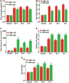
FIG. 6.
FIG. 6.
The absence of glia maturation factor (GMF) reduces oxidative stress–mediated neuronal injury of injured primary neuronal cells. Primary neuronal cells from wild type (WT) and GMF-deficient (GMF-KO) were subjected to mechanical injury and supernatants were collected from WT and GMF-KO cells. Those supernatants were used to determine oxidative stress markers such as (A) glutathione peroxidase (GPx), (B) glutathione (GSH), (C) lactate dehydrogenase (LDH), (D) prostaglandin E2 (PGE2), and (E) nitric oxide (NO) release into the culture medium at different time-points (24, 48 and 72 h) after the injury. The absence of GMF significantly decreased oxidative stress markers such as LDH, NO, GSH and PGE2, and improved antioxidant enzymes such as GSH and GPx compared with control cells. The values are given as the mean ± standard error of the mean of four experiments in each group. *p < 0.05 compared with WT and GMF-KO control. Color image is available online.

References

    1. Xiong Y., Mahmood A., and Chopp M. (2013). Animal models of traumatic brain injury. Nat. Rev. Neurosci. 14, 128–142 - PMC - PubMed
    1. Stahel P.F., Shohami E., Younis F.M., Kariya K., Otto V.I., Lenzlinger P.M., Grosjean M.B., Eugster H.P., Trentz O., Kossmann T., and Morganti-Kossmann M.C. (2000). Experimental closed head injury: analysis of neurological outcome, blood-brain barrier dysfunction, intracranial neutrophil infiltration, and neuronal cell death in mice deficient in genes for pro-inflammatory cytokines. J. Cereb Blood Flow Metab. 20, 369–380 - PubMed
    1. Hang C.H., Shi J.X., Li J.S., Wu W., and Yin H.X. (2005). Concomitant upregulation of nuclear factor-kB activity, proinflammatory cytokines and ICAM-1 in the injured brain after cortical contusion trauma in a rat model. Neurol. India 53, 312–317 - PubMed
    1. Jin W., Zhu L., Guan Q., Chen G., Wang Q.F., Yin H.X., Hang C.H., Shi J.X., and Wang H.D. (2008). Influence of Nrf2 genotype on pulmonary NF-kappaB activity and inflammatory response after traumatic brain injury. Ann. Clin. Lab. Sci. 38, 221–227 - PubMed
    1. Morrison B., 3rd, Elkin B.S., Dolle J.P., and Yarmush M.L. (2011). In vitro models of traumatic brain injury. Annu. Rev. Biomed. Eng. 13, 91–126 - PubMed

Publication types

MeSH terms

Substances