Review
. 2020 Apr;30(4):263-275.
doi: 10.1016/j.tcb.2020.01.006. Epub 2020 Feb 11.

Mitochondrial Functions in Infection and Immunity

Affiliations
Review

Mitochondrial Functions in Infection and Immunity

Varnesh Tiku et al. Trends Cell Biol. 2020 Apr.

Erratum in

Abstract

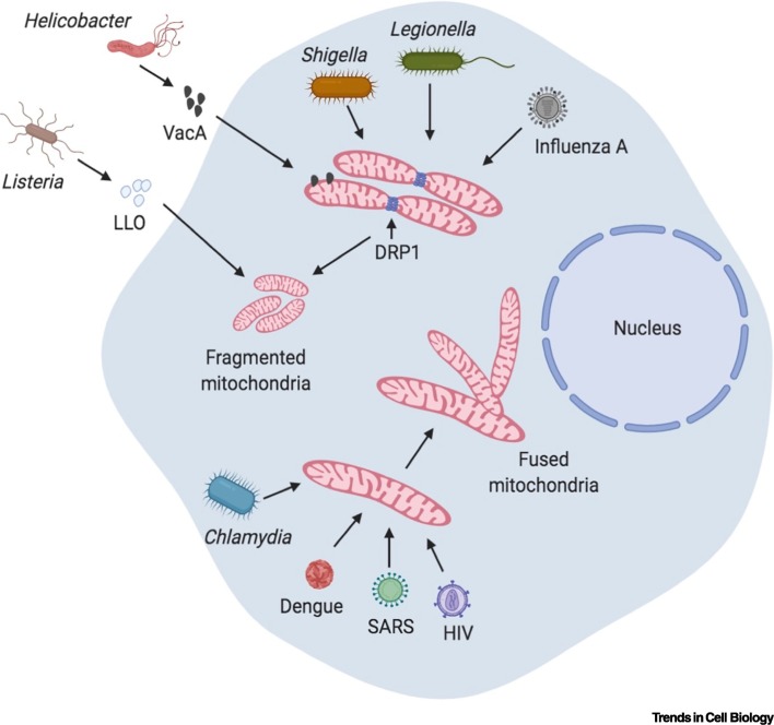
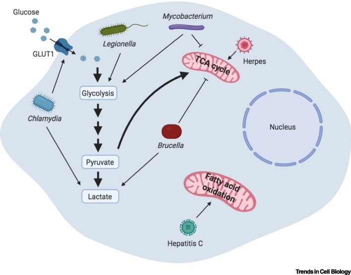
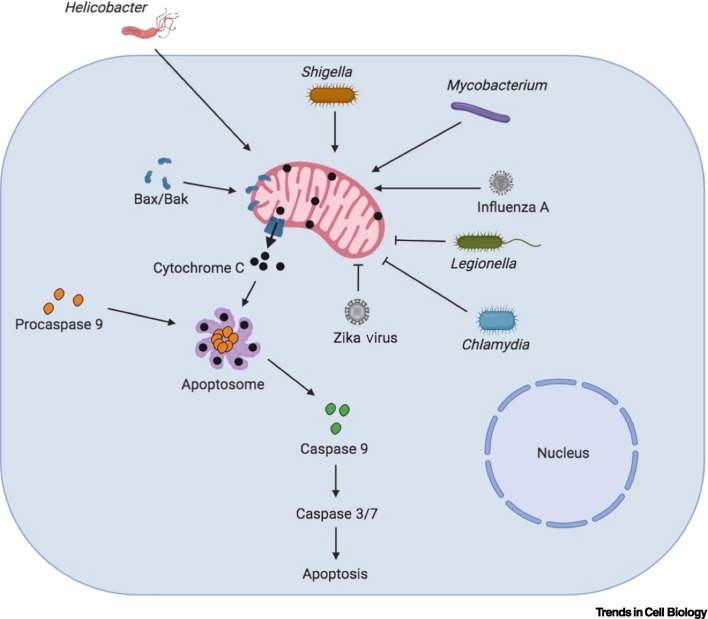
Mitochondria have a central role in regulating a range of cellular activities and host responses upon bacterial infection. Multiple pathogens affect mitochondria dynamics and functions to influence their intracellular survival or evade host immunity. On the other side, major host responses elicited against infections are directly dependent on mitochondrial functions, thus placing mitochondria centrally in maintaining homeostasis upon infection. In this review, we summarize how different bacteria and viruses impact morphological and functional changes in host mitochondria and how this manipulation can influence microbial pathogenesis as well as the host cell metabolism and immune responses.

Keywords: bacteria; cell death; innate immunity; mitochondrial metabolism; mitochondrial morphology; viruses.


Figures

Figure 1
Figure 1
Modulation of Mitochondrial Dynamics upon Infection. Bacteria and viruses induce changes in mitochondrial morphology. Listeria secretes Listeriolysin O (LLO), a pore-forming toxin that induces an unconventional form of mitochondrial fragmentation that is independent of dynamin-related protein 1 (DRP1). Vacuolating cytotoxin VacA from Helicobacter localizes to mitochondria and induces mitochondrial fragmentation, the release of cytochrome C into the cytosol, and subsequent cell death. Shigella and Legionella also induce mitochondrial fragmentation in a DRP1-dependent manner. By contrast, Chlamydia infection leads to mitochondrial fusion, which is required for its intracellular proliferation. Viruses can also modulate mitochondrial dynamics. Influenza A leads to the dissipation of mitochondrial membrane potential, which ultimately causes mitochondrial fragmentation. Dengue virus inhibits DRP1 and induces mitochondrial fragmentation, which is necessary for its replication and immune evasion. Similarly, severe acute respiratory syndrome (SARS) virus and HIV cause DRP1 degradation, thereby bringing about mitochondrial fragmentation. Created with BioRender (www.BioRender.com).
Figure 2
Figure 2
Modulation of Mitochondrial Metabolism upon Infection. Many bacteria and viruses hijack cellular metabolism for their own benefit. Mycobacterium is the best-known bacterium that influences host metabolism by enhancing aerobic glycolysis. Epithelial cells and immune cells infected with Mycobacterium exhibit a reduction in the tricarboxylic acid (TCA) cycle and a corresponding increase in the glycolytic flux. Elevated aerobic glycolysis is also observed in Mycobacterium-infected lung granulomas in animal models of infection and in patients with active tuberculosis. Legionella also promotes glycolytic flux similar to Mycobacterium, thus enhancing aerobic glycolysis. Brucella infection promotes lactate production via aerobic glycolysis, while suppressing the TCA cycle. Chlamydia enhances the levels of the glucose transporter GLUT1, leading to augmented glucose uptake and increased glycolytic flux, which is necessary for its intracellular growth. Viruses also perturb metabolic pathways for their replication and subsequent dissemination. Herpes virus induces the TCA cycle, thereby promoting mitochondrial respiration, while Hepatitis C perturbs mitochondrial fatty acid oxidation. Created with BioRender (www.BioRender.com).
Figure 3
Figure 3
Modulation of Mitochondrial Apoptosis Pathway upon Infection. B cell lymphoma 2 (Bcl-2) family proteins, including include Bcl-2-associated X protein (Bax) and Bcl-2 homologous antagonist/killer (Bak), regulate the mitochondrial cell death pathway. Bax and Bak are proapoptotic proteins that localize to mitochondria and induce outer mitochondrial membrane permeabilization (MOMP), leading to the release of proapoptotic factors, such as cytochrome C, into the cytosol, which induce cell death by activating Caspases 9, 3, and 7 via the apoptosome complex. Bacteria and viruses influence this pathway and modulate the host response. Helicobacter, Shigella, and Mycobacterium infections lead to mitochondrial disruption that stimulates the mitochondrial apoptotic machinery and causes cell death. By contrast, bacteria such as Chlamydia and Legionella block the mitochondrial cell death pathway to promote their intracellular proliferation. Different viruses also exert varied effects on the mitochondrial cell death pathway. Influenza A enhances cell death, which helps in its dissemination, while Zika virus blocks cell death. Created with BioRender (www.BioRender.com).

References

    1. Roger A.J. The origin and diversification of mitochondria. Curr. Biol. 2017;27:R1177–R1192. - PubMed
    1. Tondera D. SlP-2 is required for stress-induced mitochondrial hyperfusion. EMBO J. 2009;278:1589–1600. - PMC - PubMed
    1. Anand R. The i-AAA protease YME1L and OMA1 cleave OPA1 to balance mitochondrial fusion and fission. J. Cell Biol. 2014;204:919–929. - PMC - PubMed
    1. Cipolat S. OPA1 requires mitofusin 1 to promote mitochondrial fusion. Proc. Natl. Acad. Sci. U. S. A. 2004;101:15927–15932. - PMC - PubMed
    1. Chen H. Mitofusins Mfn1 and Mfn2 coordinately regulate mitochondrial fusion and are essential for embryonic development. J. Cell Biol. 2003;160:189–200. - PMC - PubMed