Review
. 2020 Jul:211:107540.
doi: 10.1016/j.pharmthera.2020.107540. Epub 2020 Mar 19.

Biased GPCR signaling: Possible mechanisms and inherent limitations

Affiliations
Review

Biased GPCR signaling: Possible mechanisms and inherent limitations

Vsevolod V Gurevich et al. Pharmacol Ther. 2020 Jul.

Erratum in

Abstract

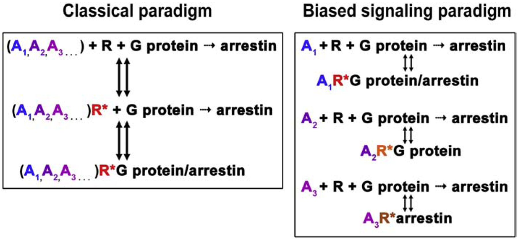
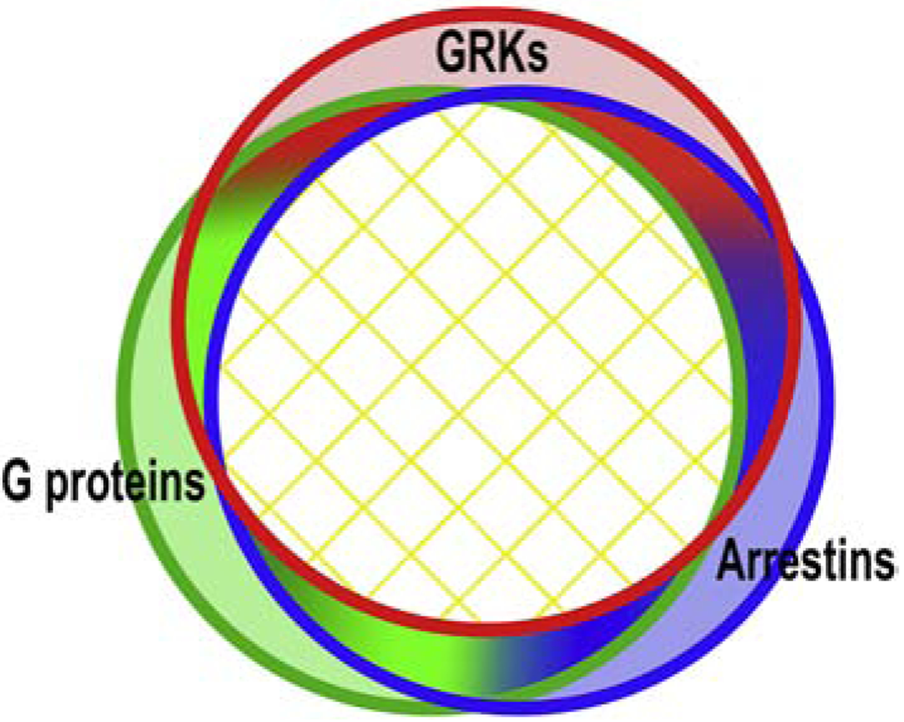
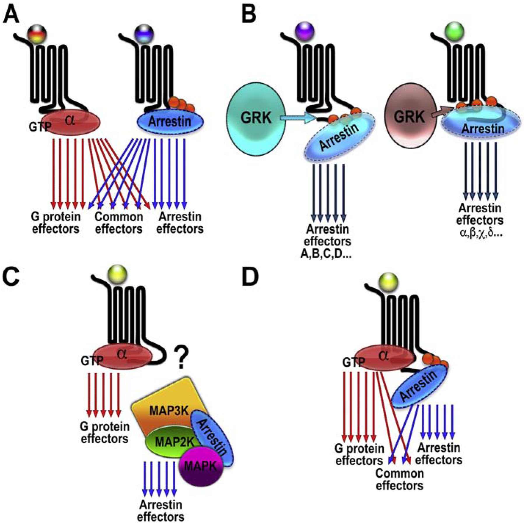
G protein-coupled receptors (GPCRs) are targeted by about a third of clinically used drugs. Many GPCRs couple to more than one type of heterotrimeric G proteins, become phosphorylated by any of several different GRKs, and then bind one or more types of arrestin. Thus, classical therapeutically active drugs simultaneously initiate several branches of signaling, some of which are beneficial, whereas others result in unwanted on-target side effects. The development of novel compounds to selectively channel the signaling into the desired direction has the potential to become a breakthrough in health care. However, there are natural and technological hurdles that must be overcome. The fact that most GPCRs are subject to homologous desensitization, where the active receptor couples to G proteins, is phosphorylated by GRKs, and then binds arrestins, suggest that in most cases the GPCR conformations that facilitate their interactions with these three classes of binding partners significantly overlap. Thus, while partner-specific conformations might exist, they are likely low-probability states. GPCRs are inherently flexible, which suggests that complete bias is highly unlikely to be feasible: in the conformational ensemble induced by any ligand, there would be some conformations facilitating receptor coupling to unwanted partners. Things are further complicated by the fact that virtually every cell expresses numerous G proteins, several GRK subtypes, and two non-visual arrestins with distinct signaling capabilities. Finally, novel screening methods for measuring ligand bias must be devised, as the existing methods are not specific for one particular branch of signaling.


Conflict of interest statement

Declaration of Competing Interest The authors declare no conflict of interest.

Figures

Fig. 1.
Fig. 1.. Classical paradigm of GPCR activation and biased signaling.
Classical extended ternary complex model (Samama, et al., 1993) of GPCR activation was based on the assumption that receptors have only two conformations, active (R*) and inactive (R). Receptors activated by any agonist (shown as differently colored A1, A2, A3) were thought to couple to G proteins, become phosphorylated by GRKs, and then bind arrestins. Arrestin binding terminated G protein-mediated signaling (Carman & Benovic, 1998). The paradigm of biased signaling is based on the idea that different agonists induce distinct active receptor conformations (shown as differently colored R*), some of which are equally good for G proteins and arrestins, whereas others differentially affect receptor interactions with these signaling partners, demonstrating bias towards G proteins or arrestins.
Fig. 2.
Fig. 2.. Conformational landscape of active GPCRs.
Active GPCRs exist in an equilibrium of multiple conformations. The great majority of active GPCR conformations are conducive to their coupling to G proteins, GRKs, and arrestins, i.e., are not biased toward a particular partner. Available data do not exclude the existence of GPCR conformations preferentially coupling to one class of these signal transducers. However, these “signaling-specific” conformations likely constitute a small fraction of the total. This simplistic diagram suggests that there might be conformations conducive to coupling to two out of three classes of these GPCR-binding proteins (gradient-filled areas). Conformations “good” for G proteins and arrestins but not for GRKs, while conceivable, do not make much biological sense: in most cases arrestin binding requires prior receptor phosphorylation. Similarly, conformations conductive to interactions with G proteins and GRKs but not arrestins are unlikely, since receptor phosphorylation by a GRK would favor arrestin binding. However, GPCRs likely do not stay in a single conformation for a long time, but oscillate among many, so the receptor might arrive at one of these G protein- + arrestin-preferring conformations after sampling the conformations “good” for GRKs, where it would be phosphorylated.
Fig. 3.
Fig. 3.. GPCR signaling.
A. G protein-biased ligand (tri-colored ball with red in the middle) facilitates the activation of numerous effectors. Arrestin-biased ligand (tri-colored ball with blue in the middle) also facilitates the activation of many effectors, some of which are the same as those activated by G protein-mediated signaling (common effectors). B. Different ligands (shown as balls of different colors) might determine which particular GRK phosphorylates the receptor. Different GRKs generate distinct patterns of receptor-attached phosphates (barcodes) which might pre-determine the direction of arrestin-mediated signaling. Two non-overlapping sets of effectors are shown. C. Arrestin-3-mediated scaffolding of ASK1-MKK4/7-JNK3 cascade does not require GPCR input. Scaffolding would only be productive when the upstream-most MAP3K is active. Whether active GPCRs play any role in the regulation of signaling by complexes scaffolded by free arrestins (like the JNK cascade shown here) remains to be determined. D. An unbiased ligand (yellow ball) can promote the formation of the super-complex, where G protein and arrestin interact with the receptor simultaneously, with the G protein engaging the inter-helical cavity and arrestin binding to the phosphorylated receptor C-terminus. This would promote simultaneous activation of the G protein- and arrestin-dependent effectors.

Similar articles

Cited by

References

    1. Ahn S, Shenoy SK, Wei H, & Lefkowitz RJ (2004). Differential kinetic and spatial patterns of beta-arrestin and G protein-mediated ERK activation by the angiotensin II receptor. J Biol Chem, 279, 35518–35525. - PubMed
    1. Ahn S, Wei H, Garrison TR, & Lefkowitz RJ (2004). Reciprocal regulation of angiotensin receptor-activated extracellular signal-regulated kinases by beta-arrestins 1 and 2. J Biol Chem, 279, 7807–7811. - PubMed
    1. Alvarez-Curto E, Inoue A, Jenkins L, Raihan SZ, Prihandoko R, Tobin AB, & Milligan G (2016). Targeted Elimination of G Proteins and Arrestins Defines Their Specific Contributions to Both Intensity and Duration of G Protein-coupled Receptor Signaling. J Biol Chem, 291, 27147–27159. - PMC - PubMed
    1. Amarandi R-M, Hjortø GM, Rosenkilde MM, & Karlshøj S (2016). Chapter Eight - Probing Biased Signaling in Chemokine Receptors In Handel TM (Ed.), Methods in Enzymology (Vol. 570, pp. 155–186): Academic Press. - PubMed
    1. Anthony DF, Sin YY, Vadrevu S, Advant N, Day JP, Byrne AM, Lynch MJ, Milligan G, Houslay MD, & Baillie GS (2011). β-Arrestin 1 inhibits the GTPase-activating protein function of ARHGAP21, promoting activation of RhoA following angiotensin II type 1A receptor stimulation. Mol Cell Biol, 31, 1066–1075. - PMC - PubMed

Publication types

MeSH terms