

Multiscale finite element modeling of mechanical strains and fluid flow in osteocyte lacunocanalicular system
- PMID: 32201360
- PMCID: PMC7354216
- DOI: 10.1016/j.bone.2020.115328
Multiscale finite element modeling of mechanical strains and fluid flow in osteocyte lacunocanalicular system
Abstract
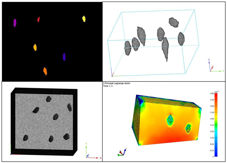
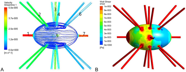
Osteocytes form over 90% of the bone cells and are postulated to be mechanosensors responsible for regulating the function of osteoclasts and osteoblasts in bone modeling and remodeling. Physical activity results in mechanical loading on the bones. Osteocytes are thought to be the main mechanosensory cells in bone. Upon load osteocytes secrete key factors initiating downstream signaling pathways that regulate skeletal metabolism including the Wnt/β-catenin signaling pathway. Osteocytes have dendritic structures and are housed in the lacunae and canaliculi within the bone matrix. Mechanical loading is known to have two primary effects, namely a mechanical strain (membrane disruption by stretching) on the lacunae/cells, and fluid flow, in the form of fluid flow shear stress (FFSS), in the space between the cell membranes and the lacuna-canalicular walls. In response, osteocytes get activated via a process called mechanotransduction in which mechanical signals are transduced to biological responses. The study of mechanotransduction is a complex subject involving principles of engineering mechanics as well as biological signaling pathway studies. Several length scales are involved as the mechanical loading on macro sized bones are converted to strain and FFSS responses at the micro-cellular level. Experimental measurements of strain and FFSS at the cellular level are very difficult and correlating them to specific biological activity makes this a very challenging task. One of the methods commonly adopted is a multi-scale approach that combines biological and mechanical experimentation with in silico numerical modeling of the engineering aspects of the problem. Finite element analysis along with fluid-structure interaction methodologies are used to compute the mechanical strain and FFSS. These types of analyses often involve a multi-length scale approach where models of both the macro bone structure and micro structure at the cellular length scale are used. Imaging modalities play a crucial role in the development of the models and present their own challenges. This paper reviews the efforts of various research groups in addressing this problem and presents the work in our research group. A clear understanding of how mechanical stimuli affect the lacunae and perilacunar tissue strains and shear stresses on the cellular membranes may ultimately lead to a better understanding of the process of osteocyte activation.
Keywords: Finite element model; Fluid flow shear stress; Lacunae; Mechanotransduction; Osteocyte; Perilacunar matrix; Strain.
Copyright © 2020 Elsevier Inc. All rights reserved.
Figures
References
-
- Franz-Odendaal TA, Hall BK, Witten PE. Buried alive: how osteoblasts become osteocytes. Developmental dynamics: an official publication of the American Association of Anatomists 2006;235: 176–190. - PubMed
-
- Castillo AB, Jacobs CR. Skeletal mechanobiology. Mechanobiology handbook 2011: 179–206.
-
- Lanyon L Osteocytes, strain detection, bone modeling and remodeling. Calcified Tissue International 1993;53: 102–107. - PubMed
-
- Carter DR. Mechanical loading histories and cortical bone remodeling. Calcified Tissue International 1984;36: 19–24. - PubMed
-
- Turner CH. Three rules for bone adaptation to mechanical stimuli. Bone 1998;23: 399–407. - PubMed
Publication types
MeSH terms
Grants and funding
LinkOut - more resources
Full Text Sources
