. 2020 May 1;295(18):6007-6022.
doi: 10.1074/jbc.RA119.011253. Epub 2020 Mar 22.

Prominins control ciliary length throughout the animal kingdom: New lessons from human prominin-1 and zebrafish prominin-3

Affiliations

Prominins control ciliary length throughout the animal kingdom: New lessons from human prominin-1 and zebrafish prominin-3

József Jászai et al. J Biol Chem. .

Abstract

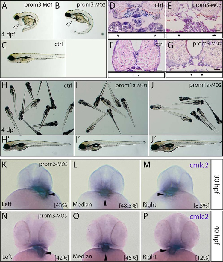
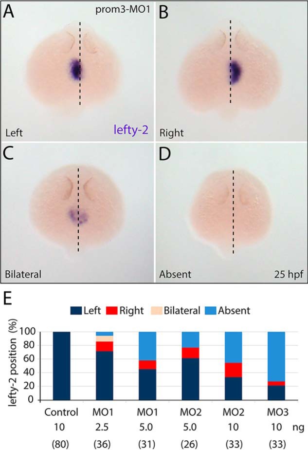
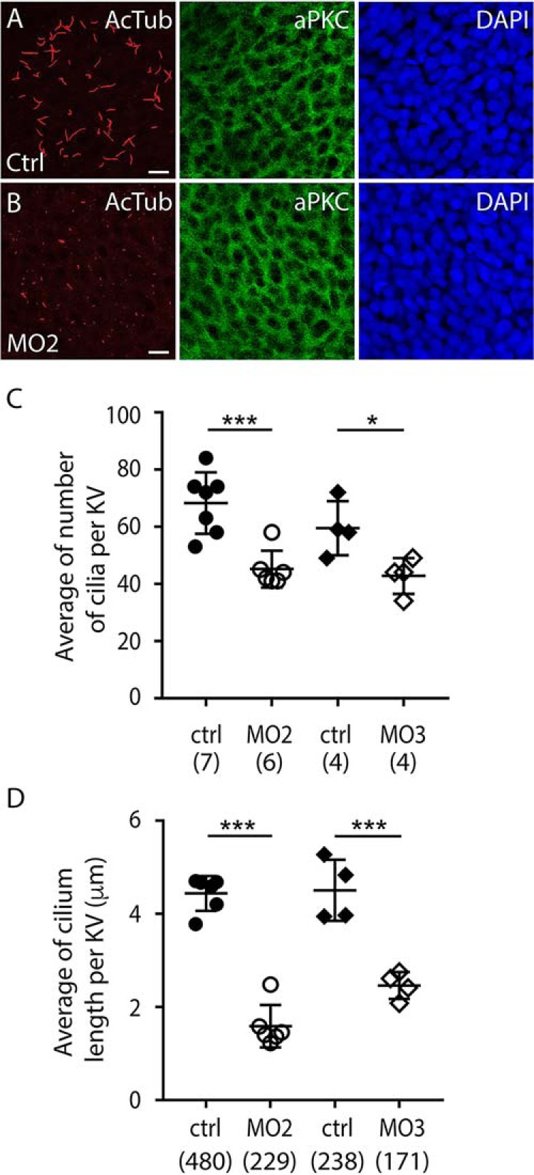
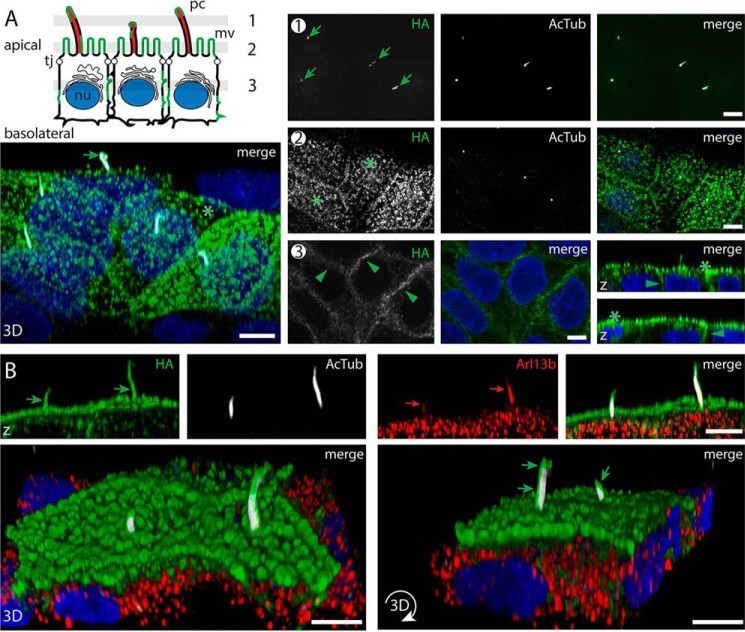
Prominins (proms) are transmembrane glycoproteins conserved throughout the animal kingdom. They are associated with plasma membrane protrusions, such as primary cilia, as well as extracellular vesicles derived thereof. Primary cilia host numerous signaling pathways affected in diseases known as ciliopathies. Human PROM1 (CD133) is detected in both somatic and cancer stem cells and is also expressed in terminally differentiated epithelial and photoreceptor cells. Genetic mutations in the PROM1 gene result in retinal degeneration by impairing the proper formation of the outer segment of photoreceptors, a modified cilium. Here, we investigated the impact of proms on two distinct examples of ciliogenesis. First, we demonstrate that the overexpression of a dominant-negative mutant variant of human PROM1 (i.e. mutation Y819F/Y828F) significantly decreases ciliary length in Madin-Darby canine kidney cells. These results contrast strongly to the previously observed enhancing effect of WT PROM1 on ciliary length. Mechanistically, the mutation impeded the interaction of PROM1 with ADP-ribosylation factor-like protein 13B, a key regulator of ciliary length. Second, we observed that in vivo knockdown of prom3 in zebrafish alters the number and length of monocilia in the Kupffer's vesicle, resulting in molecular and anatomical defects in the left-right asymmetry. These distinct loss-of-function approaches in two biological systems reveal that prom proteins are critical for the integrity and function of cilia. Our data provide new insights into ciliogenesis and might be of particular interest for investigations of the etiologies of ciliopathies.

Keywords: ADP-ribosylation factor (ARF); ADP-ribosylation factor-like protein 13B (Arl13b); CD133; Kupffer's vesicle; MDCK; cilia; ciliopathy; cilium; development; left-right asymmetry; membrane polarity; membrane protein; prominin-1 (PROM1); zebrafish.


Conflict of interest statement

The authors declare that they have no conflicts of interest with the contents of this article

Figures

Figure 1.
Figure 1.
Mutating tyrosine 819/828 of human PROM1 shortens primary cilia. A–D, polarized WT cells (MDCK), those expressing human PROM1.s1 or PROM1.s2 splice variants, or corresponding Y819/828F mutants were grown for 7 dpc (A–C) or 14 dpc (C and D) and processed for SEM (A and B) and CLSM (C and D). SEM micrographs revealed long (green) and short (red) primary cilia in cells expressing WT and mutated PROM1, respectively (A). Note the presence of very long cilia (>10 μm) in WT PROM1-positive cells (B; see also Fig. S1). Arrow, primary cilium; dashed line, origin of cilium; *, cell without primary cilium; #, artifact of SEM preparation. For the immunofluorescence analysis (C and D), cells were double-immunolabeled for PROM1 and AcTub. The length of AcTub-labeled cilia was evaluated by CLSM and classified into three categories: <3, 3–5, and >5 μm (C). The number of analyzed primary cilia is displayed in parentheses. Data (purple) obtained from cells expressing PROM1.s2 were taken from our recent study (33) and used for comparison. The percentage of total (blue) or PROM1+ (black) cells with no cilium is indicated (D). The mean and S.D. (error bars) are presented. Scale bars, 500 nm (top panels) and 2 μm (bottom panels) (A) and 1 μm (B).
Figure 2.
Figure 2.
Mutation of tyrosine 819/828 in human PROM1 reduces its interaction with Arl13b. A–D, polarized WT cells (MDCK)or those expressing human PROM1.s1 or PROM1.s2 splice variants or the corresponding Y819/828F mutants were subjected either to immunoisolation (IS) using paramagnetic beads conjugated to mAb against PROM1 (AC133) or anti-fluorescein isothiocyanate (FITC) antibody as negative control (A and B) or processed for indirect immunofluorescence followed by CLSM (C and D). For immunoisolation, the input (PROM1, 1:10; Arl13b, 1:40) and immunoisolated materials were probed for PROM1 and Arl13b by immunoblotting (A). The arrowhead and asterisks indicate plasma membrane and endoplasmic reticulum–associated species of PROM1, respectively, whereas arrows show Arl13b. Molecular mass markers (kDa) are indicated (left). The ratio of Arl13b/PROM1 (plasma membrane species only) immunoreactivities was quantified (B). 3–6 independent experiments were performed. Data are presented as mean and S.D. (error bars). Individual values are shown. **, p < 0.01; ***, p < 0.001 (two-tailed unpaired Student's t test). For immunofluorescence, cells were triple-immunolabeled for PROM1 (green), Arl13b (red), and AcTub (white). Nuclei (nu) were counterstained with DAPI (blue). The three-dimensional view (x-z orientation) was built from 38–45 x-y sections throughout the ciliary compartment (C, left panels) or x-y section (right top panels) and a composite of 5–10 x-y sections (right bottom panels) taken at the axoneme level (Ax, green bar) or at the base (red bar) of the primary cilium (C), respectively. The top view of three-dimensional reconstructions highlights Arl13 at the base of the cilium (D, dashed red line). Cells expressing either WT (left) or mutant (right) PROM1 are displayed. Green arrow, punctate PROM1 staining at the ciliary membrane of WT PROM1+ cells. Asterisk, PROM1 associated with microvillar membrane. Scale bars, 1 μm.
Figure 3.
Figure 3.
Morphological phenotypes upon prom3 knockdown. A–P, embryos were injected at the one-cell stage with distinct MO against either prom3 (A, MO1, 5 ng; B, E, and G, MO2, 10 ng; K–P, MO3, 10 ng) or prom1a (I and I′, MO1, 10 ng; J and J′, MO2, 10 ng) or with control MO (C, D, F, H, and H′, ctrl, 10 ng). At different developmental stages as indicated, specimens were either observed in a native, unfixed state (A–C and H–J), analyzed histologically with hematoxylin and eosin staining (D–G), or processed for whole-mount ISH using antisense DIG-labeled probe against cmlc2 (K–P). At early (K–M; 30 hpf) and late (N–P; 40 hpf) pharyngula periods, a randomized position (left, median, or right) of the heart tube highlighted by cmlc2 (arrowhead) is observed in prom3 morphants. Numbers in parentheses indicate the frequencies of phenotypes (30 hpf, n = 35; 40 hpf, n = 59). At 4 dpf, prom3 morphants, but neither the control nor prom1a MO-injected ones (C and H–J′), displayed severe morphological disturbances including swollen pericardium (A and B; hollow arrowhead), ventrally curved body (A and B), and an increased lumen size of the pronephric duct (E and G; outlined white surface). The surface areas of the lumen size are depicted as black blobs under the corresponding image. Orientation is as follows. A–C and H′–J′, lateral; H–J, random; K–P, anterior (dorsal toward top). Scale bars (D–G), 16 μm.
Figure 4.
Figure 4.
Simultaneous injection of two nonoverlapping MOs against prom3 results in a synergistic effect. A–F, embryos were injected at the one-cell stage with control MO (A, ctrl, 10 ng), MO2 (D, 10 ng) or a combination of MO1 + MO2 against prom3 at the doses indicated (B, C and E, F). At 58 hpf, larvae were observed in a native, unfixed state (A–D) or processed for whole-mount ISH using antisense DIG-labeled probe against cmlc2 (E and F). At the effective dose (10 ng), MO2 elicited mild to severe phenotypes (D). The simultaneous injection of one-fourth of their single effective amounts of MO1 (5 ng) and MO2 (10 ng) results in body axis distortion and fluid backup (C) similar to the severe phenotype detected upon injection of MO2 alone at the effective dose (D). Application of one-eighth of effective amounts caused no changes in body axis development (B) but often elicited L-R defects of the heart tube (F) in contrast to the normal position (situs solitus) (E). Orientation is as follows. A–D, random; E and F, anterior (dorsal toward the top). A, atrium; V, ventricle.
Figure 5.
Figure 5.
Prom3 influences molecular left-right asymmetry. A–E, embryos were injected at the one-cell stage with either one of the three MOs against prom3 (A–D, MO1 (2.5 ng); E, MO1 (2.5 and 5 ng), MO2 (5 and 10 ng), and MO3 (10 ng)) or control MO (E, 10 ng). At 25 hpf, specimens were processed for whole-mount ISH using antisense DIG-labeled lefty-2 probe (A–D). Note the randomized expression of lefty-2 (left, right, and bilateral) with regard to the median-sagittal plane (dashed line) or its absence. Shown are frequencies of the lefty-2 position (see color code) observed in prom3-morphants upon application of a given MO at various amounts (E). The number of embryos analyzed is indicated in parentheses. Orientation is as follows. A–D, dorsal (rostral toward the top).
Figure 6.
Figure 6.
Prom3 regulates the number and length of cilia in the Kupffer's vesicle. A–D, embryos were injected at the one-cell stage with control (A, C, and D, ctrl, 10 ng) and MO against prom3 (B, C, and D, MO2; C and D, MO3, 10 ng) and processed at the 8-somite stage for double immunofluorescence using anti-AcTub (red) and anti-aPKCζ (green) antibodies, counterstained with DAPI (blue), and analyzed by CLSM. The number (C) and length (D) of cilia in the Kupffer's vesicle (KV) were quantified. The total numbers of Kupffer's vesicles and cilia analyzed are indicated in parentheses. The mean and S.D. (error bars) are presented. Individual values are shown. *, p < 0.05; ***, p < 0.001. Scale bars (A and B), 10 μm.
Figure 7.
Figure 7.
Zebrafish prom3 is distributed in a nonpolarized fashion in polarized epithelial cells and co-localized with Arl13b at the primary cilium. A and B, zebrafish prom3-HA–expressing MDCK cells growing as a polarized cell monolayer (7 dpc) were either double-immunolabeled (A) for prom3-HA using anti-HA antibody (green) and anti-AcTub antibody (white) or triple-immunolabeled (B) with the detection of Arl13b (red). Nuclei (nu) were counterstained with DAPI (blue) prior to CLSM analyses. Three to four single optical x-y section planes (0.4-μm slice each) throughout the cell monolayer as illustrated in the cartoon (A, sections 1–3) revealed the presence of prom3-HA in primary cilia (pc, sections 1 and 2, green arrow) highlighted with AcTub staining and microvilli (mv, section 2, asterisk) present at the apical membrane. Prom3-HA is also detected at lateral membranes (section 3, green arrowhead). Three-dimensional views (x-z orientation (z), top panels; top-side orientations (3D), bottom panels) were built from 42 x-y sections throughout cells. The immunolabeling revealed the co-localization of prom3-HA and Arl13b in the ciliary compartment (B, green and red arrows, respectively). Note the asymmetric distribution of prom3-HA at the ciliary membrane (B, bottom panels, green arrow). Tj, tight junction. Scale bars, 5 μm.

Similar articles

Cited by

References

    1. Weigmann A., Corbeil D., Hellwig A., and Huttner W. B. (1997) Prominin, a novel microvilli-specific polytopic membrane protein of the apical surface of epithelial cells, is targeted to plasmalemmal protrusions of non-epithelial cells. Proc. Natl. Acad. Sci. U.S.A. 94, 12425–12430 10.1073/pnas.94.23.12425 - DOI - PMC - PubMed
    1. Fargeas C. A., Florek M., Huttner W. B., and Corbeil D. (2003) Characterization of prominin-2, a new member of the prominin family of pentaspan membrane glycoproteins. J. Biol. Chem. 278, 8586–8596 10.1074/jbc.M210640200 - DOI - PubMed
    1. Kania G., Corbeil D., Fuchs J., Tarasov K. V., Blyszczuk P., Huttner W. B., Boheler K. R., and Wobus A. M. (2005) Somatic stem cell marker prominin-1/CD133 is expressed in embryonic stem cell-derived progenitors. Stem Cells 23, 791–804 10.1634/stemcells.2004-0232 - DOI - PubMed
    1. Corbeil D., Karbanová J., Fargeas C. A., and Jászai J. (2013) Prominin-1 (CD133): Molecular and cellular features across species. Adv. Exp. Med. Biol. 777, 3–24 10.1007/978-1-4614-5894-4_1 - DOI - PubMed
    1. Grosse-Gehling P., Fargeas C. A., Dittfeld C., Garbe Y., Alison M. R., Corbeil D., and Kunz-Schughart L. A. (2013) CD133 as a biomarker for putative cancer stem cells in solid tumours: limitations, problems and challenges. J. Pathol. 229, 355–378 10.1002/path.4086 - DOI - PubMed

Publication types

LinkOut - more resources