

Case Reports
doi: 10.1259/bjrcr.20190044.
eCollection 2020 Mar.
Adrenal schwannoma: a case report
Affiliations
- PMID: 32201601
- PMCID: PMC7068090
- DOI: 10.1259/bjrcr.20190044
Item in Clipboard
Case Reports
Adrenal schwannoma: a case report
BJR Case Rep.
.
Abstract
Retroperitoneum is an uncommon site for Schwannoma tumors and among adrenal incidentaloma the Schwannoma is rare. This condition lacks of a specific clinical and radiological features, but correct diagnosis before pathological examination should be very important for clinical management and surgical decision. We describe a case of voluminous retroperitoneal incidental mass, that was proved to be an adrenal Schwannoma.
© 2020 The Authors. Published by the British Institute of Radiology.
Figures
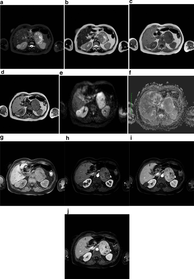
MRI examination of our patient, 61-years-old female, with left adrenal mass. (a) T2 (fat sat) weighted axial image shows voluminous and well-circumscribed solid mass in the left adrenal gland area, with heterogeneous high signal intensity, due to the presence of cystic components, also visible on (b) T2 weighted axial image. The lesion has well defined margins and fluid signal intensity areas; (c, d) T1 weighted images in phase and out of phase show the absence of signal intensity drop in opposed phase acquisitions, suggesting that intracellular fat was not present in the mass. Note also the regular profile and smooth margins of the mass; (e, f) DWI b 800 and relative ADC map axial image at the level of the adrenal mass show the presence of moderate diffusivity restriction due to lesion hypercellularity; (g) T1 (fat sat) weighted images before and (h, i, l) after paramagnetic contrast medium intravenous injection show clearly the progressive and heterogeneous contrast enhancement of the adrenal mass, with no evidence of adjacent structures infiltration. ADC, apparent diffusion coefficient; DWI, diffusion-weighted imaging.
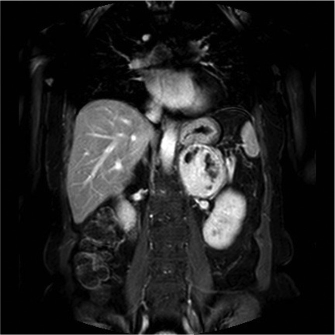
Coronal thrive acquisition after paramagnetic contrast medium intravenous injection in deleyed phase. This plane well displays that mass exhibits close relations with the superior pole of left kidney, stomach, pancreatic tail and left renal vein, aorta and diaphragmatic pillar. There is no evidence of infiltration, as confirmed later by surgery and histology.
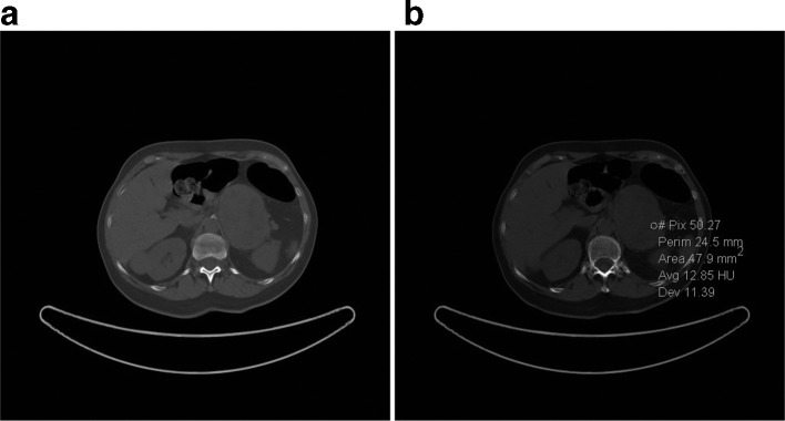
Unenhanced CT axial images show a well-circumscribed heterogeneous mass at the left adrenal level and reveals (a) intraregional cystic-like foci (13 HU) and (b) the absence of calcifications in the mass. HU, Hounsfield unit.
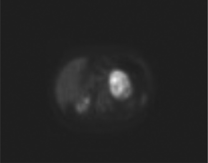
PET-FDG axial image demonstrates heterogeneous and intense FDG uptake in correspondence of the adrenal mass, suggesting a lesion with high metabolic activity. FDG, fludeoxyglucose; PET, positron emission tomography.

Post-operative unenhanced CT axial image. Surgical clips of left adrenalectomy are present. No injuries of other organ and no fluid or air collection is visible. The patient was discharged on postoperative day 5.
References
Publication types
LinkOut - more resources
Full Text Sources
