. 2020 Mar 13;6(11):eaay4697.
doi: 10.1126/sciadv.aay4697. eCollection 2020 Mar.

Global crotonylome reveals CDYL-regulated RPA1 crotonylation in homologous recombination-mediated DNA repair

Affiliations

Global crotonylome reveals CDYL-regulated RPA1 crotonylation in homologous recombination-mediated DNA repair

Huajing Yu et al. Sci Adv. .

Erratum in

Abstract

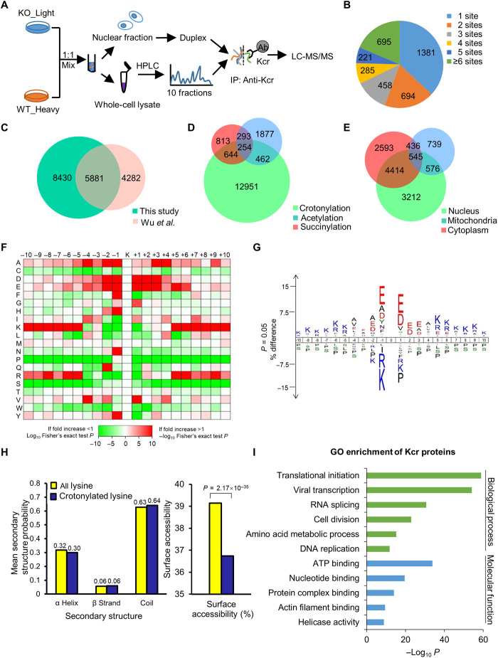
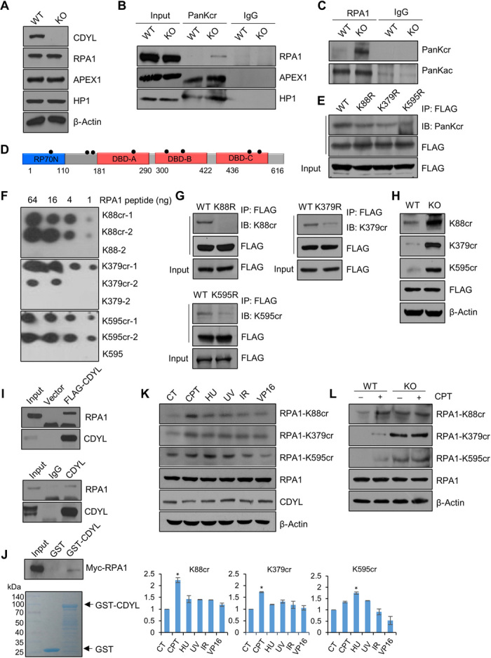
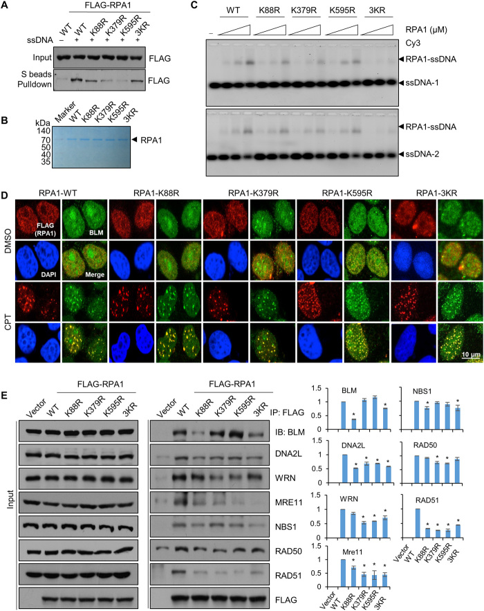
Previously, we reported that chromodomain Y-like (CDYL) acts as a crotonyl-coenzyme A hydratase and negatively regulates histone crotonylation (Kcr). However, the global CDYL-regulated crotonylome remains unclear. Here, we report a large-scale proteomics analysis for protein Kcr. We identify 14,311 Kcr sites across 3734 proteins in HeLa cells, providing by far the largest crotonylome dataset. We show that depletion of CDYL alters crotonylome landscape affecting diverse cellular pathways. Specifically, CDYL negatively regulated Kcr of RPA1, and mutation of the Kcr sites of RPA1 impaired its interaction with single-stranded DNA and/or with components of resection machinery, supporting a key role of RPA1 Kcr in homologous recombination DNA repair. Together, our study indicates that protein crotonylation has important implication in various pathophysiological processes.


Figures

Fig. 1
Fig. 1. Profiling Kcr proteome in HeLa cells.
(A) Schematic representation of experimental workflow for SILAC quantification of Kcr in WT and CDYL KO HeLa cells. (B) Pie chart showing the distribution of the number of identified Kcr sites per protein. (C) Venn diagram comparing the total numbers of Kcr sites in previous study and our study. (D) Venn diagram showing overlap between quantifiable Kcr, Kac, and Ksucc sites in HeLa cells. (E) Venn diagram showing cellular compartment distribution of Kcr proteins. (F) Motif analysis of all identified Kcr proteins. (G) Icelogo representation showing flanking sequence preferences for all Kcr sites. (H) Distribution of all lysines and crotonylated lysines in structured regions of proteins. (I) Bar graphs showing representative ontology annotations enriched with Kcr proteome.
Fig. 2
Fig. 2. Quantification analysis of crotonylome in response to CDYL KO.
(A) Histogram showing the ratio distribution of quantifiable Kcr sites between CDYL KO and WT HeLa cells. (B) Scatterplot showing the quantification of Kcr sites in relation to peptide intensities. (C) Bar graphs showing KEGG pathway associated with all identified and CDYL-regulated Kcr proteins. (D) Protein-protein interaction network of the chromatin-associated Kcr proteins based on the STRING database.
Fig. 3
Fig. 3. Characterization of CDYL-regulated Kcr of RPA1.
(A) The expression of the indicated proteins was measured by Western blotting in HeLa cells. (B) Immunoprecipitations in WT and CDYL KO HeLa cells with anti-PanKcr or anti–immunoglobulin G (IgG) followed by immunoblotting with antibodies against the indicated proteins. (C) Immunoprecipitations in WT and CDYL KO HeLa cells with anti-RPA1 or anti-IgG followed by immunoblotting with anti-PanKcr or anti-PanKac. (D) Schematic diagram of RPA1. (E) K88, K379, and K595 are major Kcr sites of RPA1 in vivo. Immunoprecipitation assays were performed in HeLa cells overexpressing indicated FLAG-tagged RPA1 constructs with anti-FLAG followed by immunoblotting (IB) with anti-FLAG or anti-PanKcr. (F) The specificity of antibodies against RPA1 K88cr, RPA1 K379cr, and RPA1 K595cr was verified by dot blot assays. The nitrocellulose membrane was spotted with the indicated amounts of uncrotonylated or crotonylated RPA1 peptides and immunoblotted with the indicated antibodies. (G) Verification of the specificity of anti–RPA1-K88cr, anti–RPA1-K379cr, or anti–RPA1-K595cr by Western blotting. Immunoprecipitation assays were performed in HeLa cells overexpressing the indicated FLAG-tagged RPA1 constructs with anti-FLAG followed by immunoblotting with the indicated antibodies, respectively. (H) The Kcr level of RPA1 is increased in CDYL KO cells. WT and CDYL KO HeLa cells were transfected with FLAG-RPA1, followed by immunoblotting with the indicated antibodies. (I) Top: Coimmunoprecipitation assays with lysates from HeLa cells overexpressing FLAG-CDYL using anti-FLAG followed by immunoblotting with antibodies against the indicated proteins. Bottom: Coimmunoprecipitation assays with lysates from HeLa cells using anti-CDYL followed by immunoblotting with antibodies against the indicated proteins. (J) Top: GST pull-down assays with GST-fused CDYL and in vitro transcribed/translated RPA1. Bottom: Coomassie brilliant blue staining of the purified GST and GST-CDYL. (K) HeLa cells were treated with UV (20 J/m2), IR (10 Gy), HU (1 mM), CPT (1 μM), VP16 (40 nM), or dimethyl sulfoxide (DMSO) for 8 hours. Western blotting was performed with the indicated antibodies. Each scale bar represents the mean ± SD for triplicate experiments. Mean data are normalized to RPA1. *P < 0.05 versus lane 1 (two-tailed unpaired Student’s t test). (L) In the presence or absence of CPT treatment, cellular extracts from WT and CDYL KO HeLa cells were immunoblotted with the indicated antibodies.
Fig. 4
Fig. 4. Kcr of RPA1 affects its ability to bind ssDNA and/or HR factors.
(A) Cellular extracts from HeLa cells overexpressing the indicated FLAG-tagged constructs were incubated with biotinylated ssDNA. Bound proteins were pulled down using streptavidin-coated beads and subjected to Western blot analysis with the indicated antibodies. (B) FLAG-tagged RPA1-WT, RPA1-K88R, RPA1-K379R, RPA1-K595R, or RPA1-3KR was expressed and purified with anti-FLAG M2 affinity gel from HEK293T cells and stained with Coomassie brilliant blue. (C) FLAG-tagged RPA1-WT, RPA1-K88R, RPA1-K379R, RPA1-K595R, or RPA1-3KR (0.2, 0.4, and 0.8 μM) was used for EMSA experiments using Cy3-labeled ssDNA substrates. Free ssDNA substrates and protein ssDNA were detected with a Typhoon FLA 9500 imager. (D) HeLa cells were treated with control or RPA1 siRNA (which targets 3′ untranslated region). Twenty-four hours later, cells were transfected with the indicated FLAG-tagged constructs. After 47 hours, cells were treated with 4 μM CPT or DMSO for 1 hour, fixed, and stained with anti-BLM and anti-FLAG. More than 60 cells per sample were analyzed at each independent experiment, and representative images are shown. (E) Coimmunoprecipitation assays with lysates from HeLa cells overexpressing the indicated FLAG-tagged constructs using anti-FLAG followed by immunoblotting with antibodies against the indicated proteins. Each scale bar represents the mean ± SD for triplicate experiments. Mean data are normalized to indicated proteins. *P < 0.05 versus WT (two-tailed unpaired Student’s t test).
Fig. 5
Fig. 5. Kcr of RPA1 enhances the function of the resection machinery in HR repair.
(A) HeLa cells were treated with RPA1 siRNA for 24 hours before cells were transfected with the indicated FLAG-tagged constructs. After 47 hours, cells were treated with 4 μM CPT or DMSO for 1 hour, fixed, and stained with anti-RAD51 and anti-FLAG. Only nuclei showing more than five colocalized foci were considered as positive. More than 60 cells per sample were analyzed at each independent experiment. Error bars indicate ±SD for the results of triplicate assays. ***P < 0.001 (two-tailed unpaired Student’s t test). (B) HeLa cells were transfected and treated with DMSO or CPT as in (A). Cells were then fixed and stained with anti-BrdU and anti-FLAG. BrdU (20 μM) was added to the medium 15 min before CPT treatment. Error bars indicate ±SD for the results of triplicate assays. **P < 0.01 and ***P < 0.001 (two-tailed unpaired Student’s t test). ns, not significant. (C) DR-GFP-U2OS cells were transfected with control or RPA1 siRNA as indicated. Twenty-four hours later, cells were transfected with HA-I–Sce I expression plasmids together with pcDNA3.1-3×FLAG-C vector or FLAG-tagged RPA1 constructs for 48 hours before they were collected and analyzed by fluorescence-activated cell sorting. Data represent the mean ± SD for triplicate experiments. ***P < 0.001 (two-tailed unpaired Student’s t test).
Fig. 6
Fig. 6. Kcr of RPA1 is critical for cell survival under DNA-damaging conditions.
(A) HeLa cells transfected with the indicated constructs were treated with DMSO, CPT, HU, or VP16. Cell viability was measured by colony formation assays. Data represent the mean ± SD for triplicate experiments. *P < 0.05, **P < 0.01, and ***P < 0.001 (two-tailed unpaired Student’s t test). (B) HeLa cells were transfected with control or RPA1 siRNA for 12 hours. The cells were then transfected with FLAG-tagged constructs and treated with 1 μM CPT for 48 hours before they were collected for annexin V and propidium iodide double staining. Cell apoptosis was determined by flow cytometry. Data represent the mean ± SD for triplicate experiments. ***P < 0.001 (two-tailed unpaired Student’s t test).

Similar articles

Cited by

References

    1. Tan M., Luo H., Lee S., Jin F., Yang J. S., Montellier E., Buchou T., Cheng Z., Rousseaux S., Rajagopal N., Lu Z., Ye Z., Zhu Q., Wysocka J., Ye Y., Khochbin S., Ren B., Zhao Y., Identification of 67 histone marks and histone lysine crotonylation as a new type of histone modification. Cell 146, 1016–1028 (2011). - PMC - PubMed
    1. Sabari B. R., Zhang D., Allis C. D., Zhao Y., Metabolic regulation of gene expression through histone acylations. Nat. Rev. Mol. Cell Biol. 18, 90–101 (2017). - PMC - PubMed
    1. Sabari B. R., Tang Z., Huang H., Yong-Gonzalez V., Molina H., Kong H. E., Dai L., Shimada M., Cross J. R., Zhao Y., Roeder R. G., Allis C. D., Intracellular crotonyl-CoA stimulates transcription through p300-catalyzed histone crotonylation. Mol. Cell 58, 203–215 (2015). - PMC - PubMed
    1. Liu X., Wei W., Liu Y., Yang X., Wu J., Zhang Y., Zhang Q., Shi T., Du J. X., Zhao Y., Lei M., Zhou J. Q., Li J., Wong J., MOF as an evolutionarily conserved histone crotonyltransferase and transcriptional activation by histone acetyltransferase-deficient and crotonyltransferase-competent CBP/p300. Cell Discov. 3, 17016 (2017). - PMC - PubMed
    1. Feldman J. L., Baeza J., Denu J. M., Activation of the protein deacetylase SIRT6 by long-chain fatty acids and widespread deacylation by mammalian sirtuins. J. Biol. Chem. 288, 31350–31356 (2013). - PMC - PubMed

Publication types