. 2020 Mar 12;6(3):703-724.
doi: 10.1016/j.chempr.2019.12.016.

Metallotexaphyrins as MRI-Active Catalytic Antioxidants for Neurodegenerative Disease: A Study on Alzheimer's Disease

Affiliations

Metallotexaphyrins as MRI-Active Catalytic Antioxidants for Neurodegenerative Disease: A Study on Alzheimer's Disease

James T Brewster 2nd et al. Chem. .

Abstract

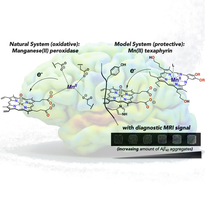
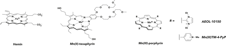
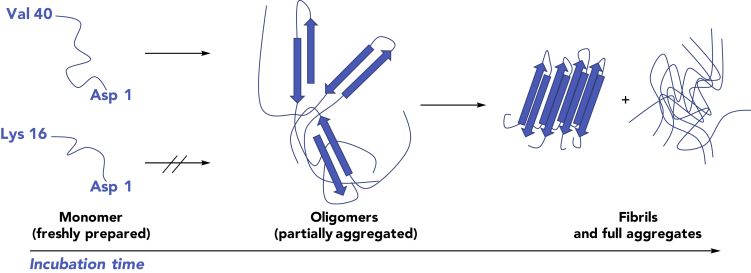
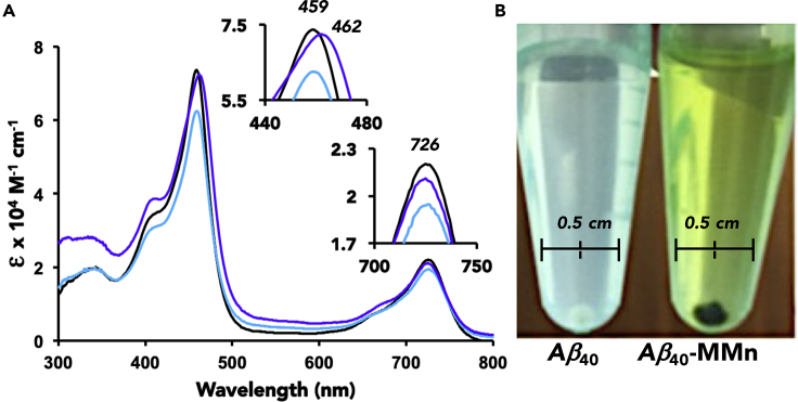
The complex etiology of neurodegeneration continues to stifle efforts to develop effective therapeutics. New agents elucidating key pathways causing neurodegeneration might serve to increase our understanding and potentially lead to improved treatments. Here, we demonstrate that a water-soluble manganese(II) texaphyrin (MMn) is a suitable magnetic resonance imaging (MRI) contrast agent for detecting larger amyloid beta constructs. The imaging potential of MMn was inferred on the basis of in vitro studies and in vivo detection in Alzheimer's disease C. elegans models via MRI and ICP-MS. In vitro antioxidant- and cellular-based assays provide support for the notion that this porphyrin analog shows promise as a therapeutic agent able to mitigate the oxidative and nitrative toxic effects considered causal in neurodegeneration. The present report marks the first elaboration of an MRI-active metalloantioxidant that confers diagnostic and therapeutic benefit in Alzheimer's disease models without conjugation of a radioisotope, targeting moiety, or therapeutic payload.

Keywords: Alzheimer’s disease; C. elegans AD models; MRI; amyloid aggregation; amyloid beta; amyloid beta modifications by ROS and RNS; expanded porphyrin; metalloantioxidant; neurodegeneration; texaphyrin.


Conflict of interest statement

The authors declare no competing interests.

Figures

None
Graphical abstract
Figure 1
Figure 1
Chemical Structures of Representative Porphyrinoids As discussed in the text proper, hemin catalyzes oxidative and nitrative damage. Manganese(II) texaphyrin (MMn) is the subject of this study. Manganese(III) tetramethylpyridinium porphyrin (MnTMPyP) is a manganese porphyrin metalloantioxidant. AEOL-10150 has successfully completed a phase I trial in ALS and healthy patients and has shown efficacy in animal models for radiation- and nerve-agent-induced toxicity. Axial ligands are omitted for clarity. See main text for further discussion of these agents.
Figure 2
Figure 2
The Aggregation State of Aβ Peptides as a Function of Incubation Time The three Aβ40 constructs utilized in this study: (left) freshly dissolved peptide is predominantly monomeric in solution, (middle) soluble oligomeric species (partially aggregated) are produced after incubation in solution, and (right) fibrils and fully soluble and insoluble aggregates are formed after prolonged incubation. Aβ16 remains monomeric in solution.
Figure 3
Figure 3
Interaction of MMn with Monomeric and Aggregated Aβ40 (A) Titration with MMn (5 μM) and monomeric Aβ40 with 0 (black), 1 (light blue), and 20 (purple) equiv and (B) photos of aggregated Aβ40 (100 μM; aged 2 months) in the absence and presence of MMn (100 μM) in PBS and after washing with PBS. The pellet shown in the right-most image was obtained by centrifugation and is in methanol.
Figure 4
Figure 4
Modification of Aggregated Aβ40 by MMn as Monitored by CD Spectroscopy and SEM Analyses CD spectra (left) of aggregated (aged for 30 days) Aβ40 (40 μM) were recorded before and after the addition of MMn (20 μM) at t = 0, 7, and 24 h. SEM photographs (right) of (A) Aβ40 aggregate, (B) an Aβ40 aggregate incubated with MMn for 24 h, and (C) monomeric Aβ40 are shown.
Figure 5
Figure 5
In Vitro MRI Analyses of MMn with Various Aβ40 Constructs (A) The longitudinal relaxation rate (R1) of MMn (140 μM) with monomeric (0.5 equiv, 70 μM), partial (1 week, 0.5 equiv, 70 μM), and aggregated (>2 months) Aβ40 (0.5 equiv, 70 μM) in comparison with MMn alone (140 μM) and in the presence of BSA (0.5 equiv, 70 μM). Dots represent individual samples; the mean (red line) and 95% confidence level for the mean (light red) are also displayed. Standard deviations are either indicated in blue or not shown if within the 95% confidence level. (B) Phantom T1-weighted MRI images of 140 μM MMn were recorded in the presence of increasing concentrations of Aβ40 aggregate (0–70 μM) in PBS at 7 T and 25°C. Samples are normalized to MMn alone, shown in the left-most box.
Figure 6
Figure 6
ICP-MS and In Vivo MRI Analyses of MMn in C. elegans AD Models Mn concentrations (A), as determined by ICP-MS, and (B) MRI analyses of MMn for pellets of wild-type (N2) and AD model (CL4176) C. elegans with and without MMn. Pellets were standardized by dissolving every 1 mg C. elegans in 100 μL HEPES and digesting in HNO3. n = 5–8 per sample, 2 replicates of each for ICP-MS measurements and n = 2–5 for MRI measurements. Dots represent individual samples; the mean (red line) and 95% confidence level for the mean (light red) are shown. Standard deviation (blue) is shown where larger than the 95% confidence level. *Two-sample t test on ICP-MS variance between MMn-wild-type (WT) and -AD models; p = 0.00018.
Figure 7
Figure 7
1H NMR Spectroscopic Study of the Hemin-Mediated Oxidation of Serotonin NMR yields were calculated by comparing the spectra of (A) serotonin (1 mM) in pH 7.4 PBS at 37°C in the presence of (B) hemin (15 μM) and an oxidative mixture containing H2O2 (1 mM) and sodium ascorbate (40 μM), (C) an H2O2-ascorbate oxidative mixture plus MnTMPyP (120 μM), and (D) an H2O2-ascorbate oxidative mixture plus MMn (120 μM) recorded after a reaction time of 120 min in all cases.
Figure 8
Figure 8
HPLC-MS Analyses of the Oxidative and Nitrative Modification of Aβ16 (A) Aβ16 incubated under hemin-nitration conditions using heme-H2O2-NaNO2 with varying concentrations of MMn. Percent composition of Aβ16 is shown in black and, as expected, increases with the [MMn]. (B) The protective effect of MMn was also tested against heme-peroxynitrite-mediated modification of Aβ16 in comparison with MnTMPyP. Sodium ascorbate (NaAsc) was added to regenerate the MMn catalyst under the reaction conditions. Reported values are averages with the corresponding standard deviation as obtained from two independent measurements.
Scheme 1
Scheme 1
Proposed Mechanism for the MMn-Based Deactivation of Hemin- and Peroxynitrite (ONOO)-Mediated Oxidation and Nitration, as well as Oxidative Decomposition of Hydrogen Peroxide (H2O2) Redox cycle of Mn(II/III) texaphyrin. Compounds I and II are generated by the reaction of hemin with H2O2. The Aβ peptide and axial ligands are omitted for clarity.
Figure 9
Figure 9
Cellular (Neuro-2A) MTT Assay Neuro-2A cell viability upon exposure to Aβ40 and mixed oxidized-nitrated Aβ40 (hemin-H2O2-NaNO2) with and without MMn, MGd, and MnTMPyP. Reported values are the average with standard deviation of three independent assays each tested four times. The Aβ40 “NO2” mix is an average of two independent assays each tested four times. *Two-sample t test on percent cell viability (MTT assay) between the Aβ40 “NO2” mix with and without MMn; p = 0.00016.

Similar articles

Cited by

References

    1. Hopkins A.L. Network pharmacology: the next paradigm in drug discovery. Nat. Chem. Biol. 2008;4:682–690. - PubMed
    1. Albert R. Scale-free networks in chemical biology. Journal of Cell Science. 2005;118:4847–4857. - PubMed
    1. Gitler A.D., Dhillon P., Shorter J. Neurodegenerative disease: models, mechanisms, and a new hope. Dis. Models Mech. 2017;10:499–502. - PMC - PubMed
    1. Wyss-Coray T. Ageing, neurodegeneration, and brain rejuvenation. Nature. 2016;539:180–186. - PMC - PubMed
    1. Prezedborski S., Vila M., Jackson-Lewis V. Neurodegeneration: what is it and where are we? J. Clin. Invest. 2003;1:3–10. - PMC - PubMed

LinkOut - more resources