. 2020;2(1):75-89.
doi: 10.33597/aimm.02-1006.

AD "Statin": Alzheimer's Disorder is a "Fast" Disease Preventable by Therapeutic Intervention Initiated Even Late in Life and Reversible at the Early Stages

Affiliations

AD "Statin": Alzheimer's Disorder is a "Fast" Disease Preventable by Therapeutic Intervention Initiated Even Late in Life and Reversible at the Early Stages

Vladimir Volloch et al. Ann Integr Mol Med. 2020.

Abstract

The present study posits that Alzheimer's disorder is a "fast" disease. This is in sharp contrast to a view, prevailing until now, that Alzheimer's Disease (AD) is a quintessential "slow" disease that develops throughout the life as one prolonged process. According to this view, beta-amyloid (Aβ) is produced and secreted solely by the beta-amyloid precursor protein (βAPP) proteolytic/secretory pathway. As its extracellular levels increase, it triggers neurodegeneration starting relatively early in life. Damages accumulate and manifest, late in life in sporadic Alzheimer's Disease (SAD) cases, as AD symptoms. In familial AD (FAD) cases, where mutations in βAPP gene or in presenilins increase production of either common Aβ isoform or of its more toxic isoforms, neurodegeneration reaches critical threshold sooner and AD symptoms occur earlier in life, mostly in late 40s and 50s. There are currently no preventive AD therapies but if they were available, according to this viewpoint it would be largely futile to intervene late in life in case of potential SAD or at mid-age in cases of FAD because, although AD symptoms have not yet manifested, the damage has already occurred during the preceding decades. In this paradigm, to be effective, preventive therapeutic intervention should be initiated early in life. The outlook suggested by the present study is radically different. According to it, Alzheimer's disease evolves in two stages. The first stage is a slow process of intracellular beta-amyloid accumulation. It occurs via βAPP proteolytic/secretory pathway and cellular uptake of secreted Aβ common to Homo sapiens, including healthy humans, and to non-human mammals, and results neither in significant damage, nor in manifestation of the disease. The second stage occurs exclusively in humans, commences shortly before symptomatic onset of the disease, sharply accelerates the production and increases intracellular levels of Aβ that is not secreted but is retained intracellularly, generates significant damages, triggers AD symptoms, and is fast. It is driven by an Aβ generation pathway qualitatively and quantitatively different from βAPP proteolytic process and entirely independent of beta-amyloid precursor protein, and results in rapid and substantial intracellular accumulation of Aβ, consequent significant neurodegeneration, and symptomatic AD. In this paradigm, a preventive therapy for AD, an AD "statin", would be effective when initiated at any time prior to commencement of the second stage. Moreover, there are good reasons to believe that with a drug blocking βAPP-independent Aβ production pathway in the second stage, it would be possible not only to preempt the disease but also to stop and to reverse it even when early AD symptoms have already manifested. The present study posits a notion of AD as a Fast Disease, offers evidence for the occurrence of the AD-specific Aβ production pathway, describes cellular and molecular processes constituting an engine that drives Alzheimer's disease, and explains why non-human mammals are not susceptible to AD and why only a subset of humans develop the disease. It establishes that Alzheimer's disease is preventable by therapeutic intervention initiated even late in life, details a powerful mechanism underlying the disease, suggests that Aβ produced in the βAPP-independent pathway is retained intracellularly, elaborates why neither BACE inhibition nor Aβ immunotherapy are effective in treatment of AD and why intracellularly retained beta-amyloid could be the primary agent of neuronal death in Alzheimer's disease, necessitates generation of a novel animal AD model capable of producing Aβ via βAPP-independent pathway, proposes therapeutic targets profoundly different from previously pursued components of the βAPP proteolytic pathway, and provides conceptual rationale for design of drugs that could be used not only preemptively but also for treatment and reversal of the early stages of the disease.

Keywords: Alzheimer’s disease; Asymmetric RNA-dependent beta-APP mRNA amplification; Mitochondrial dysfunction-related stresses; Precursor-independent generation of beta-amyloid.


Conflict of interest statement

Conflict of Interest Authors declare no conflict of interest.

Figures

Figure 1:
Figure 1:. Projected stages of the chimeric pathway of RdRp-facilitated, antisense RNA-mediated amplification of mammalian mRNA.
Top panel: Conventional, genome-originated mRNA molecule. Bottom panel: Projected stages of antisense RNA-mediated mRNA amplification. Boxed line – sense strand RNA. Single line – antisense strand RNA. “AUG” – functional translation initiation codon (could be other than “AUG”). “TCE”– 3’-terminal complementary element; “ICE”– internal complementary element, both on the antisense RNA strand. Yellow circle – helicase/modifying activity complex. Blue lines (both single and boxed) – RNA strand modified and separated from its complement by a helicase complex. Red arrowhead – position of cleavage of the chimeric intermediate. Step 1: Synthesis of antisense strand; step 2: Strand separation; step 3: Folding of antisense strand into self-priming configuration; step 4: Extension of self-primed antisense RNA; step 5: Strand separation; step 6: Cleavage of the chimeric intermediate; step 7: End-products of amplification. Note that chimeric RNA end product retains the intact coding capacity of conventional mRNA.
Figure 2:
Figure 2:. RNA-dependent mRNA amplification can result in a 5’-truncated molecule encoding C-terminal fragment of a conventionally encoded polypeptide.
Boxed line-sense strand RNA. Single line-antisense strand RNA. “AUG”- functional translation initiation codon (could be other than AUG). “TCE”–3’-terminal complementary element; “ICE”– internal complementary element, both on the antisense RNA strand. Yellow circle – helicase/ modifying activity complex. Blue lines (both single and boxed) – RNA strand modified and separated from its complement by a helicase complex. Red arrow – position of cleavage of the chimeric intermediate. Step 1: Synthesis of antisense strand; step 2: Strand separation; step 3: Folding of antisense strand into self-priming configuration; step 4: Extension of self-primed antisense RNA; step 5: Strand separation; step 6: Cleavage of the chimeric intermediate; step 7: End-products of RNA amplification. Steps 3’–7’ correspond to steps 3–7. Top panel: Conventional, genome-transcribed mRNA molecule. Middle panel: Projected stages of RNA-dependent mRNA amplification. “ICE” is located within a segment of antisense RNA corresponding to the 5’UTR of conventional mRNA; the chimeric RNA end product contains the entire coding content of conventional mRNA. Bottom panel: “ICE” is located within a segment of antisense RNA corresponding to the coding region of conventional mRNA. The amplified chimeric end product contains a 5’-truncated coding region of conventional mRNA. The translational outcome is decided by position of the first functional translation initiation codon; if in-frame, a CTF of conventional polypeptide is produced.
Figure 3:
Figure 3:. Projected topology of RNA-dependent generation of 5’-truncated mRNA encoding Aβ-containing C-terminal fragment of human beta-amyloid precursor protein.
Lowercase letters -- nucleotide sequence of the antisense RNA. Uppercase letters -- nucleotide sequence of the sense RNA. Double-stranded portions highlighted in yellow: TCE (top) and ICE (bottom) elements of the antisense RNA. Note that the TCE and ICE are separated by about 2000 nucleotides. “20112013”: nucleotide positions on the antisense RNA (starting from the complement of the AUG encoding Met1 of the βAPP) of the “uac” (highlighted in blue) corresponding to the “AUG” (highlighted in green) encoding Met671 in the βAPP mRNA. a: TCE/ICE-guided folding of the antisense βAPP RNA. 3’-terminal “c” corresponds to one of multiple transcription start sites of βAPP mRNA located 149 nucleotides upstream from its AUG initiation codon [83]; note that such folding configuration would accommodate an additional 3’C, a transcript of the capG of βAPP mRNA (not shown). b: Extension of self-primed antisense RNA into sense RNA and cleavage (red arrow; may also occur at one of the TCE/ICE mismatches), after strand separation, of the chimeric intermediate. c: Chimeric RNA end product contains 5’terminal antisense segment extending into severely 5’-truncated βAPP mRNA. Its translation initiates from the “AUG” (highlighted in green and encoding Met671 in conventional βAPP mRNA) immediately preceding the beta amyloid-encoding region.
Figure 4:
Figure 4:. The engine that drives AD: Self-propagating mutual feedback cycles of mitochondrial dysfunction-mediated overproduction of beta-amyloid and vice versa in Alzheimer’s disease.
FAD: Familial Alzheimer’s disease; SAD: Sporadic Alzheimer’s disease; Highlighted in grey: Initial stimuli of the increased production of Aβ (different in FAD and SAD); Highlighted in blue: asymmetric RNA-dependent βAPP mRNA amplification, a molecular basis of beta-amyloid overproduction in Alzheimer’s disease (note a requirement for βAPP TSS utilization discussed in main text above); Horizontal arrows: the initial Aβ overproduction cycle; Arched arrows: Mutual feedback cycles; Blue arrows: Aβ-mediated induction of mitochondrial dysfunction and, possibly, ER stress; Red arrows: Mitochondrial dysfunction (and, possibly, ER stress)-mediated asymmetric RNA-dependent amplification of βAPP mRNA resulting in overproduction of Aβ. Note that in FAD, mitochondrial dysfunction is an intrinsic component of the amyloid cascade whereas in SAD, the initial mitochondrial pathology hierarchically supersedes and triggers Aβ pathology, where self-perpetuating mutual Aβ overproduction/mitochondrial dysfunction feedback cycles are, as in FAD, a central component of the amyloid cascade. In FAD, the initial increased production of Aβ results from mutations-driven abnormal proteolysis of βAPP and occurs relatively early in life, whereas in SAD, it is compelled by an aging-dependent component; hence drastic temporal difference in the age of onset yet profound pathological and symptomatic similarity in the progression of familial and sporadic Alzheimer’s disease, reflecting mechanistically identical nature of feedback cycles in both forms of AD.
Figure 5:
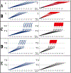
Figure 5:. Dynamics of Alzheimer’s disease in two paradigms.
Left panels: Dynamics of Aβ production; Right panels: Dynamics of neurodegeneration. Blue lines: Levels of beta-amyloid; Red lines: Extent of neurodegeneration; Black lines: Indicator lines, no significant neurodegeneration; Red blocks: Manifestation of Alzheimer’s disease. T: Threshold of symptomatic manifestation of AD (reflects levels of Aβ and consequent extent of neurodegeneration); T1: Threshold of activation of RNA-dependent βAPP mRNA amplification (reflects not only Aβ levels but also the extent of Aβ−coupled mitochondrial dysfunction and related stresses, possibly including ER stress); Numerous genetic factors such as, for example, the occurrence of various alleles of ApoE gene, as well as certain epigenetic factors, influence the age when thresholds T and T1 are reached, hence, the fanning lines. T2: Threshold of symptomatic occurrence of AD (reflects levels of Aβ and consequent extent of neurodegeneration). Panels A, B: View of the dynamics of AD prevailing until now (A: Dynamics of SAD; B: Dynamics of FAD). Levels of Aβ increase, neurodegeneration starts early and accumulates throughout the life. When threshold T is reached, AD symptoms manifest. Panels C, D: The outlook on the dynamics of Alzheimer’s disease envisioned in the present study (C: Dynamics of SAD; D: Dynamics of FAD). Levels of Aβ increase, the extent of mitochondrial dysfunction and related stresses reach threshold T1 and RNA-dependent βAPP mRNA amplification is activated. There is no significant neurodegeneration until after a lag period (when amplified RNA encoding C99 fragment of βAPP accrues and Aβ further accumulates) following the crossing of T1 threshold and activation of βAPP mRNA amplification; when the extent of neurodegeneration reaches threshold T2, AD symptoms manifest. Panel E: Dynamics of Aβ production and neurodegeneration in non-human mammals in light of the present study: T1 threshold is crossed but βAPPP mRNA is not amplified because it is not eligible for RNA-dependent amplification process. There is no significant neurodegeneration; T2 threshold is not reached, no AD symptoms manifest, no disease occurs. Note: Scenario depicted in panel (E) would occur in humans not susceptible to Alzheimer’s disease due to variations in βAPP TSSs utilization or when activation of βAPP mRNA amplification is blocked by therapeutic intervention.

Similar articles

Cited by

References

    1. Haass C, Lemere C, Capell A, Citron M, Seubert P, Schenk D, et al. The Swedish mutation causes early-onset Alzheimer’s disease by beta-secretase cleavage within the secretory pathway. Nat Med. 1995;1(12):1291–6. - PubMed
    1. Dyrks T, Dyrks E, Monning U, Urmoneit B, Turner J, Beyreuther, K. Generation of beta A4 from the amyloid protein precursor and fragments thereof. FEBS Lett. 1993;335(1):89–93. - PubMed
    1. Iizuka T, Shoji M, Kawarabayashi T, Sato M, Kobayashi T, Tada N, et al. Intracellular generation of amyloid beta-protein from amyloid beta-protein precursor fragment by direct cleavage with beta- and gamma-secretase. Biochem Biophys Res Commun. 1996;218(1):238–42. - PubMed
    1. DeStrooper B, Annaert W. Proteolytic processing and cell biological functions of the amyloid precursor protein. J Cell Sci. 2000;113(11):1857–70. - PubMed
    1. Barber RC. The genetics of Alzheimer’s disease. Scientifica (Cairo). 2012;2012:246210. - PMC - PubMed

LinkOut - more resources