. 2020 Jul 6;71(14):3999-4009.
doi: 10.1093/jxb/eraa147.

Arabidopsis ECHIDNA protein is involved in seed coloration, protein trafficking to vacuoles, and vacuolar biogenesis

Affiliations

Arabidopsis ECHIDNA protein is involved in seed coloration, protein trafficking to vacuoles, and vacuolar biogenesis

Takuji Ichino et al. J Exp Bot. .

Abstract

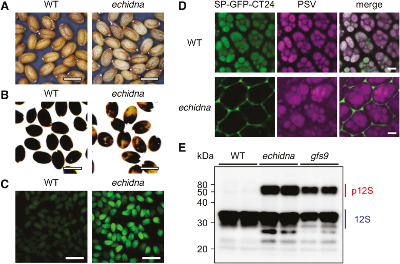
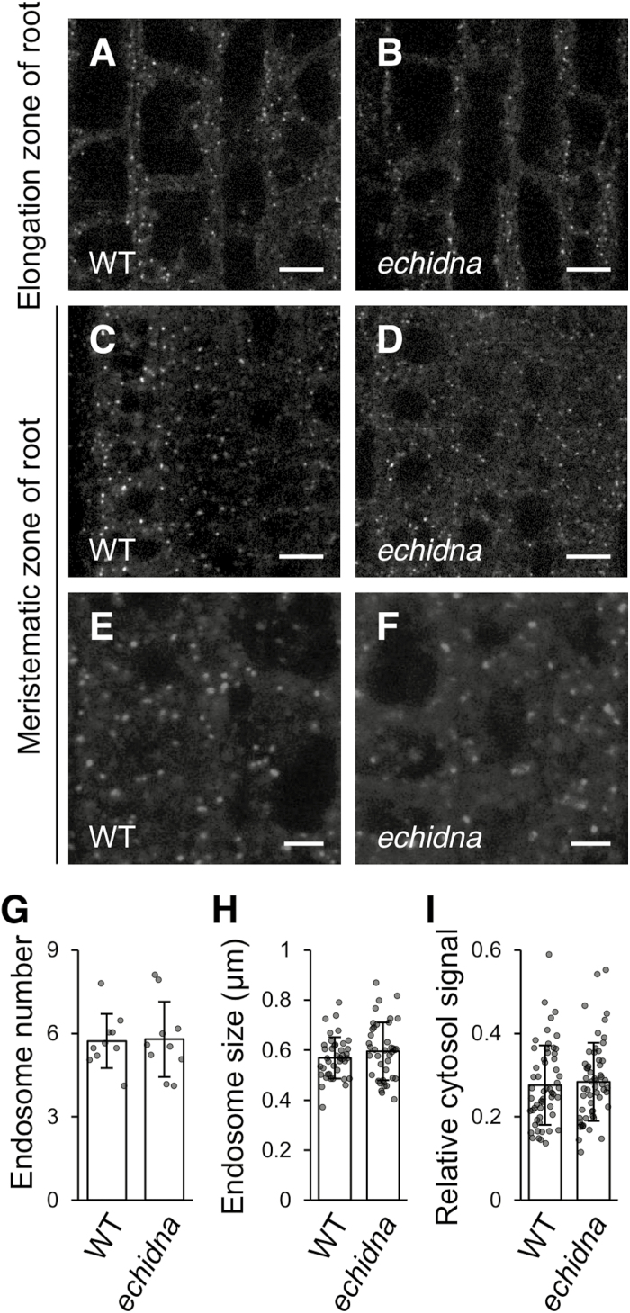
Flavonoids are a major group of plant-specific metabolites that determine flower and seed coloration. In plant cells, flavonoids are synthesized at the cytosolic surface of the endoplasmic reticulum and are sequestered in the vacuole. It is possible that membrane trafficking, including vesicle trafficking and organelle dynamics, contributes to flavonoid transport and accumulation. However, the underlying mechanism has yet to be fully elucidated. Here we show that the Arabidopsis ECHIDNA protein plays a role in flavonoid accumulation in the vacuole and protein trafficking to the vacuole. We found defective pigmentation patterns in echidna seed, possibly caused by reduced levels of proanthocyanidins, which determine seed coloration. The echidna mutant has defects in protein sorting to the protein storage vacuole as well as vacuole morphology. These findings indicate that ECHIDNA is involved in the vacuolar trafficking pathway as well as the previously described secretory pathway. In addition, we found a genetic interaction between echidna and green fluorescent seed 9 (gfs9), a membrane trafficking factor involved in flavonoid accumulation. Our findings suggest that vacuolar trafficking and/or vacuolar development, both of which are collectively regulated by ECHIDNA and GFS9, are required for flavonoid accumulation, resulting in seed coat pigmentation.

Keywords: Arabidopsis thaliana; trans-Golgi network; ECHIDNA; GREEN FLUORESCENT SEED 9; mucilage; protein sorting; seed coloration; vacuolar morphology; vacuolar trafficking; vacuole.


Figures

Fig. 1.
Fig. 1.
Seed color phenotype and missorting of vacuolar proteins in echidna mutant seeds. (A) Seeds of wild-type (WT) and echidna mutant. (B) Seeds of WT and echidna mutant after staining with p-dimethylaminocinnamaldehyde. (C) GFP fluorescence images of WT and echidna dry seeds expressing vacuolar-targeted GFP, SP-GFP-CT24. (D) Confocal microscopic images of vacuolar-targeted GFP in embryonic cotyledon cells of WT and echidna dry seeds. Vacuolar-targeted GFP fluorescent signal and autofluorescence of the protein storage vacuole (PSV) are shown. (E) Immunoblot analysis of seed storage protein in WT, echidna, and gfs9 seeds. Antibody to 12S globulin was used. The gfs9 mutant is known to be defective in protein sorting to the vacuole. p12S, precursor forms of 12S globulin; 12S, mature form of 12S globulin. Bars=0.5 mm (A, B), 1 mm (C), and 5 μm (D).
Fig. 2.
Fig. 2.
Distribution of vacuolar membrane protein VAMP711 in the echidna mutant. Confocal microscopic images of the vacuolar membrane marker mCherry-VAMP711 in wild-type (WT) (A, D, G) and echidna mutant (B, C, E, F, H, I) cells. (A–C) Immature epidermal cells of root tip; (D–F) elongated (mature) epidermal cells of seedling root; (G–I) epidermal cells of hypocotyl. Images C, F, and I are magnified views of abnormal structures in the echidna mutant. The images in B and C were observed in different seedlings; images in E and F were observed in different fields of view from the same seedling; images in H and I were observed in different fields of view from the same seedling. Bars=10 μm (A, B, D, E), 5 μm (C, F, I), and 20 μm (G, H).
Fig. 3.
Fig. 3.
ARA7-labeled late endosomes in the echidna mutant. (A–F) Confocal microscopic images of root cells from wild-type (WT) (A, C, E) and echidna mutant (B, D, F) seedlings. Late endosomes are labeled with mCherry-ARA7. (A, B) Root cells of the elongation zone; (C–F) root cells of the meristematic zone. Bars=10 μm (A–D) and 5 μm (E, F). (G) Number of ARA7-labeled late endosomes per 100 µm2 area in root cells of the elongation zone. Ten plants of each genotype were measured. Each dot represents the mean of three to five regions of interest in a single plant. Each bar graph represents the mean of 10 means ±SD. (H) Size of ARA7-labeled late endosomes in the root cells of the elongation zone. Eight plants of each genotype were measured (five endosomes per plant). Each dot represents the size of a single endosome. Each bar graph represents the mean of 40 endosomes ±SD. (I) Relative signal intensity of cytosol fluorescence, calculated as the ratio of the intensity of cytosolic signal to the intensity of punctate structures within the same cell in the root elongation zone. Fourteen plants of each genotype were measured (four cells per plant). Each dot represents the relative signal intensity of a single cell. Each bar graph represents the mean of 56 cells ±SD. P values were calculated using Student’s t-test and were as follows: P=0.91 (G), P=0.24 (H), and P=0.66 (I).
Fig. 4.
Fig. 4.
Root cap mucilage in the echidna mutant. (A) Confocal microscopic images of the root tip of the wild-type (WT) and echidna mutant stained using propidium iodide. (B) Toluidine blue-stained sections of the root tip of the WT and echidna mutant. (C) Images of the root tip of the WT and echidna mutant stained using india ink. Arrowheads indicate periplasmic mucilage. Bars=10 µm (A, B) and 50 µm (C).
Fig. 5.
Fig. 5.
Plant growth of echidna, gfs9, and the echidna gfs9 double mutant. (A) Image of 5-day-old seedlings. Bar=1 cm. (B) Primary root length of 5-day-old seedlings. The numbers of measured seedlings of wild-type (WT), gfs9, echidna, and echidna gfs9 were 56, 47, 51, and 56, respectively. (C) Image of 80-day-old fully mature adult plants. (D) Plant height of 51-day-old adult plants; 20 individuals of each genotype were measured. Data represent the mean ±SD and raw data points. Different letters above the bars indicate statistically significant differences according to Tukey’s HSD test for multiple comparisons (α=0.01).
Fig. 6.
Fig. 6.
Model of the intracellular trafficking pathway mediated by ECHIDNA. Early steps of flavonoid biosynthesis are performed on the endoplasmic reticulum (ER), and the final products (e.g. proanthocyanidins) accumulate in the vacuole. Vacuolar proteins (e.g. seed storage proteins) are also transported from the synthetic site on the ER to the final destination vacuole. GREEN FLUORESCENT SEED 9 (GFS9) on the Golgi apparatus and ECHIDNA on the trans-Golgi network (TGN) play roles in the trafficking of vacuolar proteins and flavonoids to the vacuoles. ECHIDNA also mediates the secretion of cell wall components and cuticular wax. SUPPRESSOR OF K+ TRANSPORT GROWTH DEFFECT1 (SKD1), a component of the ESCRT machinery on the multivesicular body (MVB)/late endosome (LE), has also been reported to be involved in the vacuolar trafficking of proteins and seed coat pigmentation. PM, plasma membrane.

Similar articles

Cited by

References

    1. Abrahams S, Lee E, Walker AR, Tanner GJ, Larkin PJ, Ashton AR. 2003. The Arabidopsis TDS4 gene encodes leucoanthocyanidin dioxygenase (LDOX) and is essential for proanthocyanidin synthesis and vacuole development. The Plant Journal 35, 624–636. - PubMed
    1. Abrahams S, Tanner GJ, Larkin PJ, Ashton AR. 2002. Identification and biochemical characterization of mutants in the proanthocyanidin pathway in Arabidopsis. Plant Physiology 130, 561–576. - PMC - PubMed
    1. Appelhagen I, Nordholt N, Seidel T, Spelt K, Koes R, Quattrochio F, Sagasser M, Weisshaar B. 2015. TRANSPARENT TESTA 13 is a tonoplast P3A-ATPase required for vacuolar deposition of proanthocyanidins in Arabidopsis thaliana seeds. The Plant Journal 82, 840–849. - PubMed
    1. Arganda-Carreras I, Kaynig V, Rueden C, Eliceiri KW, Schindelin J, Cardona A, Sebastian Seung H. 2017. Trainable Weka Segmentation: a machine learning tool for microscopy pixel classification. Bioinformatics 33, 2424–2426. - PubMed
    1. Baxter IR, Young JC, Armstrong G, Foster N, Bogenschutz N, Cordova T, Peer WA, Hazen SP, Murphy AS, Harper JF. 2005. A plasma membrane H+-ATPase is required for the formation of proanthocyanidins in the seed coat endothelium of Arabidopsis thaliana. Proceedings of the National Academy of Sciences, USA 102, 2649–2654. - PMC - PubMed

Publication types

Substances