. 2020 Jun 30;30(8):4581-4596.
doi: 10.1093/cercor/bhaa061.

Hippocampal Synaptic Plasticity, Spatial Memory, and Neurotransmitter Receptor Expression Are Profoundly Altered by Gradual Loss of Hearing Ability

Affiliations

Hippocampal Synaptic Plasticity, Spatial Memory, and Neurotransmitter Receptor Expression Are Profoundly Altered by Gradual Loss of Hearing Ability

Daniela Beckmann et al. Cereb Cortex. .

Abstract

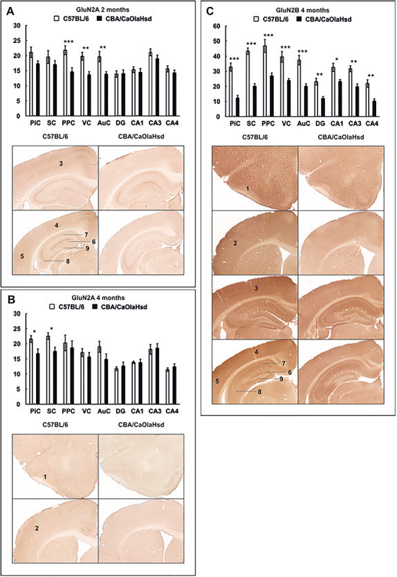
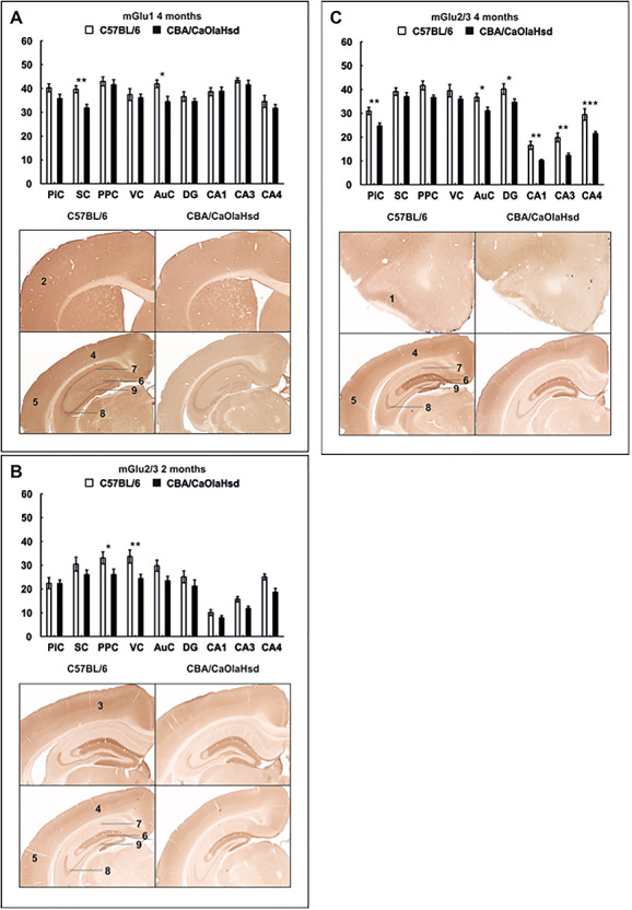
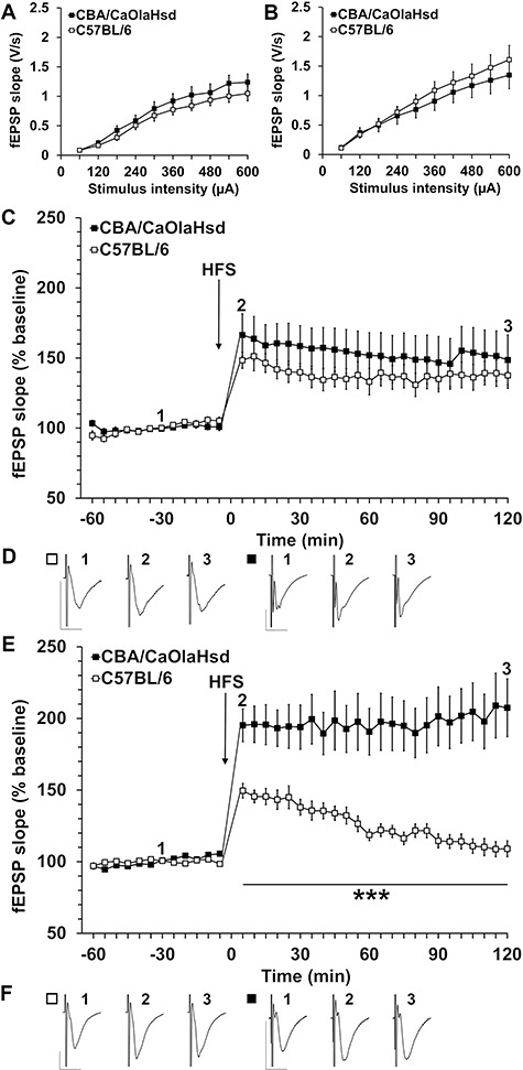
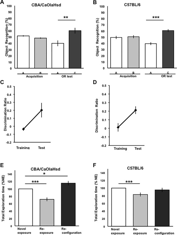
Sensory information comprises the substrate from which memories are created. Memories of spatial sensory experience are encoded by means of synaptic plasticity in the hippocampus. Hippocampal dependency on sensory information is highlighted by the fact that sudden and complete loss of a sensory modality results in an impairment of hippocampal function that persists for months. Effects are accompanied by extensive changes in the expression of neurotransmitter receptors in cortex and hippocampus, consistent with a substantial adaptive reorganization of cortical function. Whether gradual sensory loss affects hippocampal function is unclear. Progressive age-dependent hearing loss (presbycusis) is a risk factor for cognitive decline. Here, we scrutinized C57BL/6 mice that experience hereditary and cumulative deafness starting in young adulthood. We observed that 2-4 months postnatally, increases in the cortical and hippocampal expression of GluN2A and GluN2B subunits of the N-methyl-D-aspartate receptor occurred compared to control mice that lack sensory deficits. Furthermore, GABA and metabotropic glutamate receptor expression were significantly altered. Hippocampal synaptic plasticity was profoundly impaired and mice exhibited significant deficits in spatial memory. These data show that during cortical adaptation to cumulative loss of hearing, plasticity-related neurotransmitter expression is extensively altered in the cortex and hippocampus. Furthermore, cumulative sensory loss compromises hippocampal function.

Keywords: C57BL/6; GABA; NMDA receptor; hearing loss; hippocampus; metabotropic glutamate receptor.


Figures

Figure 1
Figure 1
Diagram of areas selected for immunohistochemical analysis. The areas assessed in the mouse cerebral cortex and hippocampus are shown in the histological sections (left side of each example). The specific regions examined are indicated in the schemas on the right side of each example (based on Franklin and Paxinos 2008). Markings in dark gray represent the following areas: Top left: PiC; top right: SC; bottom left: PPC; bottom right: VC (1), AuC (2), and hippocampus (3), including the DG and cornus ammonis subregions (CA1–CA3).
Figure 2
Figure 2
Expression of the GluN2A and GluN2B subunits of the NMDA receptor 2 and 4 months postnatally. Bar charts represent mean optical density of NMDA receptor subunit expression in C57BL/6 and CBA/CaOlaHsd mice 2 and 4 months postnatally. Photomicrographs highlight significantly affected areas as shown in the graphs above. (A) GluN2A expression is significantly increased in the PPC, VC, and AuC of C57Bl/6 mice at the age of 2 months. (B) GluN2A expression is significantly increased in 4-month-old C57BL/6 mice in the PiC and SC. (C) GluN2B expression is globally increased in C57BL/6 mice 4 months postnatally. Data are means ± SEM. *P < 0.05; **P < 0.01; ***P < 0.001. 1: PiC, 2: SC, 3: PPC, 4: VC, 5: AuC, 6: DG, 7: CA1, 8: CA3, and 9: CA4.
Figure 3
Figure 3
Expression of metabotropic glutamate receptors 2 and 4 months postnatally. Bar charts represent mean optical density of metabotropic glutamate receptor expression in C57BL/6 and CBA/CaOlaHsd mice at the age of 2 and 4 months. Photomicrographs highlight significantly affected areas as shown in the graphs above. (A) mGlu1 expression is significantly increased in 4-month-old C57BL/6 mice in the SC and AuC. (B) Significant increase in mGlu2/3 expression in C57BL/6 mice in the PPC and VC at the age of 2 months. (C) C57BL/6 mice show significantly increased mGlu2/3 expression in the PiC and AuC and in the hippocampus. Data are means ± SEM. *P < 0.05; **P < 0.01; ***P < 0.001. 1: PiC, 2: SC, 3: PPC, 4: VC, 5: AuC, 6: DG, 7: CA1, 8: CA3, and 9: CA4.
Figure 4
Figure 4
GABA receptor expression differs 2 and 4 months postnatally. Bar charts represent mean optical density of GABAA and GABAB expression in C57BL/6 and CBA/CaOlaHsd mice 2 and 4 months postnatally. Photomicrographs highlight significantly affected areas as shown in the graphs above. (A) Four months postnatally, GABAA is significantly decreased in the PiC, SC, and AuC and in the DG of C57BL/6 mice. (B) Optical density of GABAB receptors is significantly increased in C57BL/6 mice in the AuC and in the hippocampal subfield CA1 2 months postnatally. (C) At the age of 4 months, GABAB expression is significantly lower in the DG and CA3–4 regions of C57BL/6 mice compared with controls. Data are means ± SEM. *P < 0.05; **P < 0.01; ***P < 0.001. 1: PiC, 2: SC, 3: PPC, 4: VC, 5: AuC, 6: DG, 7: CA1, 8: CA3, and 9: CA4.
Figure 5
Figure 5
Synaptic plasticity is impaired in C57BL/6 mice. (A) Comparison of input–output relationship in 2-month-old C57BL/6 mice and control (CBA/CaOlaHsd) mice shows no significant differences in fEPSP evoked in the stimulus intensity range of 60–600 μA. (B) Comparison of input–output responses in 4-month-old C57BL/6 and control mice found no significant differences in fEPSP evoked in the stimulus intensity range of 60–600 μA (C) HFS elicits LTP in 2-month-old C57B/6 and control mice, which is not significantly different. (D) Analog examples show fEPSP recorded from 2-month-old C57BL/6 and control mice at the time points indicated in the graph shown in “C.” Vertical scale bar: 1 mV; horizontal scale bar: 10 ms. (E) HFS results in robust LTP in 4-month-old control mice. By contrast, C57BL/6 mice show a significantly impaired response to HFS. The induction and maintenance phases are significantly smaller in magnitude than that elicited in control mice. By 100 min post-HFS, evoked potentials returned to pre-HFS levels. (F) Analog examples show fEPSP recorded from 4-month-old C57BL/6 and control mice at the time points indicated in the graph shown in “E.” Vertical scale bar: 1 mV; horizontal scale bar: 10 ms.
Figure 6
Figure 6
Item-place memory, but not Object Recognition memory, is impaired in C57BL/6 mice. (A and B) Behavioral analysis revealed that CBA/CaOlaHsd mice (n = 11) and C57BL/6 mice (n = 12) performed the OR task successfully with a delay period of 24 h between phases. The mice explored both objects A and B equally during the training phase but explored the novel object C to a significantly greater extent during the test phase (CBA/CaOlaHsd: ANOVA, F(1,20) = 9.99, P = 0.4256; C57BL/6: ANOVA, F(1,22) = 45.61, P < 0.0001). (C, D) Analysis of the discrimination ratios revealed that the mice showed no preference for either object during the training phase but a strong preference for the novel object during the test phase (C: CBA/CaOlaHsd: ANOVA, F(1,20) = 7.60, P = 0.0129; D: C57BL/6: ANOVA, F(1,22) = 7.51, P = 0.0125). (E) CBA/CaOlaHsd mice acquired significant IP memory. Compared to novel exposure, the animals exhibited an habituation effect 24 h later when they were re-exposed to the same objects in the same places F(1,20) = 35.43, P < 0.0001). A further 24 h later, reconfiguration of the objects triggered an increase in exploration (ANOVA, F(1,20) = 6.79, P = 0.0178). By contrast, although C57BL/6 mice (F) exhibited an habituation effect upon object re-exposure (F(1,22) = 44.93, P < 0.0001), they exhibited no significant change in exploration behavior exposure to a spatial reconfiguration of the objects (ANOVA, F(1,22) = 0.52, P = 0.4754). Thus, the C57BL/6 mice failed to generate long-term IP memory.

References

    1. André ME, Manahan-Vaughan D. 2014. Spatial olfactory learning facilitates long-term depression in the hippocampus. Hippocampus. 23:963–968. - PubMed
    1. Attems J, Walker L, Jellinger KA. 2015. Olfaction and aging: a mini-review. Gerontology. 61(6):485–490. - PubMed
    1. Ballesteros JJ, Buschler A, Köhr G, Manahan-Vaughan D. 2016. Afferent input selects NMDA receptor subtype to determine the persistency of hippocampal LTP in freely behaving mice. Front Behav Neurosci. 8:33. - PMC - PubMed
    1. Berberich S, Jensen V, Hvalby O, Seeburg PH, Köhr G. 2007. The role of NMDAR subtypes and charge transfer during hippocampal LTP induction. Neuropharmacology. 52:77–86. - PubMed
    1. Berberich S, Punnakkal P, Jensen V, Pawlak V, Seeburg PH, Hvalby O, Köhr G. 2005. Lack of NMDA receptor subtype selectivity for hippocampal long-term potentiation. J Neurosci. 25:6907–6910. - PMC - PubMed

Publication types

Substances