Review
. 2020 May 26;60(5):2470-2483.
doi: 10.1021/acs.jcim.0c00087. Epub 2020 Mar 31.

Simulation-Based Methods for Model Building and Refinement in Cryoelectron Microscopy

Affiliations
Review

Simulation-Based Methods for Model Building and Refinement in Cryoelectron Microscopy

Thomas Dodd et al. J Chem Inf Model. .

Abstract

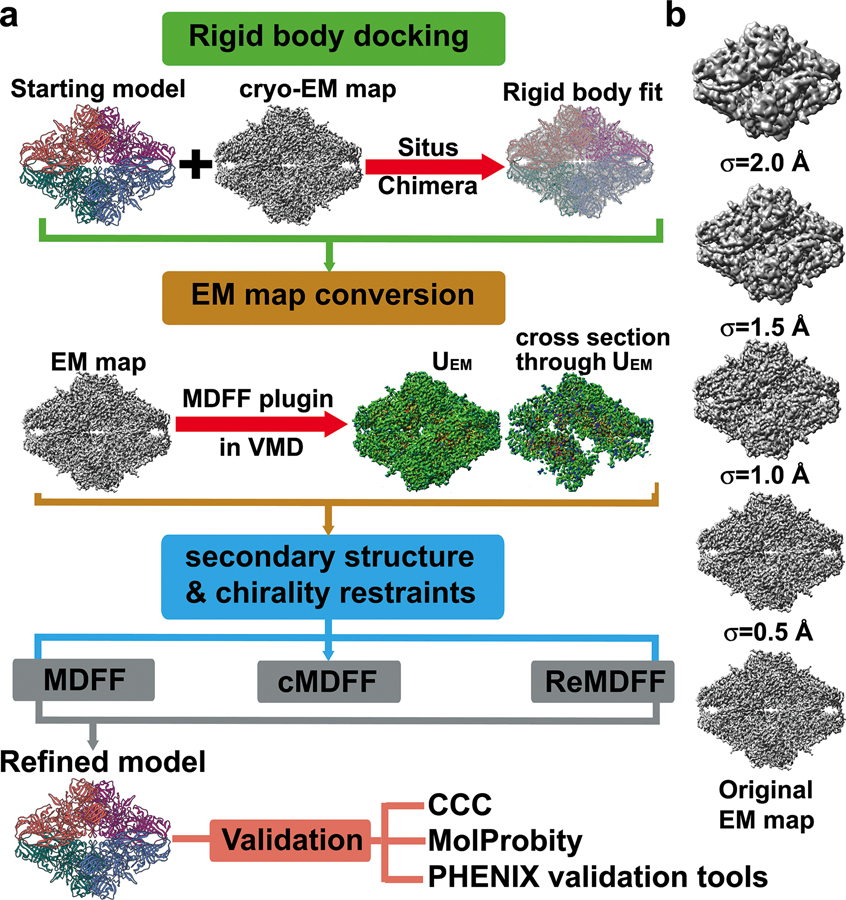
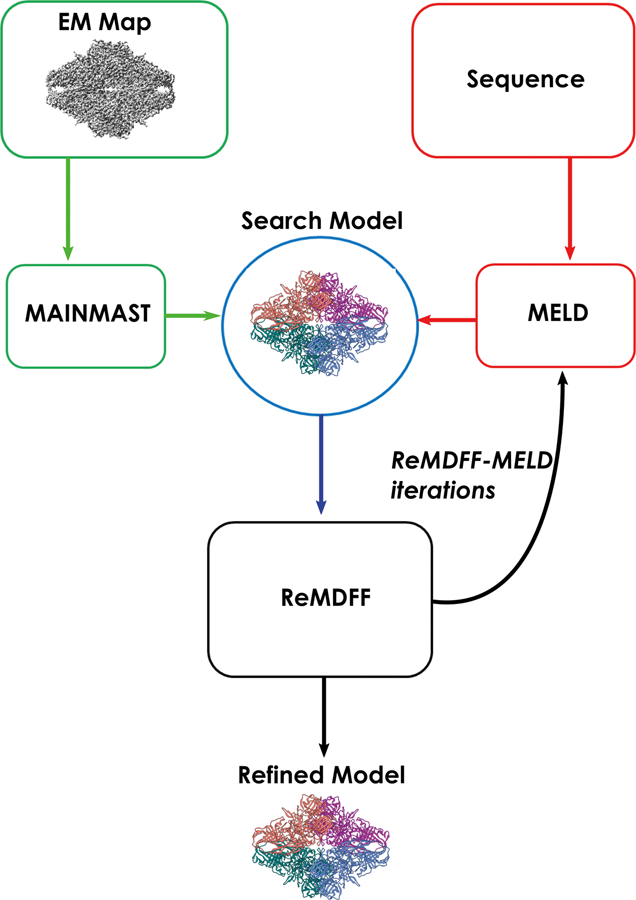
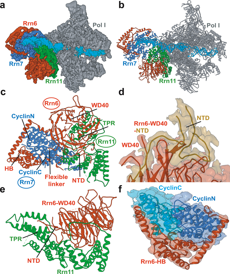
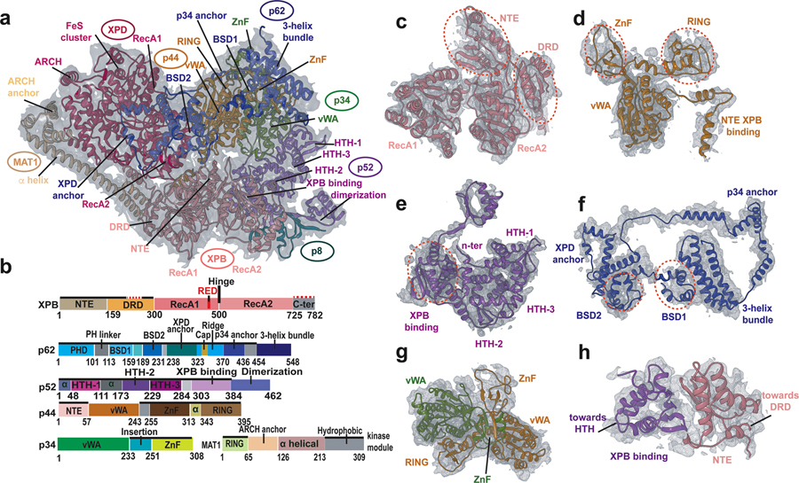
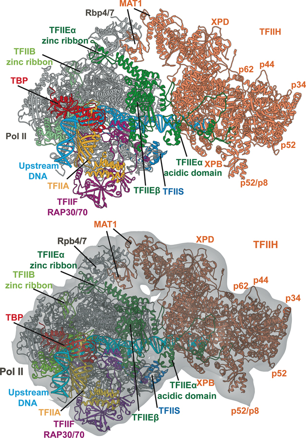
Advances in cryoelectron microscopy (cryo-EM) have revolutionized the structural investigation of large macromolecular assemblies. In this review, we first provide a broad overview of modeling methods used for flexible fitting of molecular models into cryo-EM density maps. We give special attention to approaches rooted in molecular simulations-atomistic molecular dynamics and Monte Carlo. Concise descriptions of the methods are given along with discussion of their advantages, limitations, and most popular alternatives. We also describe recent extensions of the widely used molecular dynamics flexible fitting (MDFF) method and discuss how different model-building techniques could be incorporated into new hybrid modeling schemes and simulation workflows. Finally, we provide two illustrative examples of model-building and refinement strategies employing MDFF, cascade MDFF, and RosettaCM. These examples come from recent cryo-EM studies that elucidated transcription preinitiation complexes and shed light on the functional roles of these assemblies in gene expression and gene regulation.

Keywords: RNA polymerases; cryoelectron microscopy; de novo model building; gene regulation; hybrid methods; molecular dynamics flexible fitting (MDFF); molecular modeling; transcription preinitiation complexes.


Figures

Figure 1.
Figure 1.. Simulation-based methods for flexible fitting into cryo-EM maps.
a) Flowchart representing the popular MDFF, cascade MDFF and resolution exchange MDFF methods; b) A series of Gaussian-blurred EM maps and the original unfiltered cryo-EM map used for cascade MDFF and resolution exchange MDFF. The maps illustrate the change in smoothness and level of detail of the resulting UEM potentials used for the intermediate stages of the fitting protocol.
Figure 2
Figure 2
Schematic representation of the Cryo-fold fitting protocol.
Figure 3
Figure 3. Variable local resolution as a major challenge for model building in cryo-EM.
a-c) Local resolution color mapped onto three example EM maps. The maps were chosen to showcase narrow, intermediate and wide range of local resolution; d-f) FSC curves are presented for each of the maps to illustrate how overall map resolution is determined. Notably, overall resolution of a cryo-EM map is a global property and may not be indicative of quality for specific region of the EM density.
Figure 4.
Figure 4.. Structure of Pol I preinitiation complex from cryo-EM and integrative modeling.
a) Cryo-EM reconstruction of Pol I initial transcribing complex; b) Atomistic model of Pol I initial transcribing complex; c) Atomistic model of Core Factor. The Core Factor subunits are depicted in red (Rrn6), blue (Rrn7) and green (Rrn11). Modeled structural elements include NTD, N-terminal domain; HB, helical bundle; CyclinC/N, C/N-terminal Cyclin Fold domain and TPR, tetratricopeptide repeats; d) Interface between Rrn6 HB and Rrn6 WD40 domains; e) Interface between Rrn6 WD40 and Rrn11 TPR domains; f) The two cyclin domains of Rrn7 are embedded in the Rrn6 HB protein chain.
Figure 5.
Figure 5.. Structure of transcription factor IIH from cryo-EM and integrative modeling.
a) Overall fit of the apo-TFIIH structure to the apo-TFIIH density; b) Domain organization of the TFIIH subunits highlighting newly modeled regions (solid black lines); The structural motifs were labeled for each TFIIH subunit; c–f) Selected TFIIH subunits fitted into the EM density. Domains in each subunit are indicated with red dashed circles. c) XPB; d) p44; e) p52; f) p62; g) p34 and p44; h) XPB and p52.
Figure 6.
Figure 6.. Structure of human Pol II preinitiation complex from cryo-EM and integrative modeling.
Positions of the general transcription factor subunits are indicated by color coding. The model is based on integration and comparative analysis of cryo-EM densities for human apo-TFIIH, human closed-state holo-PIC density and yeast core-PIC–TFIIH–DNA. Atomic model for the entire complex is shown with (a) and (b) without the EM envelope.

References

    1. Bai XC; McMullan G; Scheres SH How cryo-EM is revolutionizing structural biology. Trends Biochem Sci 2015, 40 (1), 49–57. - PubMed
    1. Nogales E The development of cryo-EM into a mainstream structural biology technique. Nat Methods 2016, 13 (1), 24–7. - PMC - PubMed
    1. Li X; Mooney P; Zheng S; Booth CR; Braunfeld MB; Gubbens S; Agard DA; Cheng Y Electron counting and beam-induced motion correction enable near-atomic-resolution single-particle cryo-EM. Nat Methods 2013, 10 (6), 584–90. - PMC - PubMed
    1. Yang C; Ji G; Liu H; Zhang K; Liu G; Sun F; Zhu P; Cheng L Cryo-EM structure of a transcribing cypovirus. Proc Natl Acad Sci U S A 2012, 109 (16), 6118–23. - PMC - PubMed
    1. Zhang X; Sun S; Xiang Y; Wong J; Klose T; Raoult D; Rossmann MG Structure of Sputnik, a virophage, at 3.5-A resolution. Proc Natl Acad Sci U S A 2012, 109 (45), 18431–6. - PMC - PubMed

Publication types

MeSH terms

Substances