Review
. 2020 Mar;46(2):147-168.
doi: 10.1080/1040841X.2020.1735992. Epub 2020 Mar 23.

Host receptors: the key to establishing cells with broad viral tropism for vaccine production

Affiliations
Review

Host receptors: the key to establishing cells with broad viral tropism for vaccine production

Xiaofeng Dai et al. Crit Rev Microbiol. 2020 Mar.

Abstract

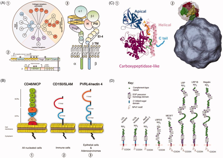
Cell culture-based vaccine technology is a flexible and convenient approach for vaccine production that requires adaptation of the vaccine strains to the new cells. Driven by the motivation to develop a broadly permissive cell line for infection with a wide range of viruses, we identified a set of the most relevant host receptors involved in viral attachment and entry. This identification was done through a review of different viral entry pathways and host cell lines, and in the context of the Baltimore classification of viruses. In addition, we indicated the potential technical problems and proposed some solutions regarding how to modify the host cell genome in order to meet industrial requirements for mass production of antiviral vaccines. Our work contributes to a finer understanding of the importance of breaking the host-virus recognition specificities for the possibility of creating a cell line feasible for the production of vaccines against a broad spectrum of viruses.

Keywords: Virus; attachment factor; entry pathway; entry receptor; susceptible cell line; vaccine production.


Figures

Figure 1.
Figure 1.
Schematic illustration on cell-based vaccine production.
Figure 2.
Figure 2.
Baltimore classification of viruses and their nucleic acid form prior to mRNA transcription.
Figure 3.
Figure 3.
Schematic representations of clathrin-mediated endocytosis and caveolin-mediated endocytosis. (A) HCV entry process illustrating the clathrin-mediated endocytosis. ①HCV binds GAGs (HS from syndecan-1 and syndecan-4) and LDLR, which have high affinity for the ApoE (apolipoprotein E). SRBI (scavenger receptor B type I) plays an important role in both binding and post-binding steps of viral entry through interaction with virion-associated lipoprotein or HCV E2. ②Binding of SRBI to HCV particles allows exposure of CD81 binding sites on HCV E2 and transfer of the virus particles to CD81. ③The virion is primed by the low-pH fusion activity of CD81 and CLDN1 and translocates to the tight junctions in order to be endocytosed. Viral internalization is dependent on clathrin-mediated endocytosis. Junction protein occludin (OCLN) can contribute to this process. TfR1, EGFR, and EphA2 (ephrin receptor A2) play a role in HCV infection at the level of glycoprotein-mediated entry, acts after CD81, and possibly are involved in the HCV particle internalization. PI3K-AKT and PI4K pathways are engaged in the late step of HCV entry. However, the molecular mechanisms need to be investigated. ④Following internalization, HCV fusion occurs in the early endosomes. Low pH environment and virion-associated cholesterol are required for the fusion process. NPC1L1 may play a role in this process via cholesterol transport. After fusion between the viral envelope and an endosomal membrane, the viral genome is released into the cytosol and replication takes place. Reprinted with permission from Zhu (2014). (B) SV40 entry process illustrating caveolin-mediated endocytosis. ①SV40 binding to the host cell is codirected by the capsid and VP2. ② The bound virus traverses the membrane and enters a caveolae. ③ The virus is endocytosed and transported in the caveolae-coated vesicles to endoplasmic reticulum.
Figure 4.
Figure 4.
Schematic representations of the structures of representative viruses that employ diverse entry pathways: non-clathrin, non-caveolin-mediated endocytosis and direct penetration. (A) HSV-1 glycoproteins and their identified cell receptors required for viral entry. The gB receptors are known to be crucial for virus attachment and membrane fusion (Fusogen), gD receptors are reported to trigger the fusion process (Fusion Trigger) and gH-gL receptors are known to regulate the fusion process with the mechanism less understood (Fusion Regulator). Reprinted with permission from Karasneh and Shukla (2011). (B) Structure of rotavirus. The three concentric capsid protein layers are the VP7 layer with VP4 spikes, the VP6 layer and the VP2 layer. The dsRNA segments are packed inside and associated to the RNA polymerase complexes VP1 and VP3.
Figure 5.
Figure 5.
Schematic representations of the structures of two major attachment receptors. (A) Schematic depiction on details of HS. HSPG is composed of HS and core proteins, with the HS structure being shown in the upper panel and a fragment amplified in the lower panel for details. HS biosynthesis is initiated by the attachment of xylose to specific serine residues in the HSPG core proteins followed by the formation of a linkage tetrasaccharide, glucuronic acid-galactose-galactose-xylose (GlcA-Gal-Gal-Xyl). Extl3 attaches the first N-acetyl-D-glucosamine (GlcNAc) residue and an enzyme complex composed of Ext1 and Ext2, alternately adds GlcA and GlcNAc to the nascent chain. The chains simultaneously undergo a series of processing reactions that begin by the removal of the acetyl groups from clusters of GlcNAc residues and substitution of the free amino groups with sulfate, catalyzed by one or more N-deacetylase-N-sulfotransferases (Ndst). The C5 epimerase (HsGlce) epimerizes the D-glucuronic acids immediately adjacent to N-sulfoglucosamine units to L-iduronic acid (IdoA). A series of O-sulfotransferases can then add sulfate. As shown in the top of the figure in red shading, the modifications occur in clusters of variable length (N-sulfated or NS domains), which are interspersed by unmodified domains (N-acetylated or NA domains). The modified domains make up binding sites for protein ligands as depicted for antithrombin, FGF and FGF receptor. (B) Schematic depiction on the global structure of two types of typical HSPGs. Syndecans are transmembrane proteins that bear HS chains distal from the plasma membrane. Some syndecans also contain ChS chains, which, by homology to syndecan-1, are located close to the membrane. Glypicans are covalently linked to a phosphatidyl inositol in the outer leaflet of the plasma membrane (GPI-linked). The HS chains are located on a likely extended protein domain near the plasma membrane. The glypican ectodomains are presumably compact and globular proteins as featured by 14 conserved cysteine residues. (C) Structure of the two common forms of SA, Neu5Ac, and Neu5Gc. (D) Structure of SA in two linkage conformations, α2,3-SA, α2,6-SA. Reprinted with permission from de Graaf and Fouchier (2014) and Byrd-Leotis et al. (2017).
Figure 6.
Figure 6.
Schematic representations of the structures of four primary internalization receptors. (A) Illustration of integrin pairing and structure. ① Integrin family member and pairing in vertebrates, as well as their primary functions. ② Integrin primary structure with prototypical αI-domain-containing integrin heterodimer as an example. Half of α integrin subunits contain the αI-domain, and all integrins contain a βI domain in the β subunit. ‘Stars’ show divalent cation-binding sites. ③ Integrin tertiary structure with prototypical αI-domain-containing integrin heterodimer as an example. Reprinted with permission from Barczyk et al. (2010). (B) Structure of receptors CD46, CD150 (SLAM) and Nectin4 (PVRL4). CD46, CD150 and Nectin4 are all transmembrane receptors sharing similar structures, and are expressed in all nucleated cells, immune cells and epithelial cells, respectively. ① CD46 structure consists of four short consensus repeats (SCRs I, II, III, IV), a serine/threonine/proline region (STPs A, B, C), a sequence of unknown significance (U), a transmembrane sequence and a cytoplasmic domain. ② CD150 contains a variable (V) domain and a constant (C2) Ig-like repeat in its extracellular domain. CD150 is the universal immune receptor for all morbilliviruses. ③ Nectin4 extracellular domain is composed of a variable domain and two C2 domains. To date, Nectin4 was shown to serve as an epithelial receptor for MV, CDV, and PPRV. Reprinted with permission from Delpeut et al. (2014). (C) Structure of TfR monomer and dimer. ① TfR monomer. A (orange) shows the apical loop (residues 312 to 328); PL (green) shows the carboxypeptidase-like loop (residues 469 to 476); C tail (blue) shows the C-terminal tail (residues 750 to 760). Reprinted with modification and permission from Bennett et al. (2000). ② One of the two apical domains (green) of a TfR dimer is shown interacting with the virus surface (grey). The other two domains shown are helical domains and carboxypeptidase-like domains of the surface-rendered TfR molecule are yellow and red, respectively. Reprinted with permission from Hafenstein et al. (2007). (D) Structure of LDLR family members in mammalian cells. All members share common motifs, including a single membrane anchor, complement-type repeats (ligand binding domains) and EGF precursor homology domains (required for acid-dependent release of ligands in endosomes). NPxY designates the four-amino-acid motif Asp-Pro-x-Tyr, which is proposed by some studies to mediate the clustering of the receptors into coated pits. O-linked sugar domains are found in some but not all LDLR species, which are considered to function as a hydrophilic spacer keeping lipophilic ligands away from the lipid bilayer of the plasma membrane. LRP refers to ‘LDL-receptor-related protein’; MEGF7 is short for ‘multiple EGF-repeat-containing protein 7’. Reprinted with permission from Nykjaer and Willnow (2002).

Similar articles

Cited by

References

    1. Agnello V, Abel G, Elfahal M, Knight GB, Zhang Q-X. 1999. Hepatitis C virus and other flaviviridae viruses enter cells via low density lipoprotein receptor. Proc Natl Acad Sci USA. 96(22):12766–12771. - PMC - PubMed
    1. Akhtar J, Shukla D. 2009. Viral entry mechanisms: cellular and viral mediators of herpes simplex virus entry. FEBS J. 276(24):7228–7236. - PMC - PubMed
    1. Aksoy P, Abban CY, Kiyashka E, Qiang W, Meneses PI. 2014. HPV16 infection of HaCaTs is dependent on beta4 integrin, and alpha6 integrin processing. Virology. 449:45–52. - PMC - PubMed
    1. Albecka A, Belouzard S, de Beeck AO, Descamps V, Goueslain L, Bertrand-Michel J, Tercé F, Duverlie G, Rouillé Y, Dubuisson J, et al. . 2012. Role of low-density lipoprotein receptor in the hepatitis C virus life cycle. Hepatology. 55(4):998–1007. - PubMed
    1. Albecka A, Laine RF, Janssen AFJ, Kaminski CF, Crump CM. 2016. HSV-1 glycoproteins are delivered to virus assembly sites through dynamin-dependent endocytosis. Traffic. 17(1):21–39. - PMC - PubMed

LinkOut - more resources