. 2020 Sep;27(9):2635-2650.
doi: 10.1038/s41418-020-0528-x. Epub 2020 Mar 18.

Inhibitor of apoptosis-stimulating protein of p53 inhibits ferroptosis and alleviates intestinal ischemia/reperfusion-induced acute lung injury

Affiliations

Inhibitor of apoptosis-stimulating protein of p53 inhibits ferroptosis and alleviates intestinal ischemia/reperfusion-induced acute lung injury

Yingchuan Li et al. Cell Death Differ. 2020 Sep.

Abstract

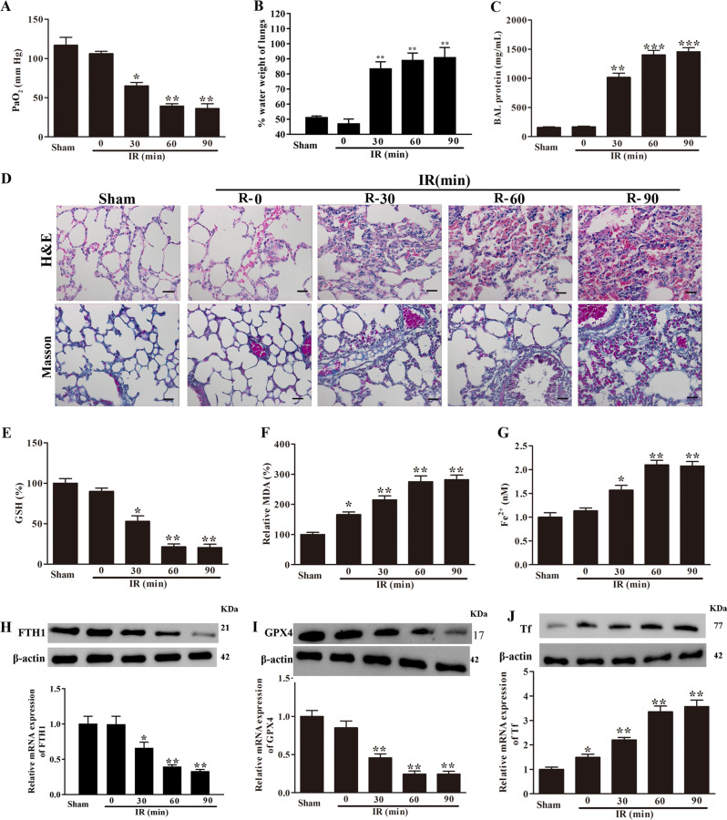
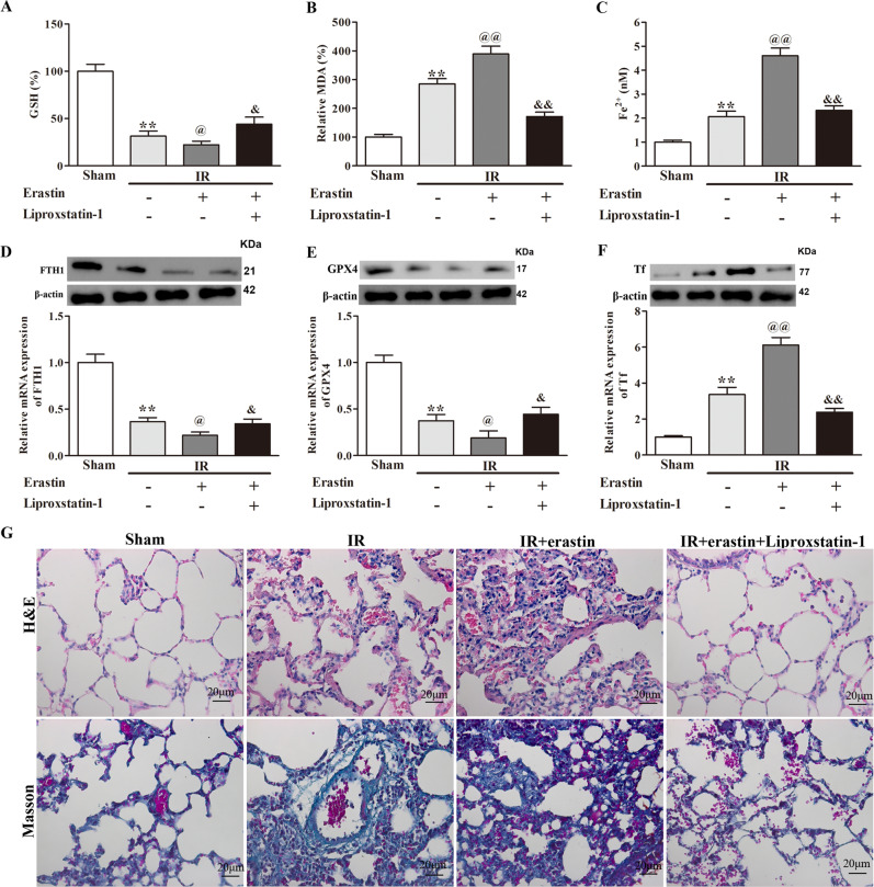
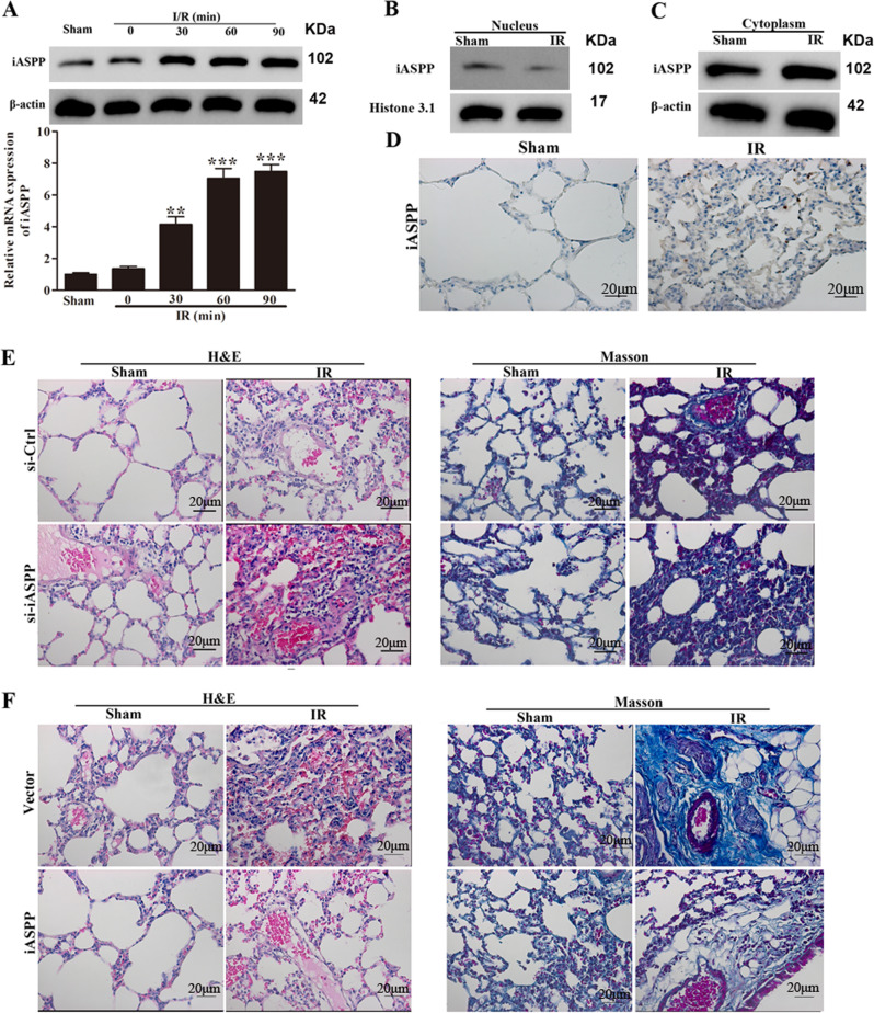
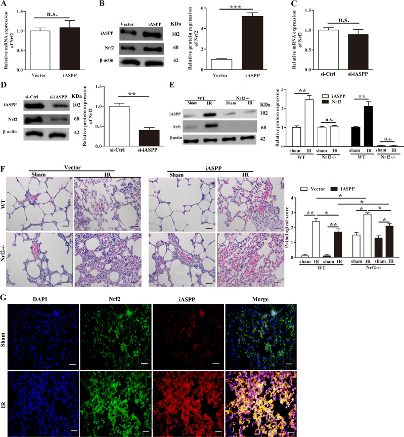
Acute lung injury (ALI) is a life-threatening disorder with high rates of morbidity and mortality. Reactive oxygen species and epithelial apoptosis are involved in the pathogenesis of acute lung injury. Ferroptosis, an iron-dependent non-apoptotic form of cell death, mediates its effects in part by promoting the accumulation of reactive oxygen species. The inhibition of ferroptosis decreases clinical symptoms in experimental models of ischemia/reperfusion-induced renal failure and heart injury. This study investigated the roles of inhibitor of apoptosis-stimulating protein of p53 (iASPP) and Nrf2 in ferroptosis and their potential therapeutic effects in intestinal ischemia/reperfusion-induced acute lung injury. Intestinal ischemia/reperfusion-induced ALI was induced in wild-type and Nrf2-/- mice. The mice were treated with erastin followed by liproxstatin-1. Ferroptosis-related factors in mice with ischemia/reperfusion-induced acute lung injury or in mouse lung epithelial-2 cells with hypoxia/regeneration (HR)-induced ALI were measured by western blotting, real-time PCR, and immunofluorescence. Ferroptosis contributed to intestinal ischemia/reperfusion-induced ALI in vivo. iASPP inhibited ferroptosis and alleviated intestinal ischemia/reperfusion-induced acute lung injury, and iASPP-mediated protection against ischemia/reperfusion-induced ALI was dependent on Nrf2 signaling. HR-induced acute lung injury enhanced ferroptosis in vitro in mouse lung epithelial-2 cells, and ferroptosis was modulated after the enhancement of intestinal ischemia/reperfusion in Nrf2-/- mice. iASPP mediated its protective effects against acute lung injury through the Nrf2/HIF-1/TF signaling pathway. Ferroptosis contributes to intestinal ischemia/reperfusion-induced ALI, and iASPP treatment inhibits ferroptosis in part via Nrf2. These findings indicate the therapeutic potential of iASPP for treating ischemia/reperfusion-induced ALI.


Conflict of interest statement

The authors declare that they have no conflict of interest.

Figures

Fig. 1
Fig. 1. Intestine I/R-induced ALI enhances ferroptosis in vivo.
C57BL/6 mice were subjected to 60 min of intestinal ischemia followed by 0, 30, 60, or 90 min of reperfusion as indicated. Sham mice were included as controls. a Arterial blood PaO2 was detected and lung water content was analyzed (b). c The protein content of BALF was measured. Results are representative of three experiments (n = 6 per group). d Representative H&E- and Masson trichrome-stained lung sections. Morphology was examined using light microscopy. Scar bar  = 20 μm. The relative values of GSH (e) and MDA (f) and the Fe2+ concentration (g) were measured. hj The ferroptosis-related proteins FTH1, GPX4, and TF were analyzed by RT-PCR and western blot analysis. *p < 0.05, **p < 0.01.
Fig. 2
Fig. 2. Erastin-induced ferroptosis promotes I/R-induced ALI injury in vivo.
C57BL/6 mice were subjected to 60 min of intestinal ischemia followed 60 min of reperfusion (I/R) as indicated, and then erastin (10 μM) was tail-injected intravenously for 20 days, followed by treatment with liproxstatin-1 (1 μM) for 20 days. Sham mice were included as controls. The relative values of GSH (a) and MDA (b) and the Fe2+ concentration (c) were measured. df The ferroptosis-related proteins FTH1, GPX4, and TF were analyzed by RT-PCR and western blot analysis. g Representative H&E- and Masson trichrome-stained lung sections. Morphology was examined using light microscopy. Scar bar = 20μm. **p < 0.01 vs. Sham; @p < 0.05 vs. IR; @@p < 0.01 vs. IR; &p < 0.05 vs. IR + Erastin; &&p < 0.01 vs. IR + Erastin.
Fig. 3
Fig. 3. The effect of iASPP knockdown or overexpression on intestinal I/R-induced ALI in WT mice.
a The relative mRNA and protein expressions of iASPP were detected in lung tissues in the sham group and I/R groups by RT-PCR and western blotting. b, c The protein expression of iASPP in the nucleus and cytoplasm following I/R (60 min/60 min) was measured by western blotting. d Representative photographs showing iASPP staining in lung tissues following I/R (60 min/60 min). Scar bar = 20 μm. e Representative H&E- and Masson trichrome-stained lung sections. Morphology was examined using light microscopy, WT mice received adenoviruses carrying si-iASPP or si-Ctrl by injection. f Representative H&E- and Masson trichrome-stained lung sections. Morphology was examined using light microscopy. WT mice received adenoviruses carrying vector control or iASPP by injection. **p < 0.01, ***p < 0.001.
Fig. 4
Fig. 4. Effect of iASPP knockdown or overexpression on ferroptosis in vivo.
ad The mRNA expression levels for FTH1, GPX4, TF, and ACSL4 following I/R (60 min/60 min) were detected by RT-PCR. WT mice received adenoviruses carrying vector control or iASPP by injection. e Relative protein expression levels of FTH1, GPX4, TF, and ACSL4 following I/R (60 min/60 min) as assessed by western blot analysis. WT mice received adenoviruses carrying vector control or iASPP by injection. fi mRNA expressions of FTH1, GPX4, TF, NQO-1, and HO-1 following I/R (60 min/60 min) were detected by RT-PCR. WT mice received adenoviruses carrying si-iASPP or si-Ctrl by injection. j Relative protein expressions of FTH1, GPX4, TF, and ACSL4 following I/R (60 min/60 min) as assessed by western blot analysis. WT mice received adenoviruses carrying si-iASPP or si-Ctrl by injection. *p < 0.05, **p < 0.01.
Fig. 5
Fig. 5. iASPP-mediated protection against I/R-induced ALI is dependent on Nrf2 antioxidative signaling in vivo.
The mRNA (a) and protein (b) expression of Nrf2 in lung tissues were detected by RT-PCR and western blot analysis, respectively. WT mice received adenoviruses carrying vector control or iASPP by injection. The mRNA (c) and protein (d) expression of Nrf2 in lung tissues were detected by RT-PCR and western blot analysis, respectively. WT mice received adenoviruses carrying si-iASPP or si-Ctrl by injection. e The protein expression levels of iASPP and Nrf2 in lung tissues were determined by western blot analysis in WT mice and Nrf2−/− mice following I/R (60 min/60 min). f Representative H&E staining of lung tissues following I/R (60 min/60 min) in WT mice and Nrf2−/− mice. WT mice and Nrf2−/− mice received adenoviruses carrying vector control or iASPP by injection. Scar bar = 20 μm. g Double immunohistochemical staining of lung tissues for Nrf2 (green) and iASPP (red). Scar bar = 20 μm. Nuclei were stained with DAPI (blue) following I/R (60 min/60 min). *p < 0.05, **p < 0.01.
Fig. 6
Fig. 6. Modulation of ferroptosis is impaired in Nrf2−/− mice after intestinal I/R.
The relative values of GSH (a) and MDA (b) and the Fe2+ concentration (c) were measured following I/R (60 min/60 min) in WT and Nrf2−/− mice. dh The mRNA expression levels for FTH1, GPX4, TF, NQO-1, and HO-1 following I/R (60 min/60 min) were detected by RT-PCR in WT mice and Nrf2−/− mice. i The protein expression levels of FTH1, GPX4, TF, NQO-1, and HO-1 following I/R (60 min/60 min) were detected by western blot analysis in WT mice and Nrf2−/− mice. *p < 0.05, **p < 0.01.
Fig. 7
Fig. 7. Nrf2 regulates I/R-induced ALI through the HIF-1α/TF signaling pathway in vivo.
a, b Representative H&E- and Masson trichrome-stained lung tissues were detected following I/R (60 min/60 min) in WT mice and Nrf2−/− mice. Morphology was examined using light microscopy. Pathological scores were assigned by an experienced pathologist. ce Concentrations of IL-1β, IL-6, and TNF-α were measured in BALF by ELISA following I/R (60 min/60 min) in WT mice and Nrf2−/− mice. f The mRNA expression levels for HIF-1α and TF were determined by RT-PCR following I/R (60 min/60 min) in WT mice and Nrf2−/− mice. g The protein expressions of HIF-1α and TF were detected by western blot analysis following I/R (60 min/60 min) in WT and Nrf2−/− mice. *p < 0.05, **p < 0.01.
Fig. 8
Fig. 8. HR-induced ALI enhances ferroptosis in vitro.
Mouse lung epithelial (MLE-2) cells were subjected to 2 h of hypoxia followed by 0, 3, 6, or 12 h of regeneration. ad The mRNA expression levels for FTH1, GPX4, NQO-1, and HO-1 were determined by RT-PCR following HR in MLE-2 cells. e The protein expression levels of FTH1, GPX4, NQO-1, and HO-1 were determined by western blot analysis following HR in MLE-2 cells. fi The mRNA expression levels for FTH1, GPX4, NQO-1, and HO-1 were determined by RT-PCR following HR (2 h/12 h) in MLE-2 cells, and then MLE-2 cells were treated with erastin (10 μM) for 24 h, followed by treatment with liproxstatin-1 (1 μM) for 24 h *p < 0.05, **p < 0.01, ***p < 0.001, @p < 0.05, &p < 0.05.
Fig. 9
Fig. 9. iASPP regulates ALI and ferroptosis through the Nrf2/HIF-1α/TF signaling pathway in vitro.
a The protein expression levels of iASPP, Nrf2, HIF-1α, and TF were determined by western blot analysis following HR in MLE-2 cells after iASPP knockdown or iASPP overexpression. b The distribution of Nrf2 was determined by examining nuclear and cytoplasmic fractions from MLE-2 cells after iASPP overexpression. c MLE-2 cells were transfected with vector or iASPP for 24 h before HR. The distribution of Nrf2 was evaluated by immunofluorescence staining. d The co-localization of iASPP (red) and Nrf2 (green) was visualized by immunofluorescence staining. Nuclei were stained with DAPI (blue) following HR (2 h/12 h). e The protein expression levels of HIF-1α and TF were determined by western blot analysis in MLE-2 cells treated with si-ctrl or si-Nrf2. fj The mRNA expression levels for FTH1, HO-1, GPX4, TF, and NQO-1 were detected by RT-PCR in MLE-2 cells treated with si-ctrl or si-Nrf2. *p < 0.05, **p < 0.01, @p < 0.05, @@p < 0.01.

References

    1. Ng CSH, Wan S, Arifi AA, Yim APC. Inflammatory response to pulmonary ischemia-reperfusion injury. Surg Today. 2006;36:205–14. doi: 10.1007/s00595-005-3124-2. - DOI - PubMed
    1. De-Perrot M, Liu M, Waddell T,S. Ischemia-reperfusion-induced lung injury. Am J Respir Crit Care Med. 2003;167:490–511. doi: 10.1164/rccm.200207-670SO. - DOI - PubMed
    1. Axelle C, Kai K, Gilliss BM, Nguyen JX, Marques MB, Marc M, et al. Platelets induce neutrophil extracellular traps in transfusion-related acute lung injury. J Clin Investig. 2012;122:2661. doi: 10.1172/JCI61303. - DOI - PMC - PubMed
    1. Ortiz MG, Mallavia B, Bins A, Headley M, Krummel MF, Looney MR. Aspirin-triggered 15-epi-lipoxin A4 regulates neutrophil-platelet aggregation and attenuates acute lung injury in mice. Blood. 2014;124:2625–34. doi: 10.1182/blood-2014-03-562876. - DOI - PMC - PubMed
    1. Rob Mac S, Mark G, Danny MA. Treatment of acute lung injury: current and emerging pharmacological therapies. Semin Respiratory Crit Care Med. 2013;34:487–98. doi: 10.1055/s-0033-1351119. - DOI - PubMed

Publication types

MeSH terms