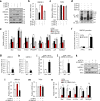
. 2020 Sep;27(9):2537-2551.
doi: 10.1038/s41418-020-0520-5. Epub 2020 Mar 23.

HPV-mediated nuclear export of HP1γ drives cervical tumorigenesis by downregulation of p53

Affiliations

HPV-mediated nuclear export of HP1γ drives cervical tumorigenesis by downregulation of p53

Sang Ah Yi et al. Cell Death Differ. 2020 Sep.

Abstract

E6 oncoprotein derived from high-risk human papillomavirus (HPV) drives the development of cervical cancer through p53 degradation. Because cervical cancer therapies to inactivate HPV or E6 protein are not available, alternative strategies are required. Here, we show that HPV-mediated nuclear export of human heterochromatin protein 1γ (HP1γ) reduces the stability of p53 through UBE2L3-mediated p53 polyubiquitination during cervical cancer progression. In general, HP1 plays a key role in heterochromatin formation and transcription in the nucleus. However, our immunostaining data showed that the majority of HP1γ is localized in the cytoplasm in HPV-mediated cervical cancer. We found that HPV E6 protein drives unusual nuclear export of HP1γ through the interaction between the NES sequence of HP1γ and exportin-1. The mutation of the NES sequence in HP1γ led to nuclear retention of HP1γ and reduced cervical cancer cell growth and tumor generation. We further discovered that HP1γ directly suppresses the expression of UBE2L3 which drives E6-mediated proteasomal degradation of p53 in cervical cancer. Downregulation of UBE2L3 by overexpression of HP1γ suppressed UBE2L3-dependent p53 degradation-promoting apoptosis of cervical cancer cells. Our findings propose a useful strategy to overcome p53 degradation in cervical cancer through the blockage of nuclear export of HP1γ.


Conflict of interest statement

The authors declare that they have no conflict of interest.

Figures
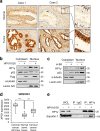
Fig. 1
Fig. 1. Aberrant nuclear export of HP1γ is induced by high-risk HPV E6 protein.
a Immunohistochemical staining of HP1γ protein in cervical tissues from normal specimens (upper) and HPV-positive cervical adenocarcinoma patients (lower). b Immunoblot analysis of cytoplasmic and nuclear extracts from C33A cells expressing Flag-HPV16 E6. c Immunoblot analysis of cytoplasmic and nuclear extracts from SiHa cells expressing siRNA of HPV16 E6. d Box-and-Whisker plot of relative XPO1 expression in cervical cancer cell lines (n = 9), normal tissues (n = 24), and cervical cancer tissues (n = 28) (GDS3233). e Immunoblot analysis of HP1γ immunoprecipitates (IP) and whole-cell lysates (WCL) from C33A cells expressing Flag-HPV16 E6.
Fig. 2
Fig. 2. Exportin-1-mediated nuclear export of HP1γ contributes to p53 degradation.
a Immunoblot analysis of HP1γ immunoprecipitates (IP) and whole-cell lysates (WCL) from HeLa cells treated with or without LMB. b Immunoblot analysis of cytoplasmic and nuclear extracts from SiHa cells treated with or without LMB. c Immunoblot analysis of SiHa cells treated with or without LMB. d SiHa cells were treated with or without LMB, followed by ChIP-qPCR analysis with a p53 antibody in the promoter regions of p53 target genes. e Immunoblot analysis and the mRNA levels of p53 target genes in SiHa cells expressing siRNA of p53 in the presence or absence of LMB. f Representation of HP1γ protein and partial CD sequence containing nuclear export signal (NES) sequences (L150 and L152) which are substituted with alanine in the mutant vector, HP1γ AA. g Immunoblot analysis of HP1γ immunoprecipitates (IP) and whole-cell lysates (WCL) from SiHa cells expressing GFP-HP1γ WT or AA mutant. h Immunofluorescence image of GFP-HP1γ WT or AA mutant expressed in HeLa (left) and SiHa (right) cells. i Immunoblot analysis of cytoplasmic and nuclear extracts from SiHa cells expressing GFP-HP1γ WT or AA mutant. j Immunoblot analysis of SiHa cells expressing GFP-HP1γ WT or AA mutant. k The mRNA levels of p53 target genes in SiHa cells expressing GFP-HP1γ WT or AA mutant. Data are presented as the mean ± SEM (n = 3). *P < 0.05; **P < 0.01; ***P < 0.001.
Fig. 3
Fig. 3. Inhibiting the nuclear export of HP1γ promotes p53 effects in cervical cancer cells.
a Immunoblot analysis of SiHa cells expressing doxycycline-induced HP1 WT or AA treated with or without doxycycline for 48 h. b Clonogenic assay of SiHa cells expressing doxycycline-induced HP1 WT or AA treated with or without doxycycline for 20 days. c–e In vivo tumorigenesis assay of SiHa cells was performed. SiHa cells expressing HP1 WT or AA were injected into the flank of nude mice. f Immunohistochemical staining of HP1γ protein in tumor tissues derived from mice injected with SiHa cells expressing HP1 WT or AA. Data are presented as the mean ± SD (n = 6). *P < 0.05; **P < 0.01; ***P < 0.001.
Fig. 4
Fig. 4. HP1 isoforms regulate UBE2L3-dependent p53 ubiquitination in cervical cancer cells.
a Model for UBE2L3-mediated p53 degradation upon high-risk HPV infection. b Pearson’s correlation analysis in different areas of cervical cancer tumors (GDS2416) (n = 33). c Immunoblot analysis of HeLa cells expressing siRNA of HP1 isoforms. d Immunoblot analysis of p53 immunoprecipitates (IP) and whole-cell lysates (WCL) from HeLa cells expressing siRNA of HP1 isoforms after MG-132 treatment. e The mRNA levels of TP53 in HeLa cells expressing siRNA of HP1 isoforms. f The mRNA levels of p53 target genes in HeLa cells expressing siRNA of HP1 isoforms. Data are presented as the mean ± SEM (n = 3). *P < 0.05, **P < 0.01, ***P < 0.001.
Fig. 5
Fig. 5. HP1γ negatively regulates p53 ubiquitination by suppressing UBE2L3 expression.
a Immunoblot analysis of HeLa cells co-expressing siRNA of HP1 isoforms. b The mRNA levels of UBE2L3 in HeLa cells co-expressing siRNA of HP1 isoforms. c The mRNA levels of TP53 in HeLa cells co-expressing siRNA of HP1 isoforms. d Immunoblot analysis of p53 immunoprecipitates (IP) and whole-cell lysates (WCL) from HeLa cells co-expressing siRNA of HP1 isoforms after MG-132 treatment. e The mRNA levels of p53 target genes in HeLa cells co-expressing siRNA of HP1 isoforms. f HeLa cells were transfected with siRNA of HP1β, followed by ChIP-qPCR analysis for HP1α and HP1γ antibodies in the promoter region of the UBE2L3 gene. g SiHa cells were treated with or without LMB, followed by ChIP-qPCR analysis with an HP1γ antibody in the promoter region of the UBE2L3 gene. h The mRNA levels of UBE2L3 in SiHa cells treated with or without LMB. i SiHa cells were transfected with GFP-HP1γ WT or AA mutant vectors, followed by ChIP-qPCR analysis with a GFP antibody in the promoter region of UBE2L3 gene. j The mRNA levels of UBE2L3 in SiHa cells expressing GFP-HP1γ WT or AA mutant. k Immunoblot analysis of HeLa cells co-expressing siRNA of HP1γ and UBE2L3. l The mRNA levels of UBE2L3 in HeLa cells co-expressing siRNA of HP1γ and UBE2L3. m The mRNA levels of TP53 in HeLa cells co-expressing siRNA of HP1γ and UBE2L3. n The mRNA levels of p53 target genes in HeLa cells co-expressing siRNA of HP1γ and UBE2L3. Data are presented as the mean ± SEM (n = 3). *P < 0.05, **P < 0.01, ***P < 0.001.
Fig. 6
Fig. 6. Overexpression of HP1γ promotes p53 signaling by directly suppressing UBE2L3 expression.
a Immunoblot analysis of HeLa cells expressing GFP-HP1γ. b The mRNA levels of UBE2L3 in HeLa cells expressing GFP-HP1γ. c Immunoblot analysis of p53 immunoprecipitates (IP) and whole-cell lysates (WCL) from HeLa cells expressing GFP-HP1γ after MG-132 treatment. d The mRNA levels of TP53 in HeLa cells expressing GFP-HP1γ. e ChIP-qPCR analysis with a p53 antibody in the promoter regions of p53 target genes in HeLa cells expressing GFP-HP1γ. f The mRNA levels of p53 target genes in HeLa cells expressing GFP-HP1γ. g Immunoblot analysis of HeLa cells expressing GFP-HP1γ. h The growth rate of HeLa expressing GFP-HP1γ. i Schematic representation of HP1γ protein and partial CD sequence containing V32. j HeLa cells were transfected with GFP-HP1γ WT or mutant (V32A, I165E, or W174A) vectors, followed by ChIP-qPCR analysis with a GFP antibody in the promoter regions of the UBE2L3 gene. k The mRNA levels of UBE2L3 in HeLa cells expressing GFP-HP1γ wild-type (WT) or V32A mutant. l Immunoblot analysis of HeLa cells expressing GFP-HP1γ WT or V32A mutant. m The mRNA levels of p53 target genes in HeLa cells expressing GFP-HP1γ WT or V32A mutant. Data are presented as the mean ± SEM (n = 3). *P < 0.05; **P < 0.01; ***P < 0.001.
Fig. 7
Fig. 7. Molecular model describing p53 regulation by HP1γ-dependent UBE2L3 suppression.
a Upon high-risk HPV infection, the E6 oncoprotein of HPV interacts with p53 and E6AP to induce polyubiquitination and proteasomal degradation of p53. Moreover, HPV infection induces exportin-1-mediated nuclear export of HP1γ, thereby allowing plenty of UBE2L3 to be expressed, which results in p53 degradation in an HPV-specific manner. b When the NES mutant HP1γ (HP1γ AA) is overexpressed artificially in cervical cancer cells, exportin-1 fails to transport HP1γ AA to the cytoplasm and HPV-specific degradation of p53 is impaired due to UBE2L3 suppression by nuclear HP1γ.

Similar articles

Cited by

References

    1. Holowaty P, Miller AB, Rohan T, To T. Natural dysplasia of the uterine cervix. J Nat Cancer Inst. 1999;91:252–8. - PubMed
    1. Dürst M, Gissmann L, Ikenberg H, zur Hausen H. A papillomavirus DNA from a cervical carcinoma and its pre valence in cancer biopsy samples from different geographic regions. Proc Natl Acad Sci USA. 1983;80:3812–5. - PMC - PubMed
    1. Smith JS, Lindsay L, Hoots B, Keys J, Franceschi S, Winer R, et al. Human papillomavirus type distribution in invasive cervical cancer and high-grade cervical lesions: a meta-analysis update. Int J Cancer. 2007;121:621–32. - PubMed
    1. Hildesheim A, Herrero R, Wacholder S, Rodriguez AC, Solomon D, Bratti MC, et al. Effect of human papillomavirus 16/18 L1 viruslike particle vaccine among young women with preexisting infection: a randomized trial. JAMA. 2007;298:743–53. - PubMed
    1. Moore DH, Tian C, Monk BJ, Long HJ, Omura GA, Bloss JD. Prognostic factors for response to cisplatin-based chemotherapy in advanced cervical carcinoma: a Gynecologic Oncology Group Study. Gynecol Oncol. 2010;116:44–9. - PMC - PubMed

Publication types

MeSH terms