Review
. 2020 Apr;17(4):335-346.
doi: 10.1038/s41423-020-0399-6. Epub 2020 Mar 19.

The pathogenic role of innate lymphoid cells in autoimmune-related and inflammatory skin diseases

Affiliations
Review

The pathogenic role of innate lymphoid cells in autoimmune-related and inflammatory skin diseases

Suqing Zhou et al. Cell Mol Immunol. 2020 Apr.

Abstract

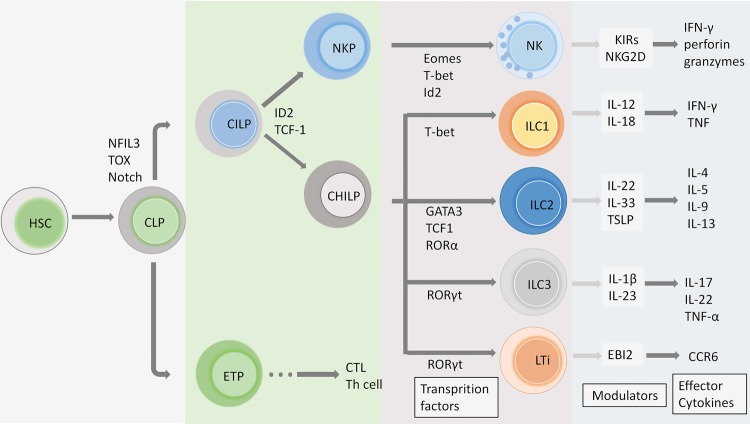
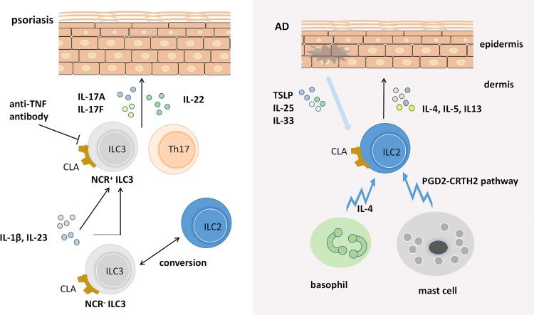
Innate lymphoid cells (ILCs), as an important component of the innate immune system, arise from a common lymphoid progenitor and are located in mucosal barriers and various tissues, including the intestine, skin, lung, and adipose tissue. ILCs are heterogeneous subsets of lymphocytes that have emerging roles in orchestrating immune response and contribute to maintain metabolic homeostasis and regulate tissue inflammation. Currently, more details about the pathways for the development and differentiation of ILCs have largely been elucidated, and cytokine secretion and downstream immune cell responses in disease pathogenesis have been reported. Recent research has identified that several distinct subsets of ILCs at skin barriers are involved in the complex regulatory network in local immunity, potentiating adaptive immunity and the inflammatory response. Of note, additional studies that assess the effects of ILCs are required to better define how ILCs regulate their development and functions and how they interact with other immune cells in autoimmune-related and inflammatory skin disorders. In this review, we will distill recent research progress in ILC biology, abnormal functions and potential pathogenic mechanisms in autoimmune-related skin diseases, including systemic lupus erythematosus (SLE), scleroderma and inflammatory diseases, as well as psoriasis and atopic dermatitis (AD), thereby giving a comprehensive review of the diversity and plasticity of ILCs and their unique functions in disease conditions with the aim to provide new insights into molecular diagnosis and suggest potential value in immunotherapy.

Keywords: ILC; SLE; atopic dermatitis; autoimmunity; psoriasis; scleroderma.


Conflict of interest statement

The authors declare no competing interests.

Figures

Fig. 1
Fig. 1
The development of ILCs. ILCs differentiate from hematopoietic stem cells (HSCs) and then generate CLPs in the bone marrow at the early stage. CLPs are a cellular source of early T cell progenitors (ETPs) and CILPs, and ILCs and NK cells share some notable parallels with T cell lineage commitment. The transcriptional factor Id2 and T cell factor 1 (TCF-1) are involved in CILP regulation and differentiation into NKPs and CHILPs. These transcription factors, such as T-bet, GATA3, and RORγt, described above, drive the generation of different ILC subsets. Downstream cytokines of the ILC group participate in orchestrating the immune network, and several modulators have been proven to regulate ILC functions in disease conditions
Fig. 2
Fig. 2
The pathogenic role of ILCs in psoriasis and AD. ILC3s are elevated in the skin of psoriasis patients. RORγt+ ILC3s secret IL-17A, IL-17F, and IL-22 and become a critical factor in promoting psoriasiform lesions. However, anti-TNF treatment can decrease the elevated level of circulating NCR+ ILC3s. Notably, ILCs can change phenotype. NCR ILC3s in healthy skin can be converted to NCR+ ILC3s when cultured with IL-1β plus IL23. In addition, ILC2s play a central role in the skin in AD in both mouse models and humans through the secretion of IL-4, IL-5, and IL-13. Emerging evidence shows that IL-25, IL-33, and TSLP are all involved in the modulation of ILC2s in inflamed skin. In addition, ILC2s interact with mast cells and basophils and participate in driving pathology in atopic dermatitis through cell interactions

References

    1. Minton K. Innate lymphoid cells: ILC diversity maintained by microbiota. Nat. Rev. Immunol. 2016;16:593. doi: 10.1038/nri.2016.101. - DOI - PubMed
    1. Klose CS, Artis D. Innate lymphoid cells as regulators of immunity, inflammation and tissue homeostasis. Nat. Immunol. 2016;17:765–774. doi: 10.1038/ni.3489. - DOI - PubMed
    1. Tait WE, Artis D. Innate lymphoid cells: Balancing immunity, inflammation, and tissue repair in the intestine. Cell Host Microbe. 2012;12:445–457. doi: 10.1016/j.chom.2012.10.003. - DOI - PMC - PubMed
    1. Tugues S, et al. Innate lymphoid cells as regulators of the tumor microenvironment. Semin. Immunol. 2019;41:101270. doi: 10.1016/j.smim.2019.03.002. - DOI - PubMed
    1. Spits H, Cupedo T. Innate lymphoid cells: Emerging insights in development, lineage relationships, and function. Annu. Rev. Immunol. 2012;30:647–675. doi: 10.1146/annurev-immunol-020711-075053. - DOI - PubMed

Publication types