Review
. 2020 Mar 19;9(3):750.
doi: 10.3390/cells9030750.

A Role for Endoplasmic Reticulum Stress in Intracerebral Hemorrhage

Affiliations
Review

A Role for Endoplasmic Reticulum Stress in Intracerebral Hemorrhage

Shaik Ismail Mohammed Thangameeran et al. Cells. .

Abstract

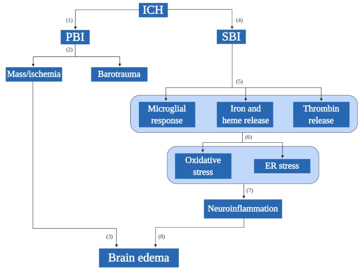
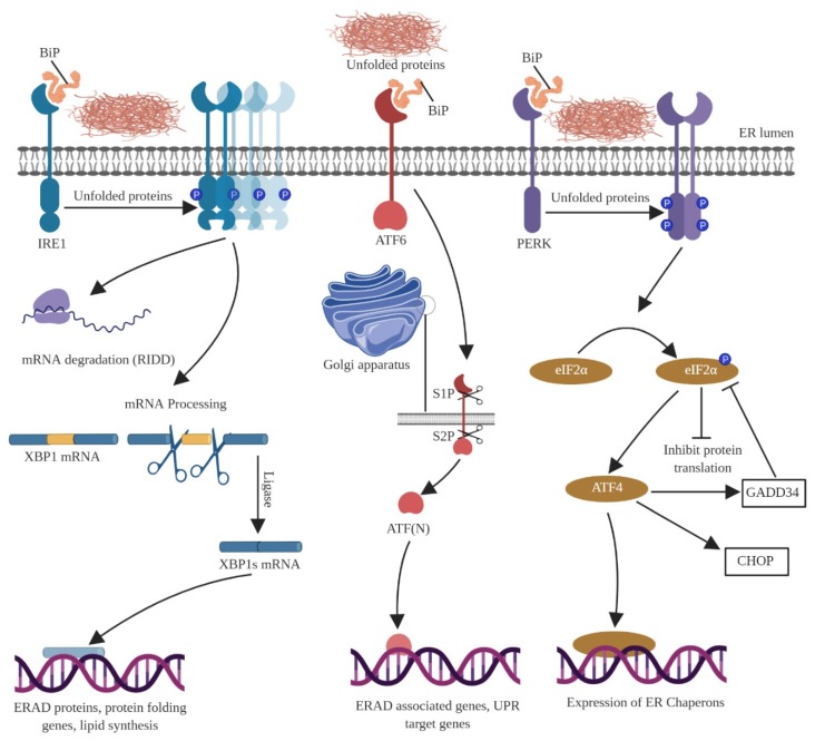
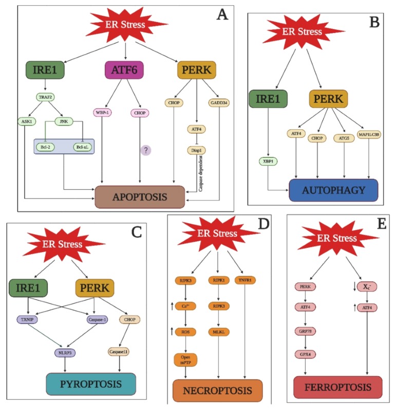
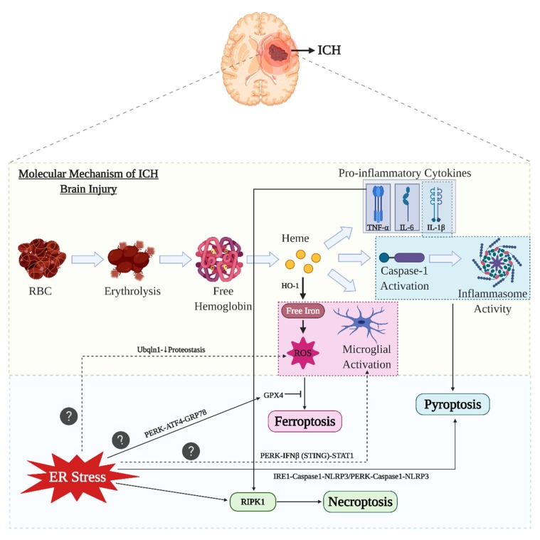
The endoplasmic reticulum (ER) is an intracellular organelle that performs multiple functions, such as lipid biosynthesis, protein folding, and maintaining intracellular calcium homeostasis. Thus, conditions wherein the ER is unable to fold proteins is defined as ER stress, and an inbuilt quality control mechanism, called the unfolded protein response (UPR), is activated during ER stress, which serves as a recovery system that inhibits protein synthesis. Further, based on the severity of ER stress, the response could involve both proapoptotic and antiapoptotic phases. Intracerebral hemorrhage (ICH) is the second most common subtype of cerebral stroke and many lines of evidence have suggested a role for the ER in major neurological disorders. The injury mechanism during ICH includes hematoma formation, which in turn leads to inflammation, elevated intracranial pressure, and edema. A proper understanding of the injury mechanism(s) is required to effectively treat ICH and closing the gap between our current understanding of ER stress mechanisms and ICH injury can lead to valuable advances in the clinical management of ICH.

Keywords: ER stress; ferroptosis; intracerebral hemorrhage; necroptosis; neuroinflammation; pyroptosis; unfolded protein response.


Conflict of interest statement

The authors declare no conflict of interest.

Figures

Figure 1
Figure 1
Pathology of intracerebral hemorrhage (ICH). Primary brain injury (PBI; 1 is the initial ictus that happens due to physical damage (2), which in turn leads to the mass effect or the ischemic effect that increases the volume of the hematoma (barotrauma). The mass/ischemic effect can directly influence the increase of the brain edema (3). The pathophysiology of ICH involves other series of steps called (4) the secondary brain injury (SBI, which is the long-term sequential aftereffects following ICH. The SBI events include (5) the release of thrombin, which acts as the initial response to repair the damaged site following which the host microglia come into play to engulf the erythrocytes. Since the initial ictus causes a clot in the site, there is an excessive release of clot components; majorly free iron and heme. (6) The course of events after SBI initiates the oxidative stress and ER stress consecutively contributing to (7) the neuroinflammation. (8) The progression of SBI events eventually leads to a brain edema. ICH: intracerebral hemorrhage, PBI: primary brain injury, SBI: secondary brain injury.
Figure 2
Figure 2
Clinical management of ICH. The steps involved in the clinical management of ICH are initial screening to analyze the clinical conditions followed by the screening procedure to understand the treatment needs of the patient. Based on the first three processes the clinical management is decided between surgical procedures or conservative management.
Figure 3
Figure 3
Cell death pathways in ICH. ICH includes all the three conventional forms of cell death (necrosis, apoptosis, and autophagy). Apart from regular cell death pathway, ICH also involves pyroptosis, necroptosis, and ferroptosis.
Figure 4
Figure 4
The molecular pathways of the unfolded protein response (UPR. During ER stress the three ER transducers (IRE1, PERK, and ATF6) tend to get activated and start the UPR mechanism to counteract the ER stress and to bring back the homeostasis. Failure to negate the ER stress will result in cell death. IRE1 undergoes oligomerization in response to unfolded proteins and the BiP gets dissociated from IRE1. IRE1 undergoes either mRNA degradation or mRNA processing with the help of XBP-1, which triggers the production of protein folding genes thereby mitigating ER stress. On the other hand, ATF6 gets into the secretory pathway inside the Golgi apparatus wherein the S1P and S2P cleave the ATF6. ATF6 fragments then translocate into the nucleus, which act as a transcription factor to generate UPR target genes. PERK first phosphorylates itself and also phosphorylates eIF2α thereby inactivating eIF2B, which further inhibits the translation of serine 51. This process leads to the translation of ATF4 producing ER chaperones.
Figure 5
Figure 5
ER stress and cell death. A depiction of ER stress related signal transduction pathways that is responsible for apoptosis, autophagy, pyroptosis, necroptosis, and ferroptosis.
Figure 6
Figure 6
ER stress and ICH-related cell death pathways. The molecular mechanisms involved after ICH namely microglial activation and ROS production directly leads to ferroptosis by inhibiting GPX4. The role for ER stress to actively participate in ferroptosis is still a question. However, the role of ER stress is demonstrated to mediate GPX4 through its PERK arm. The caspase-1 activation and inflammasome activities after ICH leads to pyroptosis. The interrelation of the ER stress with pyroptosis involves both the IRE1 and PERK arms. The proinflammatory cytokine release responsible for neuroinflammation activates RIPK1 to further induce necroptosis. There is evidence that ER stress directly activating RIPK1 thereby causing necroptosis. However, the direct relationship with all these three ICH cell death pathways with that or ER stress is still not elucidated. However, future studies should be focused on these three unconventional ICH related cell death pathways wherein ER stress plays a key role. The role of proteostasis and PERK arm is also further explained to have a relationship with oxidative stress and microglial activation.

References

    1. Xi G., Keep R.F., Hoff J.T. Mechanisms of brain injury after intracerebral haemorrhage. Lancet Neurol. 2006;5:53–63. doi: 10.1016/S1474-4422(05)70283-0. - DOI - PubMed
    1. An S.J., Kim T.J., Yoon B.W. Epidemiology, risk factors, and clinical features of intracerebral hemorrhage: An update. J. Stroke. 2017;19:3–10. doi: 10.5853/jos.2016.00864. - DOI - PMC - PubMed
    1. Hsieh F.I., Chiou H.Y. Stroke: Morbidity, risk factors, and care in Taiwan. J. Stroke. 2014;16:59–64. doi: 10.5853/jos.2014.16.2.59. - DOI - PMC - PubMed
    1. Powers W.J. Intracerebral hemorrhage and head trauma: Common effects and common mechanisms of injury. Stroke. 2010;41:S107–S110. doi: 10.1161/STROKEAHA.110.595058. - DOI - PMC - PubMed
    1. Keep R.F., Hua Y., Xi G. Intracerebral haemorrhage: Mechanisms of injury and therapeutic targets. Lancet Neurol. 2012;11:720–731. doi: 10.1016/S1474-4422(12)70104-7. - DOI - PMC - PubMed

Publication types

LinkOut - more resources