. 2020 Mar 19;10(3):517.
doi: 10.3390/ani10030517.

Calcium Electroporation of Equine Sarcoids

Affiliations

Calcium Electroporation of Equine Sarcoids

Stine K Frandsen et al. Animals (Basel). .

Abstract

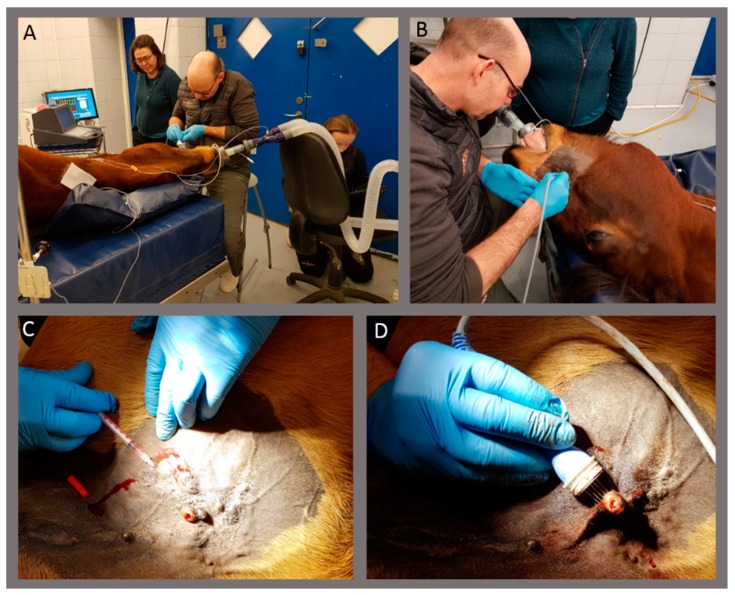
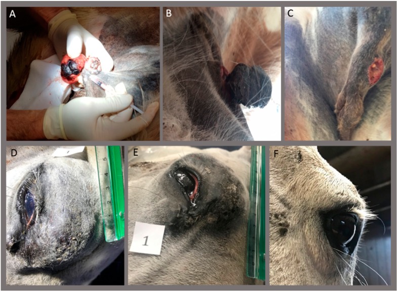
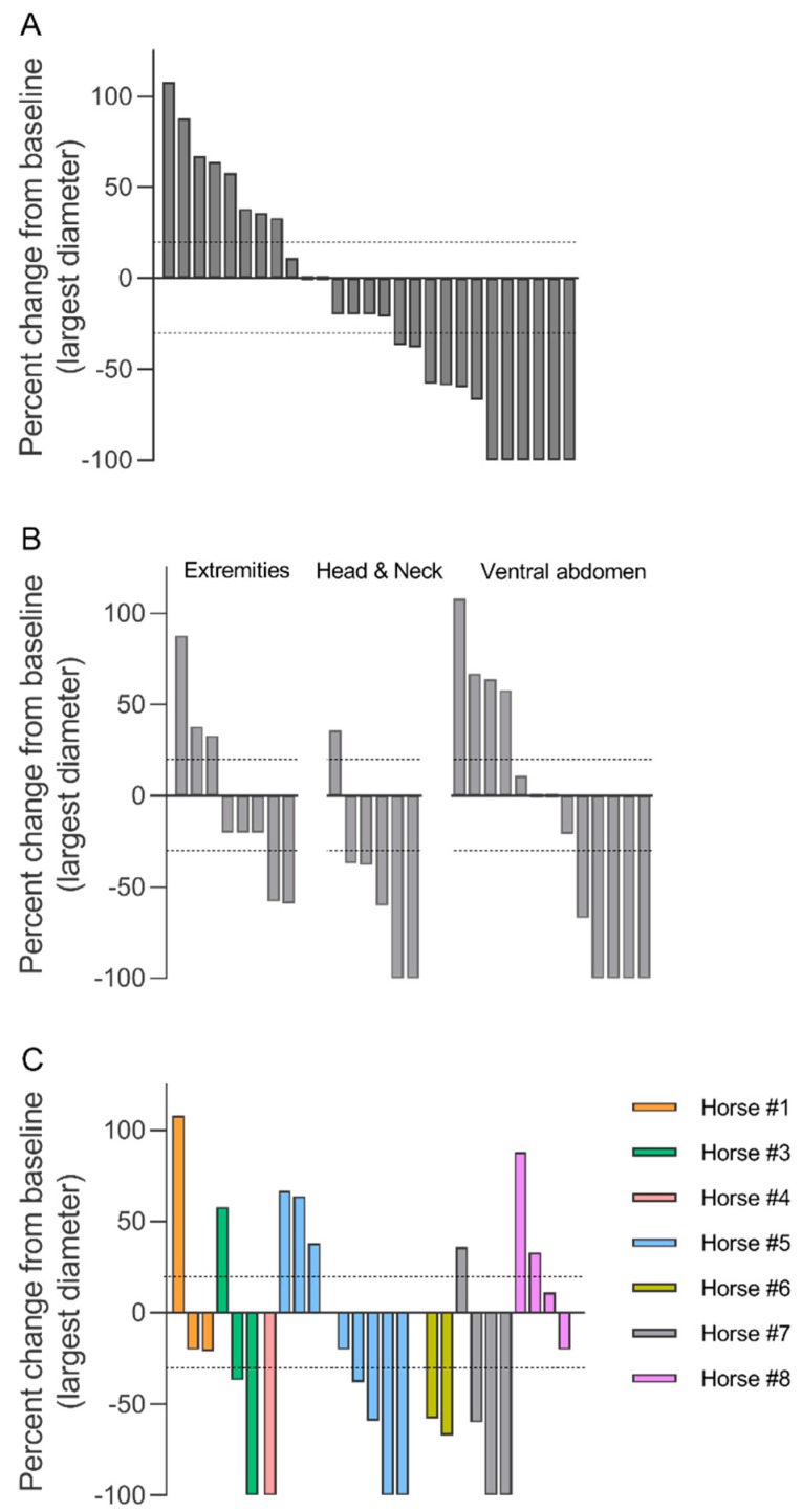
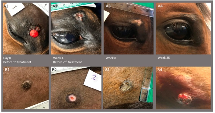
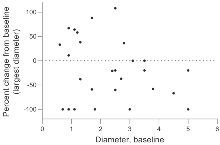
Sarcoids are common equine skin tumors where the risk of recurrence after treatment is high, and better treatment options are warranted. Calcium electroporation is a novel anti-cancer treatment where lethally high calcium concentrations are introduced into the cells by electroporation, a method where short high-voltage pulses induce transient permeabilization of the cell membrane. This study investigated the safety and long-term response of calcium electroporation on sarcoids. Thirty-two sarcoids in eight horses were included. The study suggested that calcium electroporation is a safe and feasible treatment for sarcoids, including inoperable sarcoids. Horses were treated once (2/8) or twice (6/8) under general anesthesia, where sarcoids were injected with 220 mM calcium chloride followed by electroporation with 8 pulses of 100 μs, 1 kV/cm, and 1 Hz. Biopsies were taken prior to treatment. The sarcoid size was monitored for 12-38 weeks after the first treatment. Complete response was observed in 22% (6/27) of treated sarcoids, and partial response in 22% (6/27), giving a 44% total response. Treatment efficacy did not appear to be related to location, type, or size. In all non-biopsied lesions, a complete response was seen (4/4). In conclusion, in this small study, 44% of sarcoids responded with 22% of sarcoids disappearing.

Keywords: biopsy; calcium electroporation; equine; horse; response; sarcoid.


Conflict of interest statement

A patent, “Therapeutic applications of calcium electroporation to effectively induce tumor necrosis”, has been granted and licensed (co-inventors S.K. Frandsen and J. Gehl). The sponsors had no role in the design, execution, interpretation, or writing of the study.

Figures

Figure 1
Figure 1
Images of the treatment. Horses were treated in general anesthesia (A,B). Calcium chloride solution was injected in the sarcoid (C), followed by electroporation using needle electrodes (D).
Figure 2
Figure 2
Complete response after calcium electroporation. Horse #4 (AC). Pendulating, crusted, non-pigmented sarcoid on the ventral abdomen where the base of the sarcoid was treated once with calcium electroporation (A). Four days after the treatment, the sarcoid was starting to fall off (B) and completely gone 6 days after treatment (C). No regrowth was seen afterward (38 weeks after treatment). A biopsy was performed on the top of the sarcoid, verifying the diagnose. Horse #7 (D–F). Sarcoid on the eyelid was treated twice with calcium electroporation with 8 weeks between the treatments. (D) before the first treatment and (E) before the second treatment. Complete response was observed at follow-up 32 weeks after the first treatment (F) - notice the photo is from a caudo-lateral angle. A biopsy was performed before the first and second treatment, with both biopsies verifying the diagnosis.
Figure 3
Figure 3
Sarcoid response to calcium electroporation. Percent change in the longest diameter at the last follow-up (12–38 weeks after first treatment). The response of all sarcoids (A), showing 8 sarcoids with progressive disease (above 20% increase; dotted line), 7 sarcoids with limited change (up to 30% decrease but not above 20% increase), 6 sarcoids with partial response (more than 30% decrease; dotted line), and 6 sarcoids with complete response (100% decrease). Data is also shown grouped by location of sarcoid (B) and individual horse (C).
Figure 4
Figure 4
Images of two sarcoids on horse #3 before the first treatment, before second treatment at week 4, and at follow-up 8 and 25 weeks after first treatment. Top row (A1–4) shows sarcoid placed on the eyelid with complete response (A4; only scar tissue left), and bottom row (B1–4) shows sarcoid placed on the ventral abdomen with the growth of the sarcoid 25 weeks after the first treatment (B4). Note that biopsy of the ventral abdomen sarcoid was performed before images were taken at week 4 (B2).
Figure 5
Figure 5
The sarcoid response related to sarcoid size. Percent change of sarcoid size (at the last follow-up) after calcium electroporation treatment(s) on the y-axis related to the size of the sarcoid before treatment on the x-axis. There does not seem to be a relation between sarcoid size and the likelihood of treatment success with dots evenly spread across the graph.
Figure 6
Figure 6
Histological analysis to verify the diagnosis. Light microscope images of H&E sections of fibroblastic sarcoid (40X magnification) (A) and verrucous sarcoid (20X magnification) (B).

References

    1. Bergvall K.E. Sarcoids. Vet. Clin. N. Am.-Equine. 2013;29:657–671. doi: 10.1016/j.cveq.2013.09.002. - DOI - PubMed
    1. Taylor S., Haldorson G. A review of equine sarcoid. Equine Vet. Educ. 2013;25:210–216. doi: 10.1111/j.2042-3292.2012.00411.x. - DOI
    1. Falk H., Matthiessen L.W., Wooler G., Gehl J. Calcium electroporation for treatment of cutaneous metastases; a randomized double-blinded phase II study, comparing the effect of calcium electroporation with electrochemotherapy. Acta Oncol. 2017;57:311–319. doi: 10.1080/0284186X.2017.1355109. - DOI - PubMed
    1. Frandsen S.K., Gissel H., Hojman P., Tramm T., Eriksen J., Gehl J. Direct therapeutic applications of calcium electroporation to effectively induce tumor necrosis. Cancer Res. 2012;72:1336–1341. doi: 10.1158/0008-5472.CAN-11-3782. - DOI - PubMed
    1. Frandsen S.K., Kruger M.B., Mangalanathan U.M., Tramm T., Mahmood F., Novak I., Gehl J. Normal and malignant cells exhibit differential responses to calcium electroporation. Cancer Res. 2017;77:4389–4401. doi: 10.1158/0008-5472.CAN-16-1611. - DOI - PubMed

LinkOut - more resources