

Mosquito Cell-Derived Japanese Encephalitis Virus-Like Particles Induce Specific Humoral and Cellular Immune Responses in Mice
- PMID: 32204533
- PMCID: PMC7150764
- DOI: 10.3390/v12030336
Mosquito Cell-Derived Japanese Encephalitis Virus-Like Particles Induce Specific Humoral and Cellular Immune Responses in Mice
Abstract
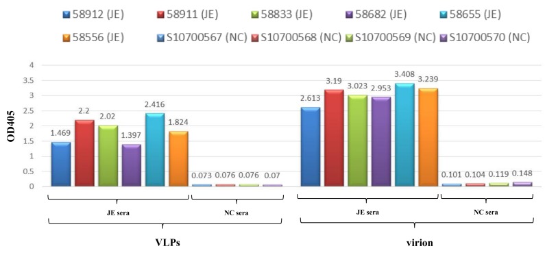
The Japanese encephalitis virus (JEV) is the major cause of an acute encephalitis syndrome in many Asian countries, despite the fact that an effective vaccine has been developed. Virus-like particles (VLPs) are self-assembled multi-subunit protein structures which possess specific epitope antigenicities related to corresponding native viruses. These properties mean that VLPs are considered safe antigens that can be used in clinical applications. In this study, we developed a novel baculovirus/mosquito (BacMos) expression system which potentially enables the scalable production of JEV genotype III (GIII) VLPs (which are secreted from mosquito cells). The mosquito-cell-derived JEV VLPs comprised 30-nm spherical particles as well as precursor membrane protein (prM) and envelope (E) proteins with densities that ranged from 30% to 55% across a sucrose gradient. We used IgM antibody-capture enzyme-linked immunosorbent assays to assess the resemblance between VLPs and authentic virions and thereby characterized the epitope specific antigenicity of VLPs. VLP immunization was found to elicit a specific immune response toward a balanced IgG2a/IgG1 ratio. This response effectively neutralized both JEV GI and GIII and elicited a mixed Th1/Th2 response in mice. This study supports the development of mosquito cell-derived JEV VLPs to serve as candidate vaccines against JEV.
Keywords: Japanese encephalitis virus; baculovirus; mosquito; virus-like particles.
Conflict of interest statement
The authors declare no conflict of interest. The funders had no role in the design of the study; in the collection, analyses, or interpretation of data; in the writing of the manuscript, or in the decision to publish the results.
Figures
Similar articles
-
Advancements in nanoparticle-based vaccine development against Japanese encephalitis virus: a systematic review.Front Immunol. 2024 Dec 20;15:1505612. doi: 10.3389/fimmu.2024.1505612. eCollection 2024. Front Immunol. 2024. PMID: 39759527 Free PMC article.
-
Enhanced immune responses against Japanese encephalitis virus using recombinant adenoviruses coexpressing Japanese encephalitis virus envelope and porcine interleukin-6 proteins in mice.Virus Res. 2016 Aug 15;222:34-40. doi: 10.1016/j.virusres.2016.05.025. Epub 2016 May 25. Virus Res. 2016. PMID: 27235810
-
Genotype I of Japanese Encephalitis Virus Virus-like Particles Elicit Sterilizing Immunity against Genotype I and III Viral Challenge in Swine.Sci Rep. 2018 May 10;8(1):7481. doi: 10.1038/s41598-018-25596-1. Sci Rep. 2018. PMID: 29748549 Free PMC article.
-
[Generation of Japanese Encephalitis Virus-like Particle Vaccine and Preliminary Evaluation of Its Protective Efficiency].Bing Du Xue Bao. 2016 Mar;32(2):150-5. Bing Du Xue Bao. 2016. PMID: 27396157 Chinese.
-
Virus-like particles: preparation, immunogenicity and their roles as nanovaccines and drug nanocarriers.J Nanobiotechnology. 2021 Feb 25;19(1):59. doi: 10.1186/s12951-021-00806-7. J Nanobiotechnology. 2021. PMID: 33632278 Free PMC article. Review.
Cited by
-
Virus-like Particle Vaccines and Platforms for Vaccine Development.Viruses. 2023 May 2;15(5):1109. doi: 10.3390/v15051109. Viruses. 2023. PMID: 37243195 Free PMC article. Review.
-
Antigenicity and immunogenicity of chikungunya virus-like particles from mosquito cells.Appl Microbiol Biotechnol. 2023 Jan;107(1):219-232. doi: 10.1007/s00253-022-12280-8. Epub 2022 Nov 25. Appl Microbiol Biotechnol. 2023. PMID: 36434113 Free PMC article.
-
Virus-like particles vaccines based on glycoprotein E0 and E2 of bovine viral diarrhea virus induce Humoral responses.Front Microbiol. 2022 Oct 31;13:1047001. doi: 10.3389/fmicb.2022.1047001. eCollection 2022. Front Microbiol. 2022. PMID: 36439839 Free PMC article.
-
Advancements in nanoparticle-based vaccine development against Japanese encephalitis virus: a systematic review.Front Immunol. 2024 Dec 20;15:1505612. doi: 10.3389/fimmu.2024.1505612. eCollection 2024. Front Immunol. 2024. PMID: 39759527 Free PMC article.
-
Fitness adaptations of Japanese encephalitis virus in pigs following vector-free serial passaging.PLoS Pathog. 2024 Aug 26;20(8):e1012059. doi: 10.1371/journal.ppat.1012059. eCollection 2024 Aug. PLoS Pathog. 2024. PMID: 39186783 Free PMC article.
References
-
- Nealon J., Taurel A.F., Yoksan S., Moureau A., Bonaparte M., Quang L.C., Capeding M.R., Prayitno A., Hadinegoro S.R., Chansinghakul D., et al. Serological Evidence of Japanese Encephalitis Virus Circulation in Asian Children From Dengue-Endemic Countries. J. Infect. Dis. 2019;219:375–381. doi: 10.1093/infdis/jiy513. - DOI - PMC - PubMed
Publication types
MeSH terms
Substances
LinkOut - more resources
Full Text Sources
Research Materials
