Review
. 2020 May;27(5):603-608.
doi: 10.1016/j.acra.2020.03.003. Epub 2020 Mar 20.

Coronavirus Disease (COVID-19): Spectrum of CT Findings and Temporal Progression of the Disease

Affiliations
Review

Coronavirus Disease (COVID-19): Spectrum of CT Findings and Temporal Progression of the Disease

Mingzhi Li et al. Acad Radiol. 2020 May.

Abstract

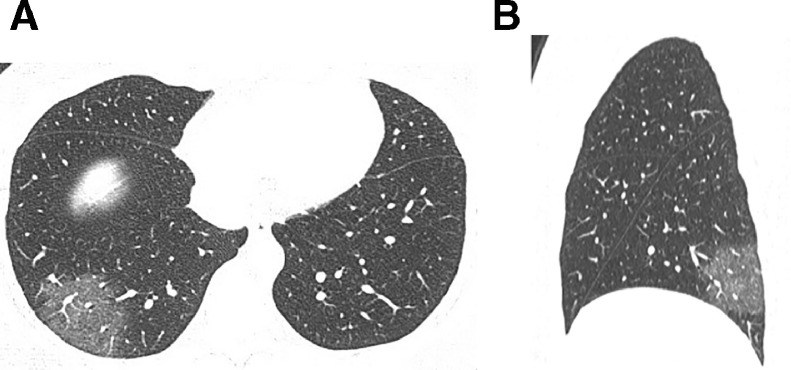
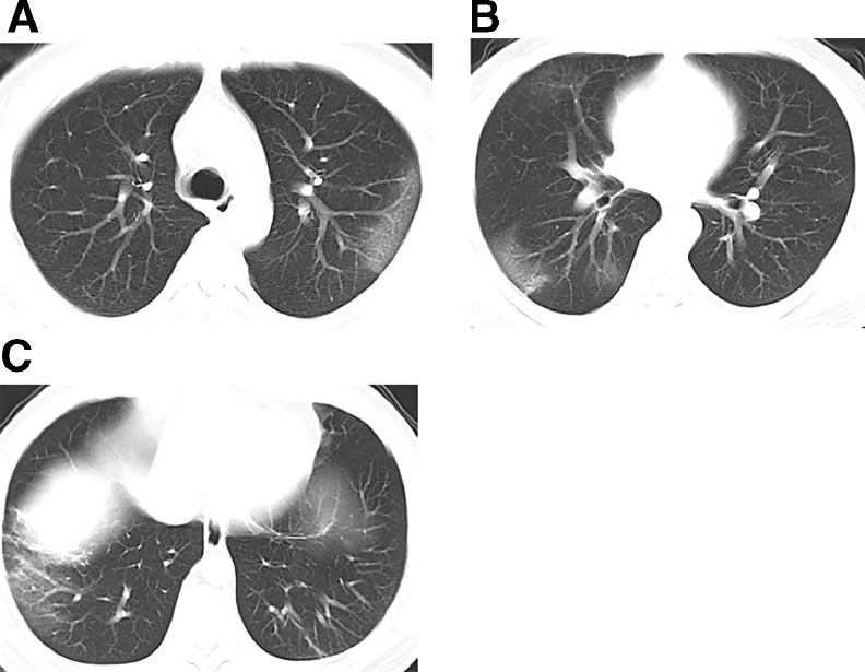
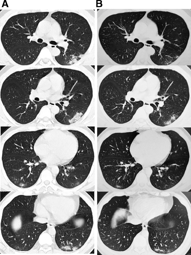
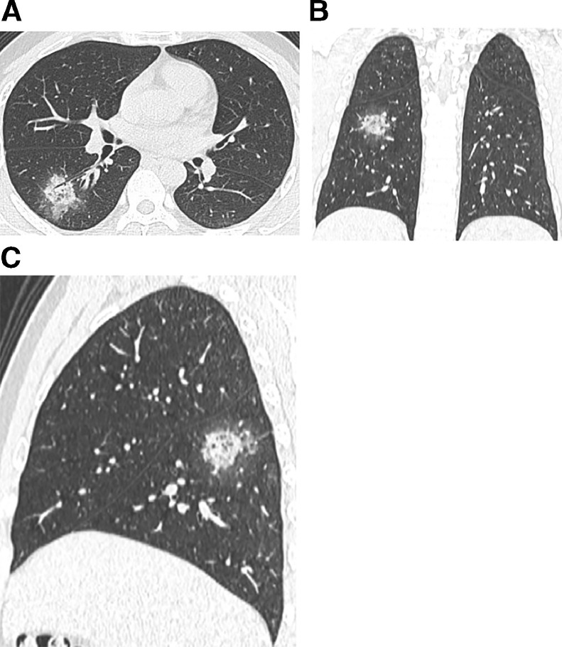
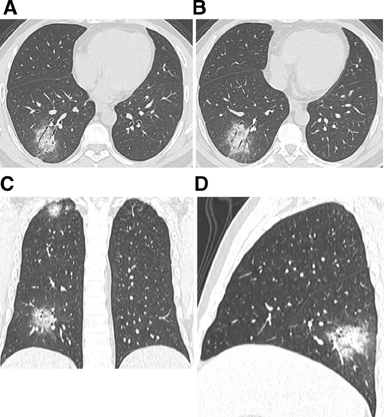
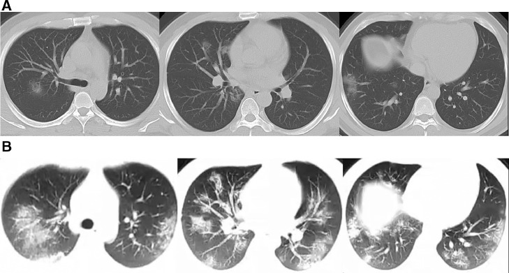
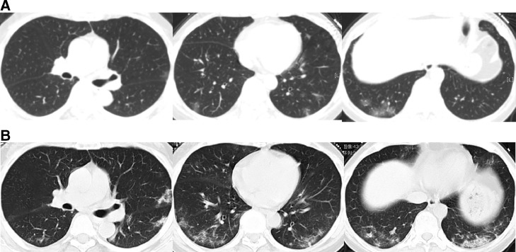
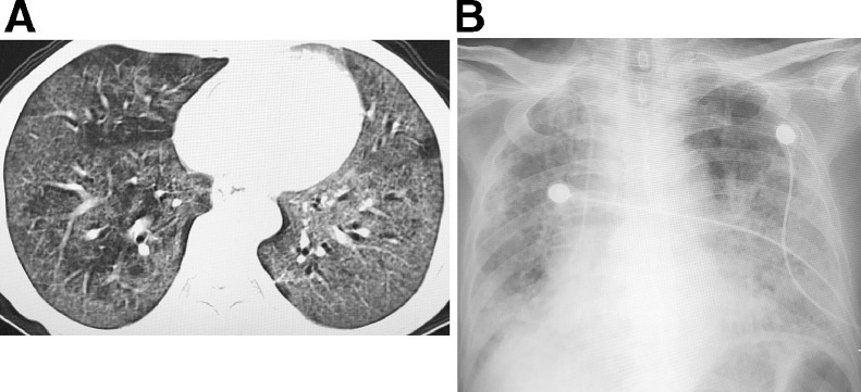
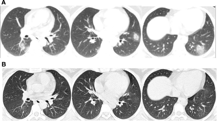
Coronavirus disease is an emerging infection caused by a novel coronavirus that is moving rapidly. High resolution computed tomography (CT) allows objective evaluation of the lung lesions, thus enabling us to better understand the pathogenesis of the disease. With serial CT examinations, the occurrence, development, and prognosis of the disease can be better understood. The imaging can be sorted into four phases: early phase, progressive phase, severe phase, and dissipative phase. The CT appearance of each phase and temporal progression of the imaging findings are demonstrated.

Keywords: 2019-nCoV; COVID-19; Coronavirus; Pneumonia; Tomography; X-ray computed.


Figures

Fig 1
Fig. 1
A 40-year-old female patient with Coronavirus disease (COVID-19) pneumonia. (A) High-resolution CT scan obtained on day 3 from the onset of symptoms shows single ground glass opacities (GGO) in the posterior basal segment of the right lower lobe, within which thickened blood vessel shadows can be observed. (B) The sagittal reconstruction shows that the lesions are located in the posterior basal segment of the right lower lobe, with a pattern of subpleural distribution.
Fig 2
Fig. 2
A 37-year-old male patient with Coronavirus disease (COVID-19) pneumonia. (A–C) Multiple patchy ground glass opacities seen in both lungs, and the lesions show subpleural distribution, inside which the thickened vascular shadows are visible.
Fig 3
Fig. 3
A 40-year-old male patient with Coronavirus disease (COVID-19) pneumonia. (A) High-resolution CT scans obtained on day 5 from onset of symptoms show multiple consolidations in the two lungs with ground glass opacities, which are distributed peripherally in both lungs. (B) Follow-up high-resolution CT scans obtained on day 14 show a reduction of the lesions in both lungs after absorption, leaving fibrous cord-like shadows.
Fig 4
Fig. 4
A 31-year-old male patient with Coronavirus disease (COVID-19) pneumonia. (A) Nodular consolidations and GGO found in the dorsal segment of the right lower lobe, and the lesions distributed along the bronchial bundle, with the thickening of the interlobular septum and intralobular septum, and halo signs seen around the nodules. (B) Coronal reconstruction. (C) Sagittal reconstruction.
Fig 5
Fig 5
A 34-year-old male patient with Coronavirus disease (COVID-19) pneumonia, who is the older brother of the previous patient (Fig 4). (A–B) Patchy high-density shadows along the bronchial bundle in the posterior basal segment of the right lower lobe, around which halo signs can be seen, and air bronchogram can be seen inside the lesions. (C) Coronal reconstruction, from which lesions can also be seen in the apical segment of the right upper lobe. (D) Sagittal reconstruction.
Fig 6
Fig. 6
A 35-year-old male patient with Coronavirus disease (COVID-19) pneumonia. (A) High-resolution CT scans obtained on day 3 from onset of the disease show multiple patchy consolidations and nodular ground-glass opacities, which are mainly distributed along bronchial bundles and subpleural regions. (B) CT images obtained on day 6 from onset of the disease show the progression of the disease, with elevated number, range and densities of the lesions in both lungs.
Fig 7
Fig. 7
A 66-year-old female patient with Coronavirus disease (COVID-19) pneumonia. (A) CT images obtained on day 4 after onset of the disease show scattered patchy ground glass opacities in both lungs, which are distributed peripherally. (B) CT images obtained on day 12 after the onset of the disease show the progression of the disease, with the number, range and densities of the lesions in both lungs increased.
Fig 8
Fig. 8
A 68-year-old female patient with Coronavirus disease (COVID-19) pneumonia. (A) On day 11 from onset of the disease, the CT images show extensive ground-glass opacities and consolidations in both lungs. (B) On day 15 after onset of the disease, the condition deteriorate further, the chest radiography show extensive consolidations in both lungs, and the patient died on day 18.
Fig 9
Fig. 9
A 35-year-old female patient with Coronavirus disease (COVID-19) pneumonia. (A) High-resolution CT scan obtained on day 4 from the onset of symptoms shows multiple nodular consolidations and ground glass opacities in both lungs, which are distributed along the bronchi bundles and the periphery of the lungs. (B) CT reexaminations on day 12 from onset of the disease show that most of the lung lesions have almost been absorbed.
Fig 10
Fig. 10
Application of artificial intelligence (AI) in COVID-19. AI can accurately segment the lesion area and calculate the volume of the lesion, besides, it can also provide follow-up function for the cases, thus enabling the doctors to quickly compare the patient's conditions and evaluate the treatment efficacy. (Color version of figure is available online.)

References

    1. Li Q, Guan X, Wu P. Early transmission dynamics in Wuhan, China, of novel coronavirus-infected pneumonia. N Engl J Med. 2020 doi: 10.1056/NEJMoa2001316. - DOI - PMC - PubMed
    1. Chen N, Zhou M, Dong X. Epidemiological and clinical characteristics of 99 cases of 2019 novel coronavirus pneumonia in Wuhan, China: a descriptive study. Lancet. 2020;395(10223):507–513. - PMC - PubMed
    1. Wu F, Zhao S, Yu B. A new coronavirus associated with human respiratory disease in China. Nature. 2020 doi: 10.1038/s41586-020-2008-3. - DOI - PMC - PubMed
    1. Zhu N, Zhang D, Wang W. A novel coronavirus from patients with pneumonia in China, 2019. N Engl J Med. 2020;382(8):727–733. - PMC - PubMed
    1. Wrapp D, Wang N, Corbett KS. Cryo-EM structure of the 2019-nCoV spike in the prefusion conformation. Science. 2020 doi: 10.1126/science.abb2507. - DOI - PMC - PubMed