

Imaging Features of Coronavirus disease 2019 (COVID-19): Evaluation on Thin-Section CT
- PMID: 32204990
- PMCID: PMC7156158
- DOI: 10.1016/j.acra.2020.03.002
Imaging Features of Coronavirus disease 2019 (COVID-19): Evaluation on Thin-Section CT
Abstract
Rationale and objectives: To retrospectively analyze the chest imaging findings in patients with coronavirus disease 2019 (COVID-19) on thin-section CT.
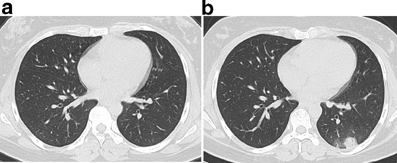
Materials and methods: Fifty-three patients with confirmed COVID-19 infection underwent thin-section CT examination. Two chest radiologists independently evaluated the imaging in terms of distribution, ground-glass opacity (GGO), consolidation, air bronchogram, stripe, enlarged mediastinal lymph node, and pleural effusion.
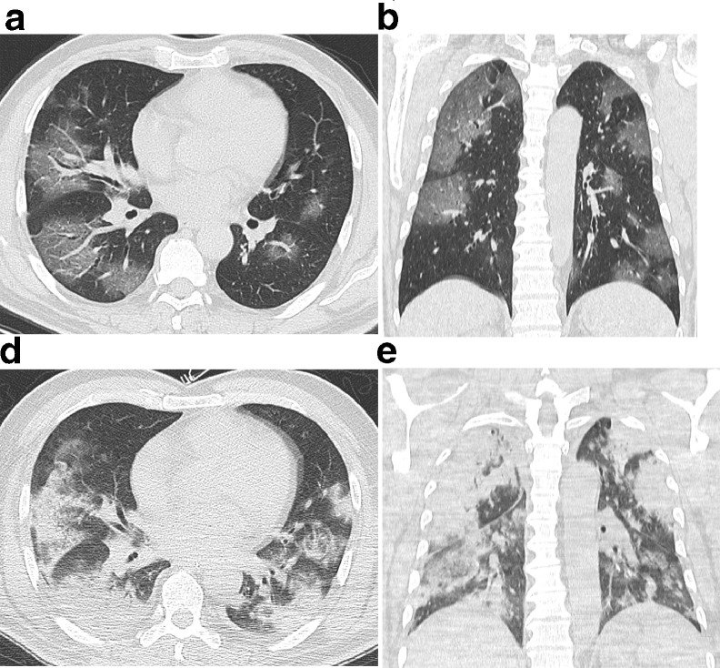
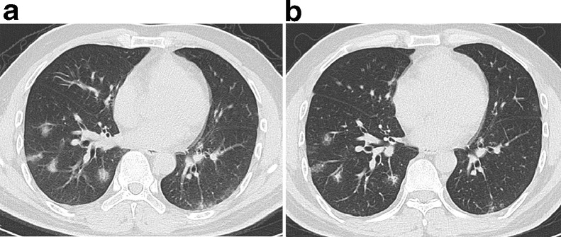
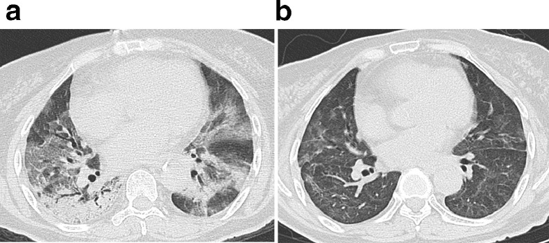
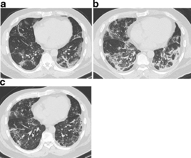
Results: Fourty-seven cases (88.7%) had findings of COVID-19 infection, and the other six (11.3%) were normal. Among the 47 cases, 78.7% involved both lungs, and 93.6% had peripheral infiltrates distributed along the subpleural area. All cases showed GGO, 59.6% of which were round and 40.4% patchy. Other imaging features included "crazy-paving pattern" (89.4%), consolidation (63.8%), and air bronchogram (76.6%). Air bronchograms were observed within GGO (61.7%) and consolidation (70.3%). Neither enlarged mediastinal lymph nodes nor pleural effusion were present. Thirty-three patients (62.3%) were followed an average interval of 6.2 ± 2.9 days. The lesions increased in 75.8% and resorbed in 24.2% of patients.
Conclusion: COVID-19 showed the pulmonary lesions in patients infected with COVID-19 were predominantly distributed peripherally in the subpleural area.
Keywords: Coronavirus; Lung; Multidetector computed tomography; Pneumonia.
Copyright © 2020 The Association of University Radiologists. Published by Elsevier Inc. All rights reserved.
Figures
Comment in
-
COVID-19 Pulmonary Involvement: Is Really an Interstitial Pneumonia?Acad Radiol. 2020 Jun;27(6):900. doi: 10.1016/j.acra.2020.04.010. Epub 2020 Apr 15. Acad Radiol. 2020. PMID: 32312654 Free PMC article. No abstract available.
References
-
- World Health Organization. Available at: https://www.who.int/emergencies/diseases/novel-coronavirus-2019. Accessed January 21, 2020.
-
- International Committee on Taxonomy of Viruses. Available at: https://talk.ictvonline.org/. Retrieved February 11, 2020.
-
- World Health Organization. Available at: https://www.who.int/news-room/detail/12-02-2020-world-experts-and-funder.... Accessed February 12, 2020.
-
- Hansell D.M., Bankier A.A., MacMahon H. Fleischner society: glossary of terms for thoracic imaging. Radiology. 2008;246:697–722. - PubMed
MeSH terms
LinkOut - more resources
Full Text Sources
