. 2020 Apr 15;40(16):3304-3317.
doi: 10.1523/JNEUROSCI.1897-19.2020. Epub 2020 Mar 23.

Interneuron NMDA Receptor Ablation Induces Hippocampus-Prefrontal Cortex Functional Hypoconnectivity after Adolescence in a Mouse Model of Schizophrenia

Affiliations

Interneuron NMDA Receptor Ablation Induces Hippocampus-Prefrontal Cortex Functional Hypoconnectivity after Adolescence in a Mouse Model of Schizophrenia

Rodrigo J Alvarez et al. J Neurosci. .

Abstract

Although the etiology of schizophrenia is still unknown, it is accepted to be a neurodevelopmental disorder that results from the interaction of genetic vulnerabilities and environmental insults. Although schizophrenia's pathophysiology is still unclear, postmortem studies point toward a dysfunction of cortical interneurons as a central element. It has been suggested that alterations in parvalbumin-positive interneurons in schizophrenia are the consequence of a deficient signaling through NMDARs. Animal studies demonstrated that early postnatal ablation of the NMDAR in corticolimbic interneurons induces neurobiochemical, physiological, behavioral, and epidemiological phenotypes related to schizophrenia. Notably, the behavioral abnormalities emerge only after animals complete their maturation during adolescence and are absent if the NMDAR is deleted during adulthood. This suggests that interneuron dysfunction must interact with development to impact on behavior. Here, we assess in vivo how an early NMDAR ablation in corticolimbic interneurons impacts on mPFC and ventral hippocampus functional connectivity before and after adolescence. In juvenile male mice, NMDAR ablation results in several pathophysiological traits, including increased cortical activity and decreased entrainment to local gamma and distal hippocampal theta rhythms. In addition, adult male KO mice showed reduced ventral hippocampus-mPFC-evoked potentials and an augmented low-frequency stimulation LTD of the pathway, suggesting that there is a functional disconnection between both structures in adult KO mice. Our results demonstrate that early genetic abnormalities in interneurons can interact with postnatal development during adolescence, triggering pathophysiological mechanisms related to schizophrenia that exceed those caused by NMDAR interneuron hypofunction alone.SIGNIFICANCE STATEMENT NMDAR hypofunction in cortical interneurons has been linked to schizophrenia pathophysiology. How a dysfunction of GABAergic cortical interneurons interacts with maturation during adolescence has not been clarified yet. Here, we demonstrate in vivo that early postnatal ablation of the NMDAR in corticolimbic interneurons results in an overactive but desynchronized PFC before adolescence. Final postnatal maturation during this stage outspreads the impact of the genetic manipulation toward a functional disconnection of the ventral hippocampal-prefrontal pathway, probably as a consequence of an exacerbated propensity toward hippocampal-evoked depotentiation plasticity. Our results demonstrate a complex interaction between genetic and developmental factors affecting cortical interneurons and PFC function.

Keywords: NMDA receptor; in vivo electrophysiology; medial PFC; mice; parvalbumin interneuron; schizophrenia.


Figures

Figure 1.
Figure 1.
Simultaneously recording of unit activity in mPFC and LFP in cortex and hippocampus allows separation of putative neocortical interneurons and pyramidal cells during different global brain states. A, Left, Safranin O staining shows an example of the electrode track left by a tetrode recording in the mPFC, and schematic of a coronal section shows tetrode positioning in the mPFC. Middle, Schematic of coronal sections showing all electrode tracks in mPFC in juvenile (left) and adult (right) mice. mPFC recordings were performed along mPFC dorsoventral axis. No differences in electrode track positioning were observed across control (Flox, black lines) and KO (red lines) mice. Right, Schematic of coronal sections showing the localization of the concentric bipolar electrodes used to record the vHP LFP (left) and the cortical field potential (right). M1, Motor cortex; cc, corpus callosum; cx, cortex. B, mPFC pyramidal cell and interneuron classification based on their electrophysiological properties. Average waveforms of isolated units classified as interneuron (green) and pyramidal (blue), including temporal parameters used in the classification process (A, half-amplitude duration; B, trough to peak time). The parameters A and B were plotted for each cell, and two clear clusters were formed and used to sort neurons into pyramidal and interneurons in juvenile (top) and adult mice (bottom). C, Top, Representative raster plot of mPFC neurons (pyramidal and interneurons) recorded simultaneously with the cortical (ctx) and hippocampal (vHP) LFP activity. Bottom, Power density analysis performed on the same signal showing slow wave (SW) activity in the cortical LFP and a spontaneous desynchronization that correlates with the emergence of theta oscillations in the vHP.
Figure 2.
Figure 2.
′Increased pyramidal and interneuron firing rate in juvenile and adult NMDAR-KO mice. A, Cumulative frequency distribution of pyramidal (left) and interneuron (right) firing rate of control and NMDAR-KO mice quantified during desynchronization periods. The curves for juvenile and adult mice are shifted to the right in the NMDAR-KO mice. B, Overall increase in PFC pyramidal and interneuron mean firing rate in the NMDAR-KO mice (two-way ANOVAs: genotype factor, pyramidal). *p < 0.0001 versus control, interneurons. *p = 0.01 versus control. Data are mean ± SEM. Number of cells are indicated inside columns from 11 juvenile control, 7 juvenile KO, 6 adult control, and 6 adult KO mice.
Figure 3.
Figure 3.
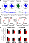
Phase-locking of mPFC neurons to local and distal rhythms is decreased in juvenile and adult NMDAR-KO mice. A, Representative examples of circular phase plots representing the occurrence of spike discharges (radial axis) as a function of LFP oscillation phase (angular axis) for 4 representative mPFC cells. Bin size 20°. Examples include pyramidal cells (blue) and interneurons (green) locked and not phase-locked to hippocampal theta or cortical γ rhythms. Red arrows indicate the resultant vector of the circular phase distribution. Red numbers indicate the radial axes for vector module. Left, First example presents a pyramidal cell phase-locked to hippocampal theta rhythm as evidenced by a nonuniform circular distribution (Rayleigh test, p < 0.001). B, Spike-triggered LFP average for each of the neurons plotted in A. STAs from phase-locked units reproduce the underlying oscillation. C, Cumulative frequency distributions of phase-locking strength to vHP theta and cortical γ oscillations for all phase-locked (Rayleigh, p < 0.05) pyramidal and interneurons from juvenile (Juv) and adult (Ad) control (C) and NMDAR-KO mice (KO). D, Phase-locking strength is represented as the average vector module for all phase-locked neurons (Rayleigh, p < 0.05) for each experimental group. Irrespective of age, neurons from KO mice show a decreased phase-locking to hippocampal theta rhythm (two-way ANOVA, genotype factor pyramidal: *p < 0.0001; interneurons: *p = 0.018). Pyramidal cells from KO mice displayed significant lower locking to cortical γ LFP oscillation at both ages (two-way ANOVA, pyramidal: genotype factor, *p < 0.0001). No changes were observed for interneurons in relation to γ rhythm (two-way ANOVA, genotype factor: p = 0.67). No significant interactions between age and genotype factors were detected by two-way ANOVAs in the statistical analysis. Data are mean ± SEM of n cells indicated in the graph bars from 11 juvenile control, 7 juvenile KO, 6 adult control, and 6 adult KO mice.
Figure 4.
Figure 4.
vHP theta oscillation is not affected in NMDAR-KO mice. A, Power spectral density analysis of LFP signals recorded from vHP in juvenile and adult control and NMDAR-KO mice during desynchronized states expressed as a percentage of the total spectral power. Data are mean ± SEM of n recorded animals. B, Power densities were calculated per band for theta oscillation (3-7 Hz). No statistically significant differences were observed across genotypes. Data are mean ± SEM. Each dot represents 1 individual animal.
Figure 5.
Figure 5.
Only adult NMDAR-KO mice show a reduced mPFC-evoked response to vHP stimulation. A, Schematic of coronal sections showing recording (mPFC; left) and stimulation (vHP; right) electrode positioning for juvenile and adult control and KO mice. B, Stimulation of vHP evoked a short-latency response in mPFC LFP (inset; red line indicates amplitude of the evoked response). mPFC stimulus-response curves evoked by stimulation of vHP show a significant decrease in response amplitude in adult, but not juvenile, KO mice compared with age-matched control mice (two-way repeated-measures ANOVAs). Adult: genotype × intensity interaction, p < 0.001. *p < 0.05 (Newman-Keuls post hoc test vs control at same stimulation intensity). Juvenile: genotype factor, p = 0.49; genotype × intensity interaction, p = 0.62. Each point represents mean ± SEM. Number of mice is indicated. Trend line indicates the best fit sigmoidal curves to population data. C, Maximum evoked mPFC response to vHP stimulation. Adult KO mice show a decreased maximal evoked response compared with age-matched control mice, although no differences were observed between control and KO juvenile mice (two-way ANOVA, genotype × age interaction). *p < 0.05 (Newman-Keuls post hoc test). Error bar indicates mean ± SEM. Each dot represents 1 individual animal.
Figure 6.
Figure 6.
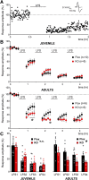
Adult KO mice present a higher susceptibility to vHP-LFS induced LTD. A, Representative experiment illustrating time course of mPFC-evoked response before and after LFS stimulation of vHP. Each dot represents amplitude of the response evoked by a test pulse delivered to vHP. Inset, Traces of mPFC-evoked responses before (black) and after (gray) LFS stimulation. The LFS protocol induces a long-lasting depression of the evoked response. Response amplitudes were normalized to mean baseline amplitude. B, mPFC response amplitude recorded from animals subjected to successive LFS protocols. Juvenile control and KO mice showed similar levels of LTD after application of successive LFS protocols (two-way repeated-measures ANOVA: genotype factor, p = 0.68; genotype × time interaction, p = 0.35). Adult KO mice presented greater levels of induced LTD than control mice (two-way repeated-measures ANOVA: genotype × time interaction, p < 0.01). *p < 0.05 (Newman-Keuls post hoc test). Each point represents mean ± SEM of n animals. C, The mPFC-evoked response is smaller after each of the LFS protocols in adult, but not in juvenile, NMDAR-KO. Statistical analysis combining genotype, age, and LFS repetition was conducted by three-way repeated-measures ANOVA (genotype × age interaction, p = 0.035). *p < 0.05 (Newman-Keuls post hoc test vs adult control). Error bar indicates mean ± SEM. Each dot represents the response measured in 1 individual animal. n.s., not significant.
Figure 7.
Figure 7.
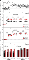
Adult KO mice exhibit normal susceptibility to vHP-HFS induced LTP. A, Representative experiment illustrating time course of mPFC-evoked response before and after HFS stimulation of vHP. Each dot represents amplitude of the response evoked by a test pulse delivered to vHP. Inset, Traces of mPFC-evoked responses before (black) and after (gray) HFS stimulation. The LTP protocol induces a long-lasting potentiation of the evoked response. Response amplitudes were normalized to mean baseline amplitude. B, mPFC response amplitude recorded from animals subjected to successive HFS protocols. A higher susceptibility to HFS in juvenile KO mice results in a significant increase in the evoked response compared with control mice after application of successive HFS pulse trains (two-way repeated-measures ANOVA: genotype × time interaction, p < 0.0001). *p < 0.05 (Newman-Keuls post hoc test vs adult control). Each point represents mean ± SEM of n animals. In contrast, adult control and KO mice showed no significant differences in LTP levels after application of successive HFS protocols (two-way repeated-measures ANOVA: genotype factor, p = 0.79; genotype × time interaction, p = 0.54). C, The mPFC-evoked response is bigger after each of the HFS protocols in juvenile, but not in adult, NMDAR-KO. Statistical analysis combining genotype, age, and LFS repetition was conducted by three-way repeated-measures ANOVA (genotype × age interaction, F(1,87) = 4.80, p = 0.036; observed power: 0.56). *p < 0.05 (Newman-Keuls post hoc vs juvenile control). Error bar indicates mean ± SEM. Each dot represents the response measured in 1 individual animal. n.s., not significant.
Figure 8.
Figure 8.
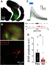
KO mice show decreased amplitude of monosynaptic EPSPs in mPFC pyramidal neurons evoked by optogenetic stimulation of vHP terminals. A, vHP microinjection of AAV2/CamKII-hChR2(H134R)-EYFP results in selective EYFP fluorescence (green) expression in transduced vHP region. Low-magnification photomicrograph shows a representative example of the vHP targeted area. B, Top, Low-power photomicrograph of a representative brain slice (300-μm-thick) used for patch-clamp recordings in mPFC after streptavidin-Cy3 staining for visualization of the neurobiotin-filled recorded neuron. Dense innervation of vHP terminals loaded with ChR2-EYGP (green channel) can be observed together with the neurobiotin-filled pyramidal neuron recorded in layer 3 of mPFC (red channel, boxed area). Bottom, Higher-magnification image of the neuron shown at top. Maximum intensity projection of series of Z stack of confocal images shows a filled cell with typical pyramidal neuron morphology, including apical dendrite and dendritic spines (inset). C, Representative example of a short latency (blue light-evoked) EPSP in an mPFC pyramidal cell during whole-cell current-clamp recording. Gray represents individual responses. Green represents average trace from 30 stimuli. Bottom, Response jitter is consistent with monosynaptic responses in both experimental groups. Each dot represents individual jitter of a recorded neuron from control (Flox: pink dots, n = 8; red dot, mean ± SEM) or mutant mice (KO: gray dots, n = 9; black dot, mean ± SEM). No significant differences between genotypes were observed (Student's t test, p = 0.51). D, EPSPs evoked by optogenetic stimulation of vHP terminals in mPFC is reduced in mutant mice (KO) compared with age-matched controls (Flox) during GABAergic blockade. EPSPs were recorded in whole-cell current-clamp configuration with 50 μm picrotoxin in the bath. *p = 0.023 (Student's t test). Error bar indicates mean ± SEM from 10 cells for flox and 8 for mutant. Each dot represents one recorded neuron (18 neurons from 14 slices from 11 animals).

Similar articles

Cited by

References

    1. Atallah BV, Bruns W, Carandini M, Scanziani M (2012) Parvalbumin-expressing interneurons linearly transform cortical responses to visual stimuli. Neuron 73:159–170. 10.1016/j.neuron.2011.12.013 - DOI - PMC - PubMed
    1. Bähner F, Meyer-Lindenberg A (2017) Hippocampal–prefrontal connectivity as a translational phenotype for schizophrenia. Eur Neuropsycho-pharmacol 27:93–106. 10.1016/j.euroneuro.2016.12.007 - DOI - PubMed
    1. Banks PJ, Burroughs AC, Barker GR, Brown JT, Warburton EC, Bashir ZI (2015) Disruption of hippocampal-prefrontal cortex activity by dopamine D2R-dependent LTD of NMDAR transmission. Proc Natl Acad Sci USA 112:11096–11101. 10.1073/pnas.1512064112 - DOI - PMC - PubMed
    1. Bartho P, Hirase H, Monconduit L, Zugaro M, Harris KB (2004) Characterization of neocortical principal cells and interneurons by network interactions and extracellular features. J Neurophysiol 112:11096–11101. - PubMed
    1. Belforte JE, Zsiros V, Sklar ER, Jiang Z, Yu G, Li Y, Quinlan EM, Nakazawa K (2010) Postnatal NMDA receptor ablation in corticolimbic interneurons confers schizophrenia-like phenotypes. Nat Neurosci 13:76–83. 10.1038/nn.2447 - DOI - PMC - PubMed

Publication types

Substances