. 2020 May 20;88(6):e00913-19.
doi: 10.1128/IAI.00913-19. Print 2020 May 20.

EirA Is a Novel Protein Essential for Intracellular Replication of Coxiella burnetii

Affiliations

EirA Is a Novel Protein Essential for Intracellular Replication of Coxiella burnetii

Miku Kuba et al. Infect Immun. .

Abstract

The zoonotic bacterial pathogen Coxiella burnetii is the causative agent of Q fever, a febrile illness which can cause a serious chronic infection. C. burnetii is a unique intracellular bacterium which replicates within host lysosome-derived vacuoles. The ability of C. burnetii to replicate within this normally hostile compartment is dependent on the activity of the Dot/Icm type 4B secretion system. In a previous study, a transposon mutagenesis screen suggested that the disruption of the gene encoding the novel protein CBU2072 rendered C. burnetii incapable of intracellular replication. This protein, subsequently named EirA (essential for intracellular replication A), is indispensable for intracellular replication and virulence, as demonstrated by infection of human cell lines and in vivo infection of Galleria mellonella The putative N-terminal signal peptide is essential for protein function but is not required for localization of EirA to the bacterial inner membrane compartment and axenic culture supernatant. In the absence of EirA, C. burnetii remains viable but nonreplicative within the host phagolysosome, as coinfection with C. burnetii expressing native EirA rescues the replicative defect in the mutant strain. In addition, while the bacterial ultrastructure appears to be intact, there is an altered metabolic profile shift in the absence of EirA, suggesting that EirA may impact overall metabolism. Most strikingly, in the absence of EirA, Dot/Icm effector translocation was inhibited even when EirA-deficient C. burnetii replicated in the wild type (WT)-supported Coxiella containing vacuoles. EirA may therefore have a novel role in the control of Dot/Icm activity and represent an important new therapeutic target.

Keywords: Coxiella burnetii; bacterial pathogenesis; host-pathogen interactions; type IV secretion system; virulence factor; virulence factors.


Figures

FIG 1
FIG 1
CBU2072 is essential for intracellular replication of C. burnetii. (A and B) Intracellular replication of C. burnetii WT (red), 2072::Tn mutant (blue), 2072::Tn pFLAG-2072 mutant (green), and 2072::Tn pFLAG-207224–165 mutant (purple) in HeLa CCL2 cells (n = 4) (A) and THP1 cells (n = 5) (B). THP1 cell data depict fold changes at 3 days postinfection. Error bars represent standard deviation. *, P < 0.05. P values were determined using an unpaired Student t test. (C) Representative confocal immunofluorescence (IF) images at 3 days postinfection for HeLa CCL2 and THP1 cells. Cells were stained with anti-LAMP1 (green), anti-C. burnetii (red), and DAPI (blue). Scale bar = 10 μm. Arrows indicate individual intracellular C. burnetii cells.
FIG 2
FIG 2
EirA is required for virulence in G. mellonella. Survival of G. mellonella following infection with C. burnetii WT (red), eirA::Tn mutant (blue), eirA::Tn pFLAG-EirA mutant (green), and eirA::Tn pFLAG-EirA24–165 mutant (purple) at 106 GE (A). A PBS control (orange outline) was also included. Results are shown as a representative of two independent biological replicates, each with 12 larvae per condition. (B) Representative confocal IF images at 3 days postinfection of G. mellonella hemocytes. Cells were stained with anti-C. burnetii (red) and DAPI (blue). Scale bar = 10 μm. Arrows indicate individual intracellular C. burnetii.
FIG 3
FIG 3
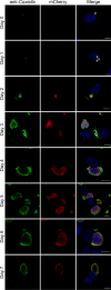
C. burnetii WT cells are able to trans-complement the eirA::Tn mutant during intracellular replication. HeLa CCL2 cells coinfected with C. burnetii WT and the eirA::Tn mutant were fixed and stained every 24 h across a 7-day infection period. Cells were stained with anti-C. burnetii (green) and DAPI (blue), while the eirA::Tn mutant was identified through transposon expression of mCherry (red). Images are representative of three independent biological replicates. Scale bar = 10 μm. Arrows indicate individual intracellular C. burnetii cells.
FIG 4
FIG 4
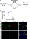
EirA is not required for intracellular viability of C. burnetii. (A) Schematic representation of the experimental procedure, in which HeLa cells were infected with C. burnetii eirA::Tn mutant and incubated for 5 days before being superinfected with either the C. burnetii WT or eirA::Tn mutant. (B) After 3 days, cells were fixed and stained, and total 568-nm fluorescence levels relative to host cell number were measured using Fiji (49) in order to quantify mCherry expressing eirA::Tn replication; n = 6. Error bars represent the standard deviation, and the P value was determined using an unpaired Student t test. (C) Confocal immunofluorescence microscopy images of representative cells stained for all C. burnetii (green), mCherry expressed by the eirA::Tn mutant (red) and DAPI (blue). Scale bar = 10 μm. Arrows indicate individual intracellular C. burnetii cells.
FIG 5
FIG 5
EirA localize to the bacterial cytoplasm and TX-100-soluble membrane fraction. (A to D) Subcellular fractionations of C. burnetii WT (A), eirA::Tn mutant (B), eirA::Tn pFLAG-EirA mutant (C), and eirA::Tn pFLAG-EirA24–165 mutant (D) were performed to observe the subcellular localization of EirA within the bacterial cell. DotB (cyto/cytoplasm), IcmK (TX-100-insoluble/OM/outer membrane), IcmD (TX-100-soluble/IM/inner membrane), and IcmX (periplasm) were used to denote specific subcellular localizations as well as whole-cell lysate (WCL). Blots are representative of three independent biological replicates. *, nonspecific bands.
FIG 6
FIG 6
Absence of EirA leads to an accumulation of some amino acids and significantly lower glycolytic and TCA cycle activities. Shown are combined metabolites detected by GC-MS and LC-MS which were significantly different in abundance between the C. burnetii eirA::Tn mutant and WT strains. Red denotes metabolites which were significantly higher in abundance in the C. burnetii eirA::Tn mutant, and blue denotes metabolites which were significantly lower in abundance in the C. burnetii eirA::Tn mutant. P < 0.05, BH-adjusted unpaired t test. Pale blue denotes metabolites which were not significant. Pale green denotes enzymes which are missing based on genome annotation data in KEGG. Purple denotes putative and defined metabolite transporters. Dotted arrows indicate pathways which have been abbreviated. Metabolite abbreviations are listed in Table S1 in the supplemental material.
FIG 7
FIG 7
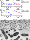
Absence of EirA does not impact susceptibility to cell membrane stressors or effect overall gross morphology in C. burnetii. C. burnetii WT and eirA::Tn, eirA::Tn pFLAG-EirA, and eirA::Tn pFLAG-EirA24–165 mutant strains were exposed to various concentrations of either ampicillin or polymyxin B at 4 days postinoculation, and the impact on bacterial replication was quantified at 24 h posttreatment by genome equivalents (GE) (A) and CFU (B) per milliliter. Fold change depicts the bacterial numbers at 24 h posttreatment relative to the numbers pretreatment. No significant differences in GE and CFU were observed based on an unpaired Student t test. (C) C. burnetii WT, eirA::Tn mutant, and eirA::Tn pFLAG-EirA mutant strains were grown for 6 days in ACCM-2 before being imaged using transmission electron microscopy (TEM). Images are representative of two technical replicates.
FIG 8
FIG 8
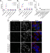
EirA is important for Dot/Icm type IV secretion system effector translocation. Plasmids encoding transcriptionally fused β-lactamase (BlaM) and T4BSS effector MceA were introduced into the C. burnetii WT and eirA::Tn mutant. These strains, along with the C. burnetii WT and eirA::Tn mutant strains, were used to infect HeLa CCL2 cells at an MOI of either 100 (red circles), 150 (blue squares), or 300 (green triangles), or at a coinfection ratio of 1:1, 1:2, or 1:5 WT to eirA::Tn mutant, respectively, where two different strains were used (A and B). At either 48 h (A) or 72 h (B) postinfection, the fluorescent β-lactamase substrate CCF2-AM was added to cells, and cleavage of the substrate was determined by calculating the ratio of fluorescence at 450 nm to 520 nm, relative to uninfected cells; n = 3. (C) Translocation assays for C. burnetii expressing BlaM-0425 were performed at 72 h postinfection with an MOI of 300. Coinfections of the WT and eirA::Tn mutant derivatives were conducted with a ratio of 1:5. (D) HeLa CCL2 cells were infected with C. burnetii strains expressing 3×FLAG-MceA and then fixed and stained at 3 days postinfection. Cells were stained with anti-FLAG (green), MitoTracker (red), and DAPI (blue). Images are representative of three independent biological replicates. Scale bar = 10 μm. Asterisks (*) indicate CCVs.

Similar articles

Cited by

References

    1. Schneeberger PM, Wintenberger C, van der Hoek W, Stahl JP. 2014. Q fever in the Netherlands – 2007–2010: what we learned from the largest outbreak ever. Med Mal Infect 44:339–353. doi:10.1016/j.medmal.2014.02.006. - DOI - PubMed
    1. Graham JG, MacDonald LJ, Hussain SK, Sharma UM, Kurten RC, Voth DE. 2013. Virulent Coxiella burnetii pathotypes productively infect primary human alveolar macrophages. Cell Microbiol 15:1012–1025. doi:10.1111/cmi.12096. - DOI - PMC - PubMed
    1. Raoult D, Marrie T, Mege J. 2005. Natural history and pathophysiology of Q fever. Lancet Infect Dis 5:219–226. doi:10.1016/S1473-3099(05)70052-9. - DOI - PubMed
    1. Hackstadt T, Williams JC. 1981. Biochemical stratagem for obligate parasitism of eukaryotic cells by Coxiella burnetii. Proc Natl Acad Sci U S A 78:3240–3244. doi:10.1073/pnas.78.5.3240. - DOI - PMC - PubMed
    1. Coleman SA, Fischer ER, Howe D, Mead DJ, Heinzen RA. 2004. Temporal analysis of Coxiella burnetii morphological differentiation. J Bacteriol 186:7344–7352. doi:10.1128/JB.186.21.7344-7352.2004. - DOI - PMC - PubMed

Publication types

LinkOut - more resources