. 2020 May 8;295(19):6344-6356.
doi: 10.1074/jbc.RA120.012525. Epub 2020 Mar 23.

Endorepellin evokes an angiostatic stress signaling cascade in endothelial cells

Affiliations

Endorepellin evokes an angiostatic stress signaling cascade in endothelial cells

Aastha Kapoor et al. J Biol Chem. .

Abstract

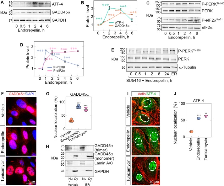
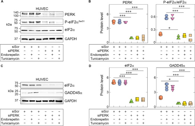
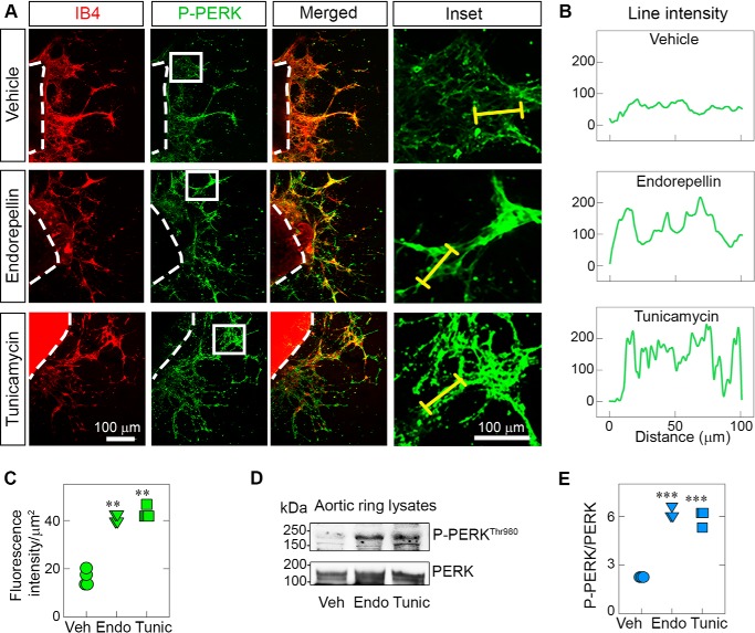
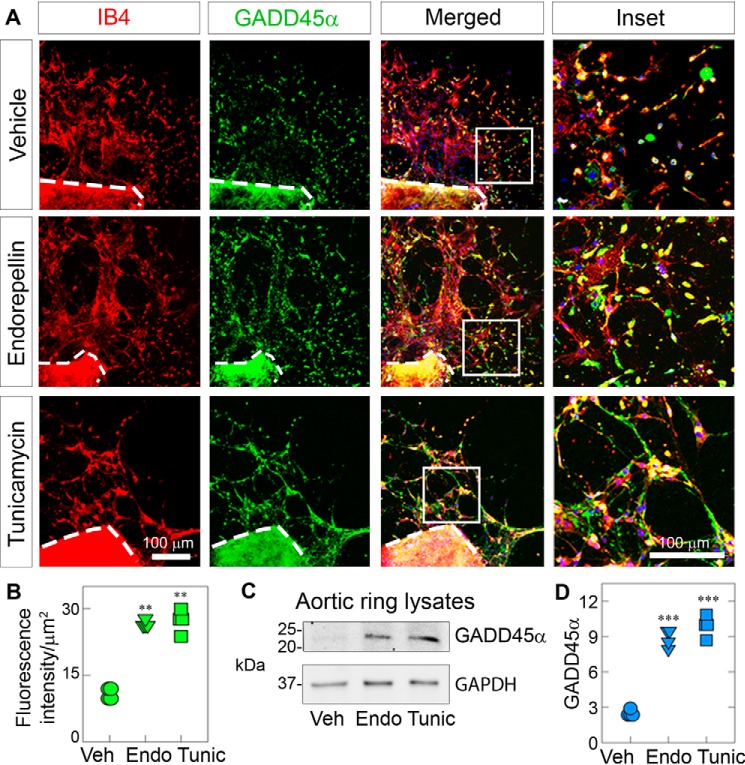
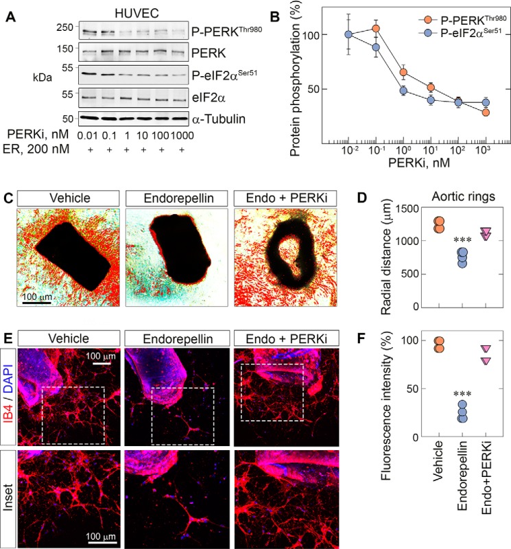
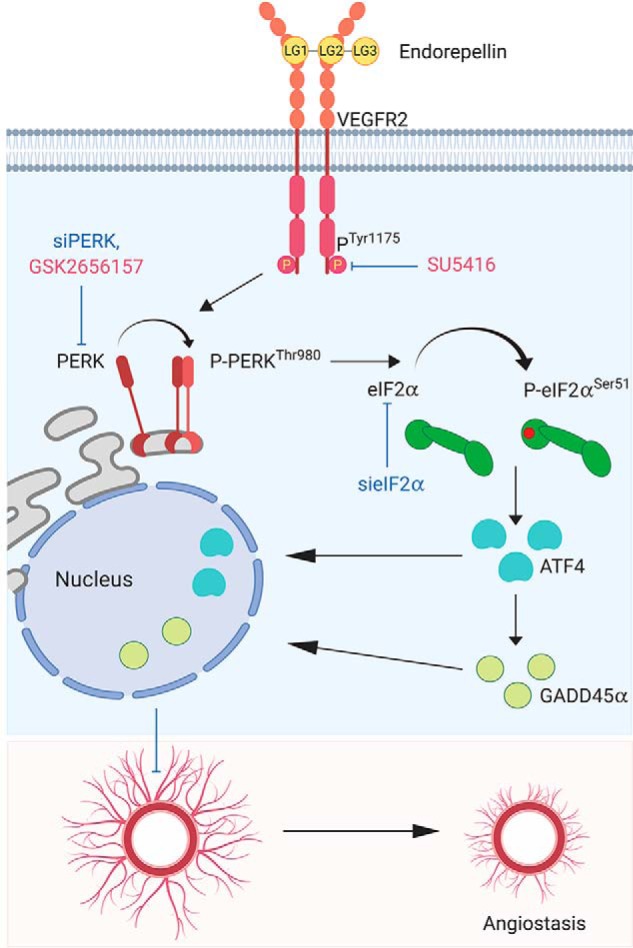
Endorepellin, the C-terminal fragment of the heparan sulfate proteoglycan perlecan, influences various signaling pathways in endothelial cells by binding to VEGFR2. In this study, we discovered that soluble endorepellin activates the canonical stress signaling pathway consisting of PERK, eIF2α, ATF4, and GADD45α. Specifically, endorepellin evoked transient activation of VEGFR2, which, in turn, phosphorylated PERK at Thr980 Subsequently, PERK phosphorylated eIF2α at Ser51, upregulating its downstream effector proteins ATF4 and GADD45α. RNAi-mediated knockdown of PERK or eIF2α abrogated the endorepellin-mediated up-regulation of GADD45α, the ultimate effector protein of this stress signaling cascade. To functionally validate these findings, we utilized an ex vivo model of angiogenesis. Exposure of the aortic rings embedded in 3D fibrillar collagen to recombinant endorepellin for 2-4 h activated PERK and induced GADD45α vis à vis vehicle-treated counterparts. Similar effects were obtained with the established cellular stress inducer tunicamycin. Notably, chronic exposure of aortic rings to endorepellin for 7-9 days markedly suppressed vessel sprouting, an angiostatic effect that was rescued by blocking PERK kinase activity. Our findings unravel a mechanism by which an extracellular matrix protein evokes stress signaling in endothelial cells, which leads to angiostasis.

Keywords: GADD45; PERK; angiogenesis; eIF2α; endothelial cell; proteoglycan; signal transduction; stress response; stress signaling.


Conflict of interest statement

The authors declare that they have no conflicts of interest with the contents of this article. The content is solely the responsibility of the authors and does not necessarily represent the official views of the National Institutes of Health

Figures

Figure 1.
Figure 1.
Endorepellin evokes phosphorylation of PERK/eIF2α and up-regulation of ATF-4/GADD45α downstream of VEGFR2. A, representative immunoblots of time course experiments of HUVECs treated with endorepellin (200 nm, ranging from 0–6 h) and probed for ATF4 and GADD45α. Data were obtained from endorepellin-treated HUVECs from three independent experiments (n = 3) and normalized to GAPDH. B, quantification of ATF4 and GADD45α from A. C, representative immunoblots of time course experiments of HUVECs treated with endorepellin (200 nm, 0–6 h) and probed for p-PERKThr980, total PERK, p-eIF2αSer51, and total eIF2α. Data were acquired from three independent experiments (n = 3) and normalized to their respective total protein levels. D, quantification of p-PERKThr980 and p-eIF2αSer51 from C. One-way ANOVA was performed on all data. E, immunoblots of time course experiments of HUVECs pretreated for 30 min with SU5416 (a VEGR2 kinase inhibitor, 30 μm), followed by addition of endorepellin ranging from 0–24 h. The last lane shows the positive control, i.e. endorepellin alone for 2 h. α-Tubulin served as the loading control. F, immunofluorescence images of HUVECs showing cytoplasmic or nuclear distribution of GADD45α (red) seen with respect to DAPI (blue) after treatment with vehicle (PBS), endorepellin (200 nm), or tunicamycin (10 μg/ml) for 4 h. Nuclear localization of GADD45α is seen as a magenta hue under endorepellin or tunicamycin treatment conditions because of the merging of red and blue tones in the nucleus. G, quantification of cells with nuclear localization of GADD45α from F. An average of 100 cells per treatment (vehicle, endorepellin, or tunicamycin) were analyzed from five independent experiments. H, representative immunoblots of cell fractionation experiments of HUVECs treated for 3 h with or without endorepellin (200 nm). The membranes were probed for lamin A/C to label nuclear fractions (Nu), GAPDH to label cytoplasmic fractions (Cy), and GADD45α. Only the nuclear fraction was enriched in the trimeric form of GADD45α, and these levels were increased by exposure to endorepellin. I, immunofluorescence images of HUVECs treated with vehicle, endorepellin, or tunicamycin for 4 h and probed for filamentous actin (red) and ATF4 (green). The nuclei are outlined by dotted lines. J, quantification of cells with nuclear localization of ATF4 from I. An average of 500 cells per treatment (vehicle, endorepellin, or tunicamycin) were analyzed from three independent experiments (n = 3). All statistical analyses were calculated via one-way ANOVA (***, p < 0.001).
Figure 2.
Figure 2.
Endorepellin-dependent PERK and eIF2α activation is essential for downstream GADD45α up-regulation. A, representative immunoblots of HUVECs pretreated with 100 pm scrambled siRNA (siScr) or siRNA targeting PERK (siPERK), followed by treatment with endorepellin (200 nm) or tunicamycin (10 μg/ml). Lysates show RNAi-mediated knockdown of PERK (siPERK) and subsequent suppression of eIF2α phosphorylation. Treatment conditions are indicated in the bottom panel. B, quantification of PERK and p-eIF2α from A, normalized on GAPDH for PERK or total protein level for P-eIF2α. Data are from three independent biological experiments (n = 3). C, representative immunoblots of HUVECs pretreated with 100 pm scrambled siRNA (siScr) or with siRNA targeting eIF2α (sieIF2α), followed by treatment with endorepellin (200 nm) or tunicamycin (10 μg/ml). Lysates show RNAi-mediated knockdown of eIF2α (sieIF2α) and subsequent suppression of GADD45α levels. D, quantification of eIF2α and GADD45α from C, normalized to GAPDH. Data are from three independent experiments (n = 3). Statistical significance was calculated via two-tailed unpaired Student's t test (**, p < 0.01). All statistical analyses were calculated via one-way ANOVA (***, p < 0.001).
Figure 3.
Figure 3.
Endorepellin activates PERK in sprouts of ex vivo aortic rings. A, representative confocal images of aortic rings following treatment with endorepellin (200 nm) or tunicamycin (10 μg/ml) for 2 h. Endothelial cells in sprouts were visualized with isolectin IB4 (red), a marker for endothelial cells, and counterstained with an antibody against P-PERKThr980 (green). Insets show magnified sprouts stained for p-PERKThr980. B, line intensity profiles of PERK (green) expression in aortic ring sprouts corresponding to the yellow line traced along the enlarged image of sprouts in the inset in A. C, quantification of the fluorescence intensity of PERK in the sprouted area from A; fluorescence intensity data are from a total of 12 vehicle (Veh)-, nine endorepellin (Endo)-, and 10 tunicamycin (Tunic)-treated aortic rings from four independent experiments (n = 4). D, representative immunoblots of three pooled, sprouted blood vessels probed for p-PERKThr980 and total PERK. E, quantification of p-PERKThr980 levels over total PERK from D. Three aortic rings were pooled per condition (vehicle, endorepellin, or tunicamycin) and repeated four times; therefore, four independent experiments were performed (n = 4). The data are derived only from newly formed sprouts, as the aortic rings were removed before solubilization. All statistical analyses were calculated via one-way ANOVA (***, p < 0.001).
Figure 4.
Figure 4.
Endorepellin up-regulates the effector protein GADD45α in aortic rings. A, representative confocal images of aortic rings following treatment with endorepellin (200 nm) or tunicamycin (10 μg/ml) for 4 h. Sprouts were labeled with IB4 (red) and GADD45α (green). Insets, magnified sprouts stained for IB4 (red), GADD45α (green), and DAPI (blue). B, quantification of the fluorescence intensity of GADD45α for the sprouted area from A. Fluorescence intensity data are from a total of 10 vehicle (Veh)-, eight endorepellin (Endo)-, and eight tunicamycin (Tunic)-treated aortic rings from four independent experiments (n = 4). C, representative immunoblots of three pooled, sprouted blood vessels probed for GADD45α. GAPDH was used as a loading control. D, quantification of GADD45α levels normalized to GAPDH from C. Data are from four independent biological experiments (n = 4), with each condition comprised of three pooled sprouts. All statistical analyses were calculated via one-way ANOVA (***, p < 0.001).
Figure 5.
Figure 5.
PERK inhibition blocks endorepellin-dependent stress signaling and angiostasis in aortic rings. A, representative immunoblots of dose-dependent experiments of HUVECs pretreated with the PERKi for 30 min, followed by endorepellin (200 nm) exposure for 1 h and 30 min. Blots were probed for p-PERK and p-eIF2α. α-Tubulin served as the loading control. B, quantification of p-PERK and p-eIF2α from A, with normalization to their respective total protein. Data are from three independent biological experiments (n = 3), plotted as mean ± S.E. One-way ANOVA was performed on the data. C, representative phase-contrast images of aortic rings treated with endorepellin or endorepellin and the PERKi for 7 days in total after sprouting. D, quantification of the radial distance of endorepellin- or endorepellin and PERKi–treated sprouts from C. Phase-contrast data are from 12 vehicle-, 10 endorepellin-, and 10 endorepellin and PERKi–treated rings from four independent experiments (n = 4). E, representative confocal images of aortic rings treated with endorepellin or endorepellin and PERKi–treated rings. Rings were labeled with IB4 (red) and DAPI (blue). F, quantification of fluorescence intensity of the sprouted area from E. Confocal data are from eight vehicle-, eight endorepellin-, and nine endorepellin and PERKi–treated rings from four independent biological experiments (n = 4). All statistical analyses were calculated via one-way ANOVA (***, p < 0.001).
Figure 6.
Figure 6.
Working model elucidating the mechanism of endorepellin-based stress activation via VEGFR2 and subsequent inhibition of angiogenesis. See text for details.

References

    1. Sparro G., Galdenzi G., Eleuteri A. M., Angeletti M., Schroeder W., and Fioretti E. (1997) Isolation and N-terminal sequence of multiple forms of granulins in human urine. Protein Expr. Purif. 10, 169–174 10.1006/prep.1997.0726 - DOI - PubMed
    1. Zoeller J. J., McQuillan A., Whitelock J., Ho S.-Y., and Iozzo R. V. (2008) A central function for perlecan in skeletal muscle and cardiovascular development. J. Cell Biol. 181, 381–394 10.1083/jcb.200708022 - DOI - PMC - PubMed
    1. Costell M., Gustafsson E., Aszódi A., Mörgelin M., Bloch W., Hunziker E., Addicks K., Timpl R., and Fässler R. (1999) Perlecan maintains the integrity of cartilage and some basement membranes. J. Cell Biol. 147, 1109–1122 10.1083/jcb.147.5.1109 - DOI - PMC - PubMed
    1. Cohen I. R., Grässel S., Murdoch A. D., and Iozzo R. V. (1993) Structural characterization of the complete human perlecan gene and its promoter. Proc. Natl. Acad. Sci. U.S.A. 90, 10404–10408 10.1073/pnas.90.21.10404 - DOI - PMC - PubMed
    1. Handler M., Yurchenco P. D., and Iozzo R. V. (1997) Developmental expression of perlecan during murine embryogenesis. Dev. Dyn. 210, 130–145 10.1002/(SICI)1097-0177(199710)210:2<130::AID-AJA6>3.0.CO;2-H - DOI - PubMed

Publication types

MeSH terms

LinkOut - more resources