. 2020 May 11;202(11):e00064-20.
doi: 10.1128/JB.00064-20. Print 2020 May 11.

Fis Contributes to Resistance of Pseudomonas aeruginosa to Ciprofloxacin by Regulating Pyocin Synthesis

Affiliations

Fis Contributes to Resistance of Pseudomonas aeruginosa to Ciprofloxacin by Regulating Pyocin Synthesis

Yuqing Long et al. J Bacteriol. .

Abstract

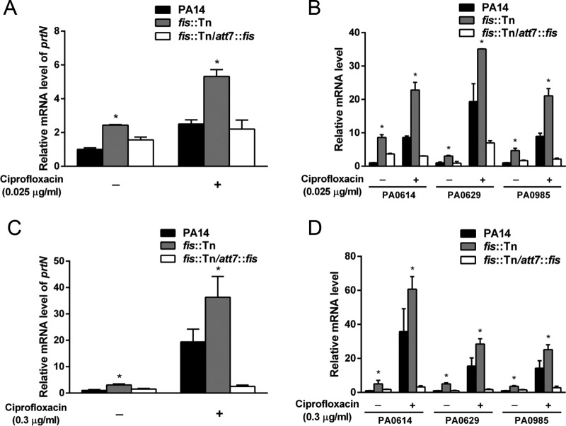
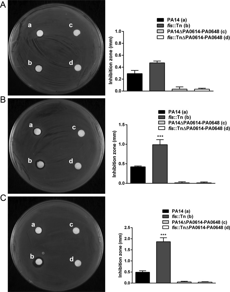
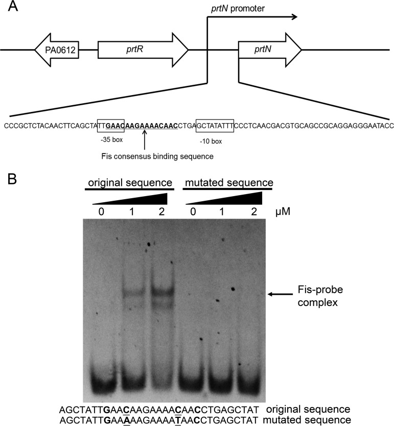
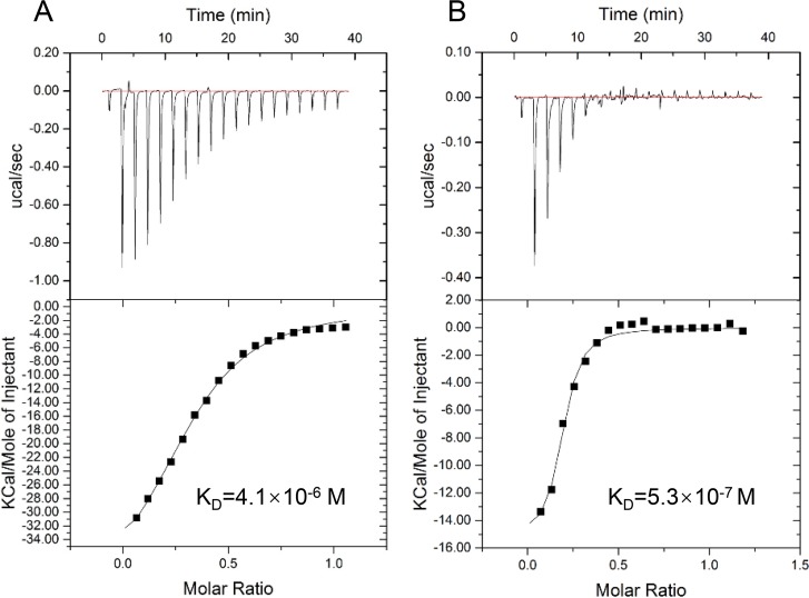
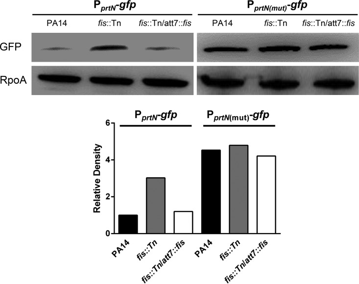
Factor for inversion stimulation (Fis) is a versatile DNA binding protein that plays an important role in coordinating bacterial global gene expression in response to growth phases and environmental stresses. Previously, we demonstrated that Fis regulates the type III secretion system (T3SS) in Pseudomonas aeruginosa In this study, we explored the role of Fis in the antibiotic resistance of P. aeruginosa and found that mutation of the fis gene increases the bacterial susceptibility to ciprofloxacin. We further demonstrated that genes related to pyocin biosynthesis are upregulated in the fis mutant. The pyocins are produced in response to genotoxic agents, including ciprofloxacin, and the release of pyocins results in lysis of the producer cell. Thus, pyocin biosynthesis genes sensitize P. aeruginosa to ciprofloxacin. We found that PrtN, the positive regulator of the pyocin biosynthesis genes, is upregulated in the fis mutant. Genetic experiments and electrophoretic mobility shift assays revealed that Fis directly binds to the promoter region of prtN and represses its expression. Therefore, our results revealed novel Fis-mediated regulation on pyocin production and bacterial resistance to ciprofloxacin in P. aeruginosaIMPORTANCEPseudomonas aeruginosa is an important opportunistic pathogenic bacterium that causes various acute and chronic infections in human, especially in patients with compromised immunity, cystic fibrosis (CF), and/or severe burn wounds. About 60% of cystic fibrosis patients have a chronic respiratory infection caused by P. aeruginosa The bacterium is intrinsically highly resistant to antibiotics, which greatly increases difficulties in clinical treatment. Therefore, it is critical to understand the mechanisms and the regulatory pathways that are involved in antibiotic resistance. In this study, we elucidated a novel regulatory pathway that controls the bacterial resistance to fluoroquinolone antibiotics, which enhances our understanding of how P. aeruginosa responds to ciprofloxacin.

Keywords: Fis; Pseudomonas aeruginosa; pyocin; resistance.


Figures

FIG 1
FIG 1
Expression levels of pyocin-related genes. The overnight bacterial cultures were diluted 100-fold into fresh LB medium with or without 0.025 μg/ml ciprofloxacin and grown to an optical density at 600 nm (OD600) of 1.0 at 37°C. For the treatment with 0.3 μg/ml ciprofloxacin, the bacteria were grown in LB to an OD600 of 1.0, followed by incubation with the antibiotic for 30 min at 37°C. Total RNA was isolated from the bacteria. The relative mRNA levels of prtN (A, C) and PA0614, PA0629, and PA0985 (B, D) were determined by real-time PCR. rpsL was used as the internal control. The results shown represent data from three independent experiments with similar results. Error bars represent standard deviations. *, P < 0.05 compared by Student's t test to PA14 or the complemented strain.
FIG 2
FIG 2
Production levels of pyocins. Indicated strains were grown in LB (A) or treated with 0.025 μg/ml (B) and 0.3 μg/ml (C) ciprofloxacin. The filtered and concentrated bacterial supernatant was spotted on a filter paper that was laid on a lawn of the indicator strain PAK. The size of the inhibition zone was calculated by subtracting the diameter of the filter paper from the diameter of the inhibition circle. The results shown represent data from three independent experiments with similar results. Error bars represent standard deviations. ***, P < 0.001 compared by Student's t test to PA14 or the complemented strain.
FIG 3
FIG 3
Fis directly binds to the prtN promoter region. (A) Diagram of the genetic organization and putative promoter regions of prtN. The arrow of each open reading frame represents the transcriptional direction. The −10 and −35 regions of the prtN promoter are shown in boxes. The potential Fis binding sequence is underlined. (B) Sequences of the probes used in the electrophoretic mobility shift assay (EMSA) and the consensus Fis binding sequence are shown. Underlined letters represent mutated nucleotides. Purified Fis protein was incubated with the fragment with original or mutated sequence for 30 min at 25°C. The position of the Fis-probe complex is indicated.
FIG 4
FIG 4
Binding affinities of Fis and PrtR to the prtN promoter region. The binding affinities were measured by isothermal titration microcalorimetry. Shown is isothermal titration microcalorimetry of Fis at 37 μM (A) or of PrtR at 33 μM (B) binding to 0.2 mM DNA probe at 25°C. KD, binding affinity.
FIG 5
FIG 5
Expression levels of the gfp transcriptional fusions. Protein levels of the gfp gene driven by the original or mutated prtN promoter. PA14, the fis::Tn mutant, and the complemented strain carrying PprtN-gfp or PprtN(mut)-gfp were grown to an OD600 of 1.0. The levels of GFP were determined by Western blotting. RpoA was used as the loading control. The band intensities were quantified with ImageJ software and normalized to the corresponding RpoA band intensities.

Similar articles

Cited by

References

    1. Govan JR, Deretic V. 1996. Microbial pathogenesis in cystic fibrosis: mucoid Pseudomonas aeruginosa and Burkholderia cepacia. Microbiol Rev 60:539–574. doi:10.1128/MMBR.60.3.539-574.1996. - DOI - PMC - PubMed
    1. Stover CK, Pham XQ, Erwin AL, Mizoguchi SD, Warrener P, Hickey MJ, Brinkman FS, Hufnagle WO, Kowalik DJ, Lagrou M, Garber RL, Goltry L, Tolentino E, Westbrock-Wadman S, Yuan Y, Brody LL, Coulter SN, Folger KR, Kas A, Larbig K, Lim R, Smith K, Spencer D, Wong GK, Wu Z, Paulsen IT, Reizer J, Saier MH, Hancock RE, Lory S, Olson MV. 2000. Complete genome sequence of Pseudomonas aeruginosa PAO1, an opportunistic pathogen. Nature 406:959–964. doi:10.1038/35023079. - DOI - PubMed
    1. Pukatzki S, Kessin RH, Mekalanos JJ. 2002. The human pathogen Pseudomonas aeruginosa utilizes conserved virulence pathways to infect the social amoeba Dictyostelium discoideum. Proc Natl Acad Sci U S A 99:3159–3164. doi:10.1073/pnas.052704399. - DOI - PMC - PubMed
    1. Moradali MF, Ghods S, Rehm BH. 2017. Pseudomonas aeruginosa lifestyle: a paradigm for adaptation, survival, and persistence. Front Cell Infect Microbiol 7:39. doi:10.3389/fcimb.2017.00039. - DOI - PMC - PubMed
    1. Michel-Briand Y, Baysse C. 2002. The pyocins of Pseudomonas aeruginosa. Biochimie 84:499–510. doi:10.1016/s0300-9084(02)01422-0. - DOI - PubMed

Publication types

MeSH terms