. 2020 Mar 19:20:87.
doi: 10.1186/s12935-020-1158-6. eCollection 2020.

MYC-regulated lncRNA NEAT1 promotes B cell proliferation and lymphomagenesis via the miR-34b-5p-GLI1 pathway in diffuse large B-cell lymphoma

Affiliations

MYC-regulated lncRNA NEAT1 promotes B cell proliferation and lymphomagenesis via the miR-34b-5p-GLI1 pathway in diffuse large B-cell lymphoma

Chong-Sheng Qian et al. Cancer Cell Int. .

Abstract

Background: LncRNA NEAT1 has been identified as a tumour driver in many human cancers. However, the underlying mechanism of lncRNA NEAT1 in diffuse large B-cell lymphoma (DLBCL) progression is unclear.

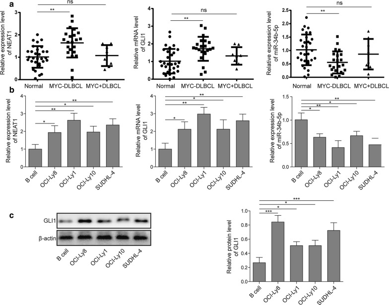
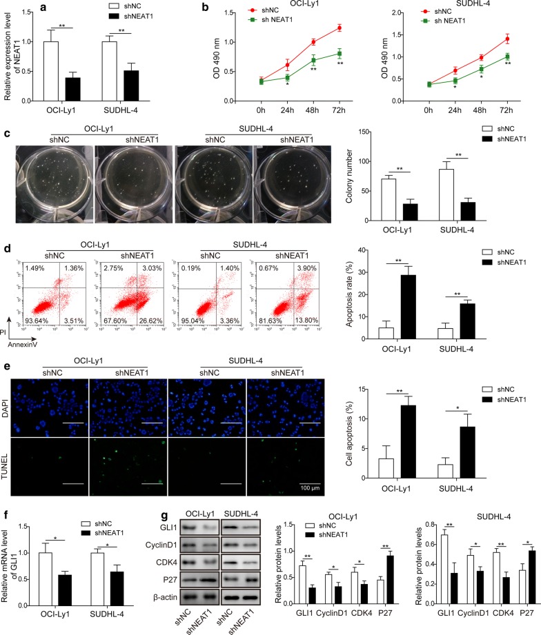
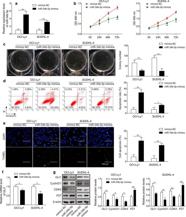
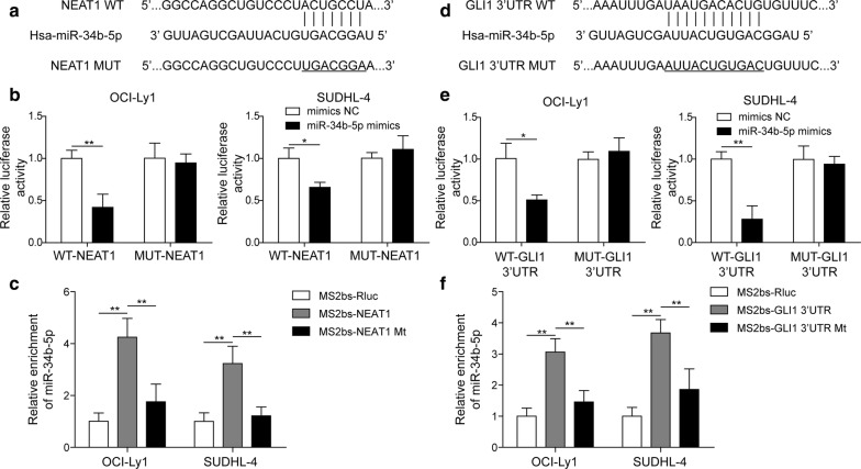
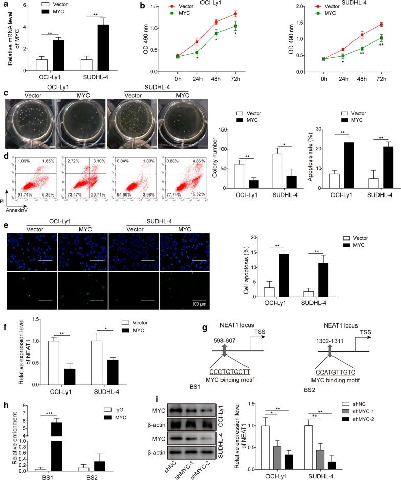
Methods: The expression levels of NEAT1, GLI1 and miR-34b-5p were detected by RT-qPCR and Western blotting in DLBCL tissues and cell lines. MTT and colony formation assays were performed to examine cell proliferation, while annexin-V staining and TUNEL assays were performed to measure cell apoptosis. The effect of NEAT1, GLI1 and miR-34b-5p on cell cycle-associated proteins was evaluated by Western blotting. Dual-luciferase reporter and RNA immunoprecipitation (RIP) assays were employed to investigate the interaction between NEAT1 and miR-34b-5p or GLI1 and miR-34b-5p. Moreover, chromatin immunoprecipitation (ChIP) was performed to demonstrate the interaction between MYC and NEAT1.

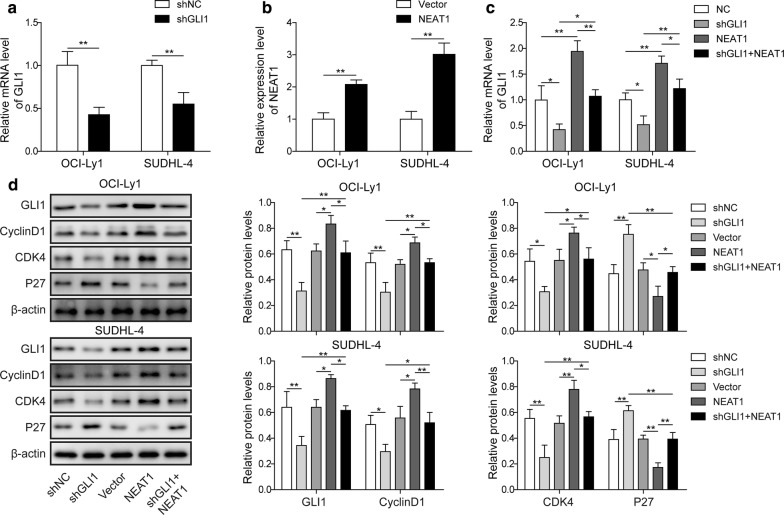
Results: NEAT1 and GLI1 were upregulated while miR-34b-5p was downregulated in DLBCL tissues and cell lines compared to normal controls. Knockdown of NEAT1 or overexpression of miR-34b-5p inhibited cell proliferation but promoted cell apoptosis. Overexpression of NEAT1 reversed GLI1-knockdown induced attenuation of cell proliferation. In other words, NEAT1 acted as a competing endogenous RNA (ceRNA), regulating the miR-34b-5p-GLI1 axis, further affecting the proliferation of DLBCL. Moreover, MYC modulated NEAT1 transcription by directly binding to the NEAT1 promoter.

Conclusion: We revealed that MYC-regulated NEAT1 promoted DLBCL proliferation via the miR-34b-5p-GLI1 pathway, which could provide a novel therapeutic target for DLBCL.

Keywords: Cell proliferation; Diffuse large B-cell lymphoma; GLI1; LncRNA NEAT1; MYC.


Conflict of interest statement

Competing interestsThe authors declare that they have no conflict of interests.

Figures

Fig. 1
Fig. 1
LncRNAs NEAT1 and GLI1 were upregulated while miR-34b-5p was downregulated in DLBCL tissues and cell lines. a The levels of NEAT1, GLI1 and miR-34b-5p were detected by RT-qPCR in tissues from DLBCL patients with MYC rearrangement (n = 8), patients without MYC rearrangement (n = 22) or healthy donors (n = 30). **P < 0.01 vs. normal, T-test. b The levels of NEAT1, GLI1 and miR-34b-5p were measured by RT-qPCR in the DLBCL cell lines OCI-Ly1, OCI-Ly8, OCI-Ly10, SUDHL-4 and in normal B cells. *P < 0.05 and **P < 0.01 vs. normal B cells, T-test. c Immunoblotting and quantification of GLI1 (and β-actin as loading control) in DLBCL cell lines OCI-Ly1, OCI-Ly8, OCI-Ly10, SUDHL-4 and in normal B cells. *P < 0.05 and ***P < 0.001 vs. normal B cells, T-test
Fig. 2
Fig. 2
Knockdown of lncRNA NEAT1 suppressed cell proliferation and facilitated cell apoptosis in DLBCL. a RT-qPCR for NEAT1 expression in NEAT1 knockdown cells (shNEAT1) or negative control cells (shNC). **P < 0.01, ns = not significant vs. shNC, T-test. b Cell viability was measured in NEAT1 knockdown cells (shNEAT1) or negative control cells (shNC) with MTT assay. *P < 0.05, **P < 0.01 vs. shNC, T-test. c Colony formation assays were performed in NEAT1 knockdown cells (shNEAT1) or negative control cells (shNC). A representative image of the colony formation assay in OCI-Ly1 and SUDHL-4 cells is shown (left), and the total number of colonies per plate was counted (right). **P < 0.01 vs. shNC, T-test. d Flow cytometry analysis of annexin-V/PI staining of apoptotic cells following NEAT1 knockdown in OCI-Ly1 and SUDHL-4 cell lines. A representative image of FACS staining in OCI-Ly1 and SUDHL-4 cells is shown on the left, and the statistical data are shown on the right. **P < 0.01 vs. shNC, T-test. e TUNEL staining for OCI-Ly1 and SUDHL-4 shNEAT1 cell lines and their shNC lines. Representative images of TUNEL staining (left) and the statistical data (right) are shown. *P < 0.05, **P < 0.01 vs. shNC, T-test. f The mRNA level of GLI1 was assessed in OCI-Ly1 and SUDHL-4 shNEAT1 cell lines and their shNC lines by RT-qPCR. *P < 0.05 vs. shNC, T-test. g Immunoblotting and quantification of GLI1, cyclin D1, CDK4 and p27 (and β-actin as loading control) in OCI-Ly1 and SUDHL-4 shNEAT1 cell lines and their shNC lines. *P < 0.05, **P < 0.01 vs. shNC, T-test
Fig. 3
Fig. 3
Overexpression of miR-34b-5p attenuated cell proliferation and accelerated cell apoptosis in DLBCL in vitro. a The miR-34b-5p levels were measured by RT-qPCR after miR-34b-5p mimic or mimic NC transfection into OCI-Ly1 and SUDHL-4 cell lines. **P < 0.01 vs. mimic NC, T-test. b Cell viability was detected by MTT assay in OCI-Ly1 and SUDHL-4 cell lines transfected with miR-34b-5p mimic or mimic NC. *P < 0.05, **P < 0.01 vs. mimic NC, T-test. c Colony formation assays were performed in OCI-Ly1 and SUDHL-4 cell lines transfected with miR-34b-5p mimic or mimic NC. A representative image (left) is shown, and the total number of colonies per plate were counted (right). **P < 0.01 vs. mimic NC, T-test. d Flow cytometry analysis of apoptotic cells by annexin-V/PI staining following miR-34b-5p mimic or mimic NC in OCI-Ly1 and SUDHL-4 cell lines. A representative image of FACS staining is shown on the left, and the statistical data are shown on the right. *P < 0.05 and **P < 0.01 vs. mimic NC, T-test. e TUNEL staining in miR-34b-5p mimic or mimic NC. A representative image of TUNEL staining (left) and the statistical data (right) are shown. **P < 0.01 vs. mimic NC, T-test. f The mRNA level of GLI1 was assessed by RT-qPCR in OCI-Ly1 and SUDHL-4 lines transfected with miR-34b-5p mimic or mimic NC. *P < 0.05 vs. mimic NC, T-test. g Immunoblotting and quantification of GLI1, Cyclin D1, CDK4, p27 (and β-actin as loading control) in OCI-Ly1 and SUDHL-4 cells transfected with miR-34b-5p mimic or mimic NC. *P < 0.05, **P < 0.01, vs. mimic NC. T-test
Fig. 4
Fig. 4
Overexpression of lncRNA NEAT1 reverses the suppression of cell proliferation by knockdown of GLI1. a RT-qPCR to determine GLI1 expression in GLI1 knockdown cells (shGLI1) or negative control cells (shNC). **P < 0.01 vs. shNC, T-test. b RT-qPCR to determine NEAT1 expression in NEAT1-overexpressing cells (NEAT1) or negative control cells (Vector). **P < 0.01 vs. Vector, T-test. c RT-qPCR for GLI1 expression in GLI1 knockdown cells (shGLI1), NEAT1 overexpression cells (NEAT1), GLI1 knockdown plus NEAT1 overexpression cells (shGLI1 + NEAT1) and negative control cells (NC). *P < 0.05, **P < 0.01, T-test. d Immunoblotting for GLI1, Cyclin D1, CDK4 and p27 (and β-actin as loading control) in the cell lines described in c. The quantification of each protein is shown. *P < 0.05, **P < 0.01, T-test
Fig. 5
Fig. 5
LncRNA NEAT1 acted as ceRNA, sponging miR-34b-5p to regulate GLI1 expression. a Diagram showing that StarBase predicts the interaction between lncRNA NEAT1 and miR-34b-5p. b The effect of wt-NEAT1 or mut-NEAT1 on miR-34b-5p expression detected by dual-luciferase reporter assay. *P < 0.05, **P < 0.01, vs. Control, T-test. c Interaction between wt-NEAT1 or mut-NEAT1 and miR-34b-5p was detected by RIP. **P < 0.01, vs. MS2bs-Rluc or MS2bs-NEAT1 Mt, T-test. d TargetScan predicts the binding region between miR-34b-5p and the GLI1 3′-UTR. e Dual-luciferase reporter assay was performed to analyse the influence of miR-34b-5p on wt-GLI1 3′-UTR or mut-GLI1 3′-UTR. *P < 0.05, **P < 0.01, vs. Control, T-test. f Interaction between miR-34b-5p and GLI1 3′-UTR was detected by RIP. **P < 0.01, vs. MS2bs-Rluc or mut-GLI1 3′-UTR Mt, T-test
Fig. 6
Fig. 6
MYC modulated DLBCL proliferation through regulating NEAT1 transcription by binding to the promoter. a RT-qPCR to determine MYC expression in MYC overexpression cells (MYC) or negative control cells (Vector). **P < 0.01, vs. Vector, T-test. b MTT assay to determine cell proliferation in MYC-overexpressing cells (MYC) or negative control cells (Vector). *P < 0.05, **P < 0.01, vs. Vector, T-test. c Colony formation assay was performed in MYC-overexpressing cells (MYC) or negative control cells (Vector). A representative image of the colony formation assay in OCI-Ly1 and SUDHL-4 cells is shown (left), and the total number of colonies per plate was counted (right). *P < 0.05, **P < 0.01, vs. Vector, T-test. d Flow cytometry analysis of apoptotic cells by annexin-V/PI staining following MYC overexpression in OCI-Ly1 and SUDHL-4 cell lines. A representative image of FACS staining is shown on the left, and the statistical data are shown on the right. **P < 0.01 vs. Vector, T-test. e TUNEL staining in MYC-overexpressing cells. A representative image of TUNEL staining (left) and statistical data (right) are shown. **P < 0.01 vs. Vector, T-test. f RT-qPCR to determine NEAT1 expression in MYC-overexpressing cells (MYC) or negative control cells (Vector). *P < 0.05, **P < 0.01, vs. Vector, T-test. g Known or potential binding sites of MYC on the promoter of NEAT1 were examined by JASPAR. h CHIP assay was performed to detect the interaction of MYC on BS1 or BS2. ***P < 0.001, vs. IgG, T-test. i Western blot to determine MYC levels (left panel) and ChIP assay to determine enrichment of MYC on the promoter of NEAT1 (right panel) were performed in MYC knockdown cell lines (shMYC-1, shMYC-2) and negative control cell lines (shNC); *P < 0.05, **P < 0.01, vs. shNC, T-test

References

    1. Morin RD, Mendez-Lago M, Mungall AJ, Goya R, Mungall KL, Corbett RD, et al. Frequent mutation of histone-modifying genes in non-Hodgkin lymphoma. Nature. 2011;476(7360):298–303. doi: 10.1038/nature10351. - DOI - PMC - PubMed
    1. Sehn LH, Gascoyne RD. Diffuse large B-cell lymphoma: optimizing outcome in the context of clinical and biologic heterogeneity. Blood. 2015;125(1):22–32. doi: 10.1182/blood-2014-05-577189. - DOI - PubMed
    1. Winkle M, Kluiver JL, Diepstra A, van den Berg A. Emerging roles for long noncoding RNAs in B-cell development and malignancy. Crit Rev Oncol Hematol. 2017;120:77–85. doi: 10.1016/j.critrevonc.2017.08.011. - DOI - PubMed
    1. Yong W, Yu D, Jun Z, Yachen D, Weiwei W, Midie X, et al. Long noncoding RNA NEAT1, regulated by LIN28B, promotes cell proliferation and migration through sponging miR-506 in high-grade serous ovarian cancer. Cell Death Dis. 2018;9(9):861. doi: 10.1038/s41419-018-0908-z. - DOI - PMC - PubMed
    1. Ji Y, Wang M, Li X, Cui F. The long noncoding RNA NEAT1 targets miR-34a-5p and drives nasopharyngeal carcinoma progression via Wnt/β-catenin signaling. Yonsei Med J. 2019;60(4):336–345. doi: 10.3349/ymj.2019.60.4.336. - DOI - PMC - PubMed

LinkOut - more resources